Advances in Life Sciences

p-ISSN: 2163-1387    e-ISSN: 2163-1395

2012;  2(6): 156-169

doi:10.5923/j.als.20120206.03

The Nature of Mildly Deleterious Mutations Eliminated upon Sexual Reproduction in Meiosis

Chubykin V. L.

Vavilov Institute of General Genetics, Russian Academy of Sciences; Gubkina 3, Moscow, 119991, Russia

Correspondence to: Chubykin V. L., Vavilov Institute of General Genetics, Russian Academy of Sciences; Gubkina 3, Moscow, 119991, Russia.

Email:

Copyright © 2012 Scientific & Academic Publishing. All Rights Reserved.

Abstract

Mildly deleterious mutations (MDMs) with incomplete dominance, which decrease the viability of the progeny, apparently play a significant role in the evolution of sexual reproduction. In particular, they are thought to be eliminated in meiosis. The nature of MDM remains unclear. By studying the accumulated MDMs in Drosophila strains carrying various meiotic mutations (c(3)G17, mei-P22, mei-W68, mei-41, mei-218), we found that impair the formation of DNA breaks is more effective in accumulation of MDMs. The relationship between survival and generation number upon MDMs accumulation suggests that MDMs interaction corresponds to their synergistic epistasis. The viability in progeny after meiosis in heterozygotes with chromosome with accumulated MDMs and normal chromosome, and in heterozygotes with independently accumulated MDMs chromosomes was shown to be restored. Our results support the hypothesis that MDMs have epigenetic nature. It is proposed that: during the life cycle “mutant” variants of the formation of structural and functional loop domains appear in the chromosomes; these variants are normally corrected in meiosis; an abnormal loop alters the activity of many genes (~17), increasing (+) or decreasing (–) it. The hybrids with chromosomes carrying independently accumulated MDMs partially restore viability due to complementary interaction of + and – genes.

Keywords: Sex Evolution, Mildly (Slightly) Deleterious Mutations, Meiosis, Chromosome Loop Domains, Epigenetic Mutations, Drosophila Melanogaster

Cite this paper: Chubykin V. L., The Nature of Mildly Deleterious Mutations Eliminated upon Sexual Reproduction in Meiosis, Advances in Life Sciences, Vol. 2 No. 6, 2012, pp. 156-169. doi: 10.5923/j.als.20120206.03.

1. Introduction

Diploidy, multicellular organization, the increase in the genome size and the structural complexity of organisms are associated with the development of sexual reproduction. The evolutionary advantage of sexual reproduction (which is in fact less effective and energetically less beneficial) over asexual one is not quite understood[1],[2]. More than 20 hypotheses have been advanced to explain this phenomenon[3]. In the evolution of sex, mildly deleterious mutations (MDMs), the effects of which are intermediate between neutral and deleterious, are thought to play a significant role. MDMs in homozygous state result in mortality of only part of the progeny. Moreover, they are semidominant and manifest in heterozygotes and these properties of MDMs provide their inheritance and maintenance in populations[1-5]. A high rate of appearance of new MDMs makes evolution with accumulation of advantageous mutations impossible[1]. Herman Muller was the first to theoretically demonstrate (later it was documented experimentally) that in asexual organisms, the gene pool is slowly but consistently degraded owing to the accumulation of MDMs (“Muller’s ratchet”)[4],[5]. The MDM number in such organisms increases with decreasing population size and increasing complexity of the genome (increasing number of genes). According to Muller, asexual populations, in spite of the mutation pressure, exist because of the simplicity of their organization (small genome size), extremely large population sizes, and strong stabilizing selection, rapidly eliminating MDM carriers, which are replaced by mutation-free clones. In this connection, an alternative explanation is that MDMs are eliminated in sexual reproduction[6],[7]. The mechanism of the elimination and its association with meiosis are unclear. Hypotheses highlighting the role of recombination (generation of MDM-free recombinant forms) in effective elimination of MDMs and, consequently, in reducing the mutational load of sexual populations[3],[5], hold only on unlikely condition of constant environmental changes, when in each generation new genotypes with high fitness would be required[1]. Indeed, the axiom on the evolutionary role of recombination has a serious defect, which lies in recombination itself. The high recombination rate, which is actually observed in nature, would destroy a beneficial gene combination not later than in the following generations. A simple reshuffling of not eliminated MDMs would have no effect on the progeny viability at the population level, which is the same proportion of the progeny as before recombination.
In papers devoted to the study of MDMs, less discussed issue is their nature. Is MDM the "classical" mutations with changes in the nucleotide sequences of structural genes, or there are changes in the regulation of their activity? It is unknown. Incomplete dominance is used for classification of regulatory mutations and is associated with changes in the activity of structural genes[8]. Based on this MDM property, Mukai[9] suggested that MDMs are located in noncoding regions. Another suggestion connects reduce the viability of offspring with numerous insertions of mobile elements[10], which affect gene activity. Finally, it was concluded about the absence of MDMs upon mutagenic exposure[11].
The method of MDM accumulation in Drosophila, consisting in disturbance of main meiotic processes in an individual chromosome pair, confirms the role of meiosis in their elimination. A circuit method of MDM registration and accumulation in Drosophila was first proposed by Muller in 1928[12]. This method involves suppression of meiotic pairing and recombination in the examined chromosomes in many generations. With this aim, only heterozygotes with the corresponding chromosomes carrying multiple inversions and transpositions (balancers and crossover suppressors) were employed for reproduction. Chromosomes with multiple rearrangements are usually lethal in homozygous state. The main sign of the MDM manifestation and accumulation in Drosophila is mortality of organism from embryo to eclosion of imago from pupae[13]. In adult flies, MDMs affect individual adaptation to the environment. In such experiments, a reduction in viability and partial mortality of the progeny increases in 20[14], 40[15], 250[16] generations. These MDMs appear practically in each individual in the progeny at a rate an order of magnitude higher than that of recessive lethals and manifest in heterozygotes with the coefficient of dominance 0.2-0.5[15],[17].
Our approach was to study the progeny viability in the strains of Drosophila carrying recessive meiotic mutations (mei-mutations) maintained using balancer chromosomes or transmitted from father to son. In this case, selection for reproduction of only heterozygotes with balancers is not required for MDM accumulation. Mei-mutations in homozygote do not affect viability of their carriers. They only disturb meiosis, thus affecting fertility (due to the formation of abnormal gametes) and promote MDM preservation and accumulation in the progeny of small laboratory populations. The absence of recombination in male meiosis upon only paternal X-chromosome inheritance also promotes MDM accumulation.
The purpose of this paper is to clarify the nature and meсhanism of MDM elimination at meiosis.

2. Materials and Methods

2.1. Fly strains

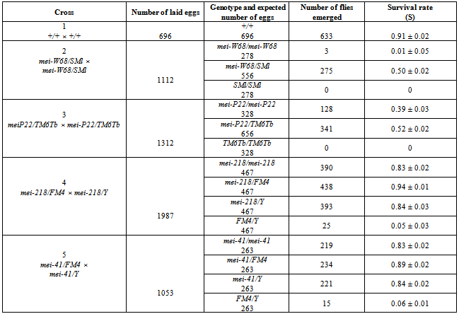
Flies were reared at 25°C on standard medium. We used the following strains of Drosophila melanogaster (the abbreviated designation is given):
(1) st[1] c(3)G17 [1] ca[1]/ TM2 ri Ubx[130] e[s] ca[1] (c(3)G17/TM2); (2) st[1] c(3)G17 [1] ca[1]/TM3 y+ri[1] p[p] sep bx[34e] e[s] Sb[sbd-1] Ser[1] (c(3)G17/TM3); (3) sp[2];st[1] c(3)G17 [1] ca[1]/ TM1 Me[1] kni[ri-1] Sb[sbd-1] (c(3)G17/TM1); (4) ru[1] h[1] th[1] st[1] cu[1] sr[1] e[s] ca[1]/ TM6 Hu[1] e[1] Tb[1] ca[1] ] (rucuca/TM6 Tb); (5) h[1] th[1] st[1] cu[1] sr[1] e[s] Pr[1] ca[1]/ TM6B Bri[1] Tb[1] (TM6B Bri Tb); (6) ru[1] h[1] th[1] st[1] cu[1] sr[1] e[s] ca[1]; (7) y[1]; al[1] dp[1] b[1] pr[1] cn[1] mei-W68[L1]/In(2LR) SM1 al[2] Cy cn[2] sp[2] (Cy suppressed) (mei-W68/SM1); (8) y[1] w[1]/Dp(1;Y) y[+]; mei-P22[P22]; sv[spa-pol] (mei-P22/); (9) Dp(1;1) sc[V1] y[1] mei-41[1] car[1] y[+]/C(1)DX y[1] f[1] bb[-]/Y (not y[+]) (mei-41/); (10) Dp(1;1} sc[VI] y[1] mei-218[1] car[1] y[+]/C(1)DX y[1] f[1] bb[-]/Y (mei-218/); (11) l[21pn]/FM4 y[31d] sc[8] dm[1] B[1] (/FM4); (12) Df(2L)A267, b cn bw/In(2LR)O Cy dp pr cn (/CyO); (13) wild type (Oregon R) (+/+).
The information on the genome, mutations, and balancers of D. melanogaster is presented in the manual by Lindsley and Grell[18] and at http://flybase.bio.indiana.edu.

2.2. The Method of Examining the Viability of the Heterozygous Parent’S Progeny

The crossing of heterozygous parents excludes the effects of recessive mei-mutations in the progeny (Fig. 1).
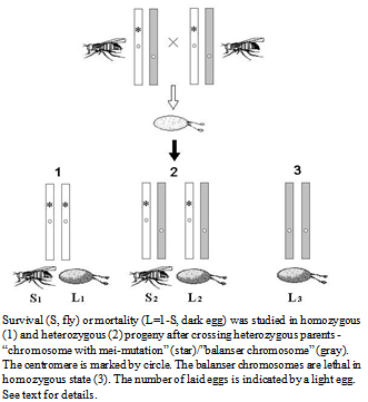
Figure 1. Scheme of studying the effect of MDMs accumulation on the viability of the progeny
To estimate viability, each virgin female was mated with two males in a vial with standard nutrient medium. In total, 30 to 50 females were used in every cross. After 24 h, all flies were transferred to a bottle with the medium. The laid eggs were counted each 3-4 h. Typically, the bottles were replaced three to four times. The number of laid eggs in the experiments varied approximately from 400 to 2000. Then, the numbers of pupae and hatched flies of different phenotypes were recorded. The viability (S) was measured as the proportion of hatched flies with a particular genotype (and, accordingly, phenotype) in the number of laid eggs with the same genotype. The number of eggs with the given genotype was determined as the proportion in the total number of laid eggs, using the ratio of genotype classes in the progeny of the cross. The progeny mortality (L) was estimated as the proportion of eggs that did not develop to the adult stage in the total number of eggs laid after the cross (L=1-S). Crosses are listed in the first column of Tables 1 and 3.

2.3. Counting the Number of Generations, Accumulating MDMs

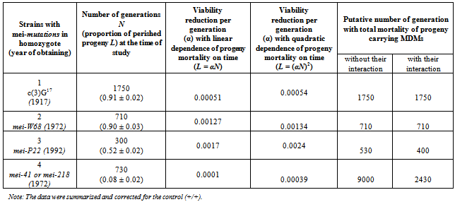
The number of generations (N), during which there were violations of the pairing and recombination of chromosomes during meiosis under the influence of different mutations, was calculated. Strains with mutations were maintained with crossover suppressors from the time of their registration or production to the time of the experiments. The strains carrying mei-с(3)G17, mei-W68, mei-P22, mei-41 and mei-218 are maintained since 1917[19],1972[20], 1992[21], 1972[22] respectively. Taking into account the duration of the Drosophila life cycle and the practice of maintaining strains in laboratory (18 days), consequently, the approximate number of generations elapsed to the time of analysis were different for different strains: 1750 for mei- с(3)G17, 710 for mei-W68, 300 for mei-P22, 730 for mei-41 and mei-218.

2.4. Viability of Progeny in Strains and Their Hybrids Carrying Mei-Mutation C3)G17

Progeny viability was examined in three laboratory strains of D. melanogaster (1-3), carrying meiotic mutation c(3)G17. Laboratory strain 2 (c(3)G17/TM3) is maintained since 1985; it was derived from strain 1 (c(3)G17/TM2) by substitution of the balancer. Strain 2 was supplied by I.D. Alexandrov (United Institute of Nuclear Research, Dubna, Russia). Strain 3 (c(3)G17/TM1) was provided by the Bloomington Drosophila Stock Center, previously, it was kept in the Caltech Stock Center, approximately up to 1970 (http//www.flybase.edu). In addition, we examined viability of the hybrid progeny, homozygous for the mei-mutation and produced by crossing flies of different strains (1 × 2, 1 × 3 and 2 × 3). To facilitate phenotypic marking of the progeny, we transferred the studied chromosome (carrying mutation c(3)G17) to heterozygote with a new balancer chromosomes; the flies were taken in the experiment during three generations. In some cases the reproduction was conducted for about 20 and 50 generations. We used strains 4 and 5 as a source of new balancer chromosomes and strain 6 for generating crossover chromosomes (females st c(3)G17 ca/ru h th st cu sr e ca) containing different regions with accumulated MDMs ( th st cu c(3)G17 (?) ca, ru h st c(3)G17 (?) sr e ca). Strain 13 was used as control. Strains 4–6, 13 were also provided by the Bloomington Stock Center.

2.5. Comparative Analysis of Viability in Strains Carrying mei-mutations c(3)G17, mei-P22, mei-W68, mei-41, mei-21

Progeny viability was examined in D. melanogaster strains 7-10, homozygous at various mei-mutations (mei-W68, mei-P22, mei-41, mei-218). Strains 7 and 9 were provided by A.T.C. Саrpenter. In the strain 8 with mutation mei-Р22, the balancer chromosome was at some point lost, and this mutation is currently maintained in homozygote (http://flybase.org).
Strains 8 and 10 were supplied by the Bloomington Stock Center. Strains 4 and 11 were used as a source of new balancer chromosomes.

2.6. Mortality of Hybrid Progeny with Chromosomes From Strains Carrying Different Mei-Mutations with Accumulated MDMs

Strains 7, 8, 2 -, containing the mei-mutations W68, P22 and c(3)G17 respectively were used. Strains 4 and 12 were used as a source of new balancer chromosomes. We examined the mortality of the following hybrid progeny: (a) c(3)G17/ mei-P22; (b) mei-W68/+, c(3)G17/+ and (c) mei-W68/+, mei-P22/+. In addition, mortality of the progeny was studied in heterozygotes c(3)G17/rucuca, c(3)G17/TM6Tb, mei-P22/TM6Tb and mei-W68/SM1.

3. Results and Discussion

3.1. Viability of the Progeny in Strains and Their Hybrids Carrying Mei-mutation c(3)G17

In this section of paper we refer to some historical research data. Since the average value of life cycle duration estimated previously[13],[23] was somewhat lower, here it was standardized for comparing different strains.
The first meiotic mutation, с(3)G17, was found in a natural population in 1917 and has been since then maintained in laboratory strains. Mei-mutation с(3)G17 in autosome 3 (3-57.4; 89A5) disturbs the formation of synaptonemal complex (SC), suppresses recombination in homozygous females[24],[25], and, which is worth mentioning, enhances recombination in heterozygotes with the normal chromosome[26]. Note also the exclusive maintenance of autosome 3 in all strains in heterozygote at balancers and the absence of chromosome pairing and crossing over in homozygotes due to the mei-mutation.
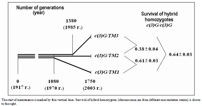
Figure 2. History of strains carrying mei-mutation c(3)G17
All three c(3)G17 - strains (1-3) practically totally lacked homozygous progeny (1 – 0.0001 ± 0.0001, 2 – 0.00005 ± 0.00004, 3 – 0) which was represented only by balancer heterozygotes (1 -0.66 ± 0.04, 2 – 0.68 ± 0.04, 3 – 0.70 ± 0.03)[13],[23]. Thus, a labour-consuming study of adaptation of the flies with accumulated MDMs to the environment in this case was excluded. We first noted a very small number of homozygotes in a strain carrying the c(3)G17 mutation in 1997, when a few homozygotes were recorded in the progeny[27]. The character of the dependence of the progeny viability on the generation number upon meiosis suppression can be “restored” from the results of viability of hybrids between the initial strain and a dated derivate, typically maintained with another balancer. This is caused by independent random appearance and rapid fixation of MDMs in small populations, which are represented by laboratory strains. MDMs accumulated in the initial strain are shared or homologous (Fig. 2).
In view of the history of the strains, the viability estimates for interstrain hybrids (Fig. 2) are in agreement with the assumption of the quadratic relationship between survival S (L = 1 - S, mortality of progeny) and generation number N upon MDM accumulation (Fig. 3).
This relationship is described empirically by the equation S = 1 – (α ·N)2, where α is the reduction in viability resulting from the MDM appearance in one generation. . Considering the lethality of the progeny in wild-type strain +/+ (0.09 ± 0.02), . The factual data correspond to the splitting of the strains from the initial stock in 1970 and 1985, i.e., after their maintenance together for 1010 and 1430 generations, respectively. The above relationship suggests that MDMs interaction corresponds to their synergistic epistasis.
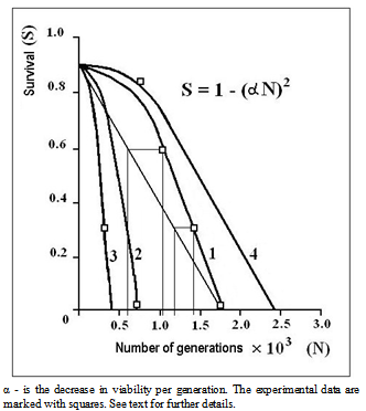
Figure 3. Putative quadratic relationship between the progeny viability S and the number of generations N that accumulate MDMs in strains carrying mei-mutations c(3)G17 (1), W68 (2), P22 (3), 41 or 218 (4)
Calculations show that with a linear relationship (additive effect, S = 1 – αN, α = 5.1∙10-4 and accordingly N = (1 – S)/α), the strains would have split from the original stock respectively in 1956 and 1975 (after 550 and 1180 generations), which was not the case (Fig. 3). Our conclusion on the MDM interaction was indirectly supported by the results reported for Drosophila by other authors: it was shown that the effect on the progeny viability and coefficient of dominance increase with the number of generations, in which MDM accumulation occurs[16],[28].
To assess the accumulation of MDMs in different region of metacentric autosome 3 and the effect of meiosis on their manifestation, we constructed strains with non-crossover and reciprocally recombinant chromosomes by a double exchange (in different arms) between a chromosome from strain 2 carrying the с(3)G17 mutation and a chromosome from strain 6 with normal viability (Fig. 4). Our results show that MDMs located in the middle, pericentromeric part of chromosome 3 are nearly twice as efficient in mortality as those from the distal euchromatic regions. The mortality of homozygous progeny (taking into account the control) per a physical unit of chromosome region length, calculated according to[29] in pericentromeric and distal chromosome regions, respectively, is (0.89 – 0.21)/ (89 + 55 + 81) = 0.68/225 ≈ 0.0030 and (0.89- 0.59)/ (88 + 120) = 0.30/208 ≈ 0.0014 (in Fig. 4, sizes of the chromosome regions are given in arbitrary units above the chromosome schemes). Probably, chromosome recombination as such does not play a significant role in decreasing the pressure of these mutations, since after normal meiosis non-crossover chromosomes partially (0.23 ±0.04) restore viability, which was previously zero (P>0.999). In this case, gene conversion is not excluded. On the other hand, the presence in the control population of MDMs that result is nearly 0.1 mortality in the progeny may be explained by their localization in chromosome regions with less effective pairing and corresponding meiotic processes, namely, in pericentromeric regions.
The substitution of the balancer chromosome in the strain carrying mei-mutation с(3)G17 for a new balancer proved to result in a partial (from 0 ± 0.02 to 0.27 ± 0.03) restoration of viability (P>0.999) of с(3)G17/с(3)G17 homozygotes during the first 20-30 generations (Fig. 5). Further maintenance of the strain on the same balancer for 20 generations led to decline in viability to the former level (Fig. 5). This phenomenon, albeit long known to drosophila genetics, has not been studied.
Apparently, chromosome recombination as such does not play a significant role in decreasing the MDM pressure, since recombinants of a structurally normal chromosome with multiple inversions in the balancer do not survive because of deletions. In our case, gene conversion (intragenic recombination), also initiated by DNA breaks, successfully occurs[30-32].
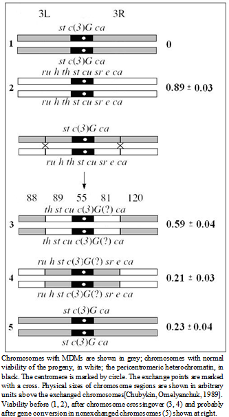
Figure 4. Viability of homozygous progeny before and after mei-recombination in heterozygotes st c(3)G17 ca/ru h th st cu sr e ca

3.2. Comparative Analysis of Viability of the Progeny in Strains Carrying Mei-mutations c(3)G17, mei-P22, mei-W68, mei-41, mei-218

To evaluate the role of other meiotic events that promote MDM accumulation, we examined the accumulation of MDMs in strains having other mei-mutations: mei-W68 (2-94; 56D9) and mei-P22 (3-21.5; 65E9). Both these mutations disturb the formation of double-strand DNA breaks without changing SC[21],[33]. In addition, we studied strains carrying mei-mutations in the X chromosome (mei-218 (1-56.2, 15D6) and mei-41 (1-54.2; 14C3)), disrupting repair of DNA breaks appearing during meiosis[20]. We note that mutations at the time of their generation and description did not affect the viability of their carriers in either homozygote or heterozygote. The effect of mei-mutations on MDM accumulation in a laboratory strain is possible only in homozygotes for recessive mei-mutations. This condition was met in laboratory strains carrying mutations с(3)G17, mei-W68 and mei-P22.
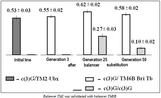
Figure 5. Effect of balancer replacement on the viability of heterozygotes and homozygotes for mei-mutation c(3)G17 depending on the number of generations
Table 1. The survival rate of progeny in the strains carrying mutations mei-W68 and mei-P22 in the autosomes 2 and 3, respectively, and mutations mei-218 and mei-41 in the X-chromosome
     
Table 2. Characteristics of strains carrying different mei-mutations
     
The results of estimating progeny viability in crosses of mei-W68 and mei-P22 heterozygotes are presented in Fig. 3 and Table 1. The viability of the progeny homozygous for mei-mutations - is 0.01 ± 0.05 (mei-W68/mei-W68) and – is 0.39 ± 0.03 (mei-P22/mei-P22). Taking into account the presence of MDM in wild-type strain Oregon R (0.91 ± 0.02 progeny survives), the progeny mortality in mei-mutation homozygotes in one generation varied among the strains. In the case of MDM interaction, i.e., according to the quadratic dependence of progeny mortality on time, the viability reduction per generation (α) was as follows: 0.00054 in strain с(3)G17, 0.00134 in strain mei-W68 and 0.0024 in strain mei-P22 (Fig. 3, Table 2).
The highest efficiency in the MDM accumulation was observed in the strain carrying the mei-P22 mutation. For example, the efficiency of viability decline per generation in this strain was more than four times higher than in the с(3)G17 strain (0.0024/0/00054 ≈ 4.44). This is explained by partial maintenance of this mutation in homozygote. In that case, MDMs can accumulate in the whole genome. On the other hand, if a mutation is maintained with a balancer, normal meiotic processes take place, in heterozygous individuals in all chromosomes except the one carrying the mei-mutation and paired with the balancer. It is exactly in this chromosome the MDMs are accumulated. Strictly speaking, we can compare only two strains with the identical conditions of maintaining mei-mutations in autosomes on a balancer, с(3)G17 и mei-W68.
Based on this comparison, we can conclude that changes in the topological heterochromatin structure caused by double-strand DNA breaks, controlled by gene MEI-W68, in comparison to disruption of SC formation, controlled by gene MEI- c(3)G17, play a significant role in controlling the MDM rate and accumulation - the efficiency of their accumulation was more than two times higher (α c(3)G17 / α W68 = 0.00134/0.00054 ≈ 2.5, Table 2). It is not clear whether such DNA breaks occur in homozygous c(3)G17 mutants. Apparently, the answer to this question is no, because the SC formation in these mutants is disturbed. However, we found homologous chromosome pairing in half of their oocytes[27], which does not exclude this possibility.
X-chromosomes carrying mutations mei-218 and mei-41 showed exclusively paternal inheritance (Drosophila males SC and chromosome recombination are undefined). Females carried one chromosome with two sets of the X-chromosome genetic material, i.e., acrocentric compound C(1)DX. Thus, in this case, as in the c(3)G17 strain, we recorded the effect of the complete absence of the SC and recombination rather than the mutations. The viability of the progeny with accumulated MDMs in strains carrying mutations mei-41 and mei-218 in Х-chromosomes is presented in Fig. 3 and Table 1. In both strains, the same proportion of females (0.83 ± 0.02) and males (0.84 ± 0.03) survived (P<<0.95). Taking into account the progeny viability in the wild-type strain (0.91 ± 0.02), the proportion of viability reduction in homozygous females mei-41/mei-41 and mei-218/mei-218 and hemizygous males mei-41/Y and mei-218/Y per generation was identical in both strains. Corrected for the gene interaction, this parameter (α) constituted 0.00039 (Table 2). The difference of this estimate from that in the c(3)G17 strain, (nearly 1.4-fold, 0.00054/0.00039) is apparently explained by the difference in physical size between the X chromosome and autosome 3 or by specific features of sex chromosome functioning in Drosophila.

3.3. Mortality of the Hybrid Progeny With Chromosomes from the Strains Carrying different Mei-Mutations with Accumulated MDMs

The MDM effect on viability of hybrids containing heterozygous (from different strains) chromosomes with MDMs in autosome 3 (cis position) with c(3)G17/ mei-P22 was examined (Fig. 6a). We also examined hybrids with combination of autosomes 2 and 3 with MDMs from strains with meiotic mutations c(3)G17, mei-P22 and mei-W68 in heterozygote with a chromosome that conferred normal viability (trans position), mei-W68/+, c(3)G17/+ (Fig. 6b) and mei-P22/+, c(3)G17/+ (Fig. 6c). The results produced in different crosses are presented and analyzed in Fig. 6 and Table 3.
The mortality of the progeny of the wild-type strain +/+ (0.09 ± 0.03) and homozygotes for multiple phenotypic markers rucuca/rucuca (0.11 ± 0.02) showed practically no difference (P<<0.95). The high mortality of progeny rucuca/TM6Tb (0.42 ± 0.03 and 0.47 ± 0.03 in different crosses) suggests that the balancer chromosomes, in addition to lethal mutations, carry MDMs]. However, the equal mortality of the heterozygous progeny with c(3)G17/rucuca and c(3)G17/TM6Tb (0.57 ± 0.03 and 0.57 ± 0.03 respectively, Table 3) cast doubt on this assumption. Nevertheless, these results indicate that there is no difference between the mortality of heterozygous progeny with chromosomes carrying accumulated MDMs in balancers and with structurally normal chromosomes, which was established earlier in classical studies[14],[15].
Table 3. The mortality rate of progeny with different genotypes, including hybrids
     
Figure 6 schematically presents the hybrids and the progeny mortality in case of changed gene activity in all chromosomes with independently accumulated MDMs, expected if they are not allelic (arrows 1) and the experimental data (arrows 2).
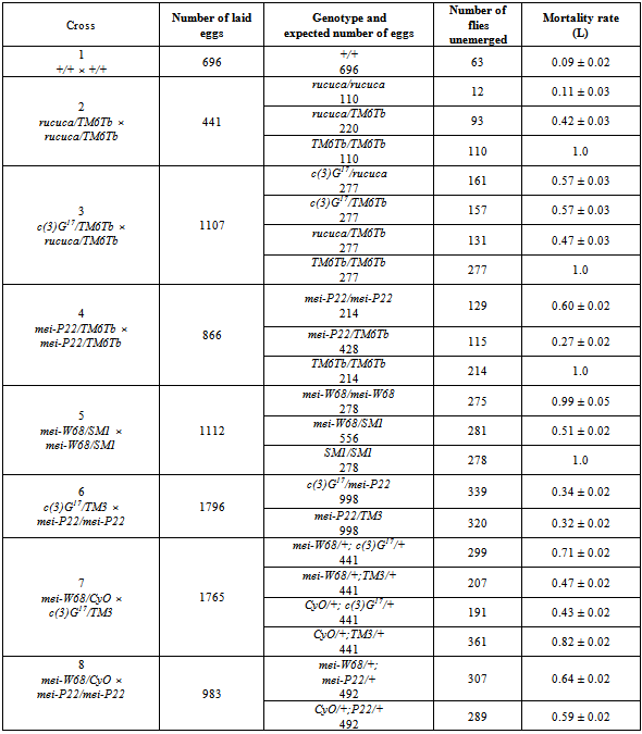
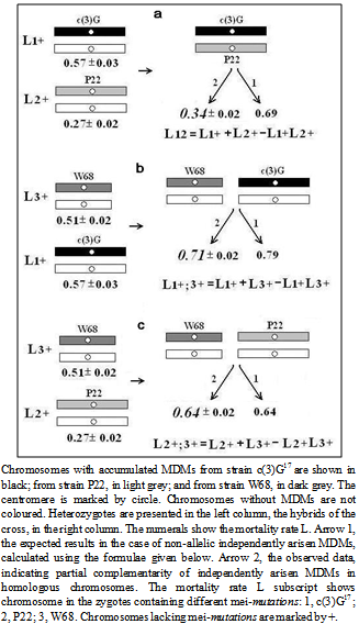
Figure 6. Mortality rate of heterozygotes and hybrid progeny carrying homologous (a) and nonhomologous (b, c) chromosomes with independently accumulated MDMs in different mei-mutant strains
The mortality rates of the hybrid progeny c(3)G17/ mei-P22 (a), mei-W68/+, c(3)G17/+ (b) and mei-W68/+, mei-P22/+ (c) are presented in Fig. 6 (marked by arrow 2) and in Table 3. These experimental results were unexpected. Collectively they indicate altered activity of genes in all chromosomes with independently accumulated MDMs. The surprising result was a significant decrease in mortality of two hybrids relative to the expected value, which was observed at a different extent (Fig. 6, arrow 1). The survival frequency of the progeny with genotype (a) c(3)G17/ mei-P22 was higher then expected by about twice (0.69/0.34 = 2.03), with genotype (b) mei- W68/+, c(3)G17/+, by 1.11 (0.79/0.71), while the progeny with genotype (c) mei-W68/+, mei-P22/+ show the expected viability (0.64/0.64). Upon independent MDM accumulation, these results are unlikely, if we assume that gene mutate. The MDM frequency P in the chromosome is calculated as
(1).
Where N is the number of generations accumulating MDM, q is the proportion of the genes in the chromosome in the total gene number G = 13767, k is the coefficient of relative MDM accumulation efficiency, and U is the number of MDMs generated per generation. The frequency of coincidence (complementation) of MDMs in hybrids containing heterozygous (from different strains) chromosomes with independently accumulated MDMs in autosome 3 (c(3)G17/mei-P22) is equal to the product Р(1) for Drosophila strains with different meiotic mutations P c(3)G17 ∙PP22. The values of these parameters are presented at http://flybase.bio.indiana.edu and in[29]. According to these data, the proportion of genes in autosome 3 is ≈ 0.38. Parameter k was estimated as the ratio of the rates of progeny mortality per generation (α) in strains carrying mutations mei-P22 и c(3)G17 (0.0024/0.00054 ≈ 4,4).
(2)
If we take the maximum U value, earlier theoretically estimated in Drosophila for recombination suppression[11] (one gene mutates per zygote), then the probability of complementation is 0.0018.
The chromosomal cis-trans test of independently arisen MDMs showed functional “complementarity” of ~ 0.5 mutations manifested as restoration of the normal phenotype in heterozygotes. What is the minimum number of genes that should simultaneously mutate in this case? Using equation (2), we obtain 0.0018 U2= 0.5 or U ≈ 17. This suggests that a gene cluster simultaneously mutates in the chromosomes. The term complementarity is taken in quotation marks because, apparently, the reduced viability is caused not by gene mutations, but by alteration of structural chromosomal segments that are far larger than genes.

3.4. The Nature of MDM

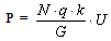
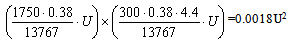
The nature of MDMs is still unclear. The MDM expression is similar to the position-effect variegation - all progeny carries MDMs but only part of the progeny perishes. This similarity in mutation expression also suggests that MDM impair the gene activity regulation. We have shown that normal meiosis in heterozygote of chromosomes with accumulated MDMs and structurally normal, MDM-free chromosomes restores viability both in recombinant and in non-crossover chromosomes with accumulated MDMs (Fig. 4). Chromosomal recombination as such apparently does not significantly reduce MDM pressure, since we cannot exclude the involvement of gene conversion (intragenic recombination), which is also initiated by DNA breaks [30-32]. This is evidenced by the data on partial viability restoration during 20 generations by changing the balancer (Fig. 5). The MDM elimination in meiosis is problematic to explain on the basis of gene nature of MDMs, changing DNA sequence, since the probability of their reverse mutation is very low. All facts listed above suggest that MDMs in some manner change gene expression rather than changing DNA.
In contrast to the effect of mutation c(3)G17, the mei-mutations examined in this study (mei-W68, mei-P22) do not disturb homologous chromosome pairing and the SC formation. These mutations affect the initiation of recombination[21],[33] by means of impairing the generation of DNA breaks, produced by topoisomerases after the SC formation in Drosophila oocytes. It is known that DNA breaks, releasing structural tension in the packaged chromatin, promote its reorganization and accessibility in structural modification[34] (for instance, in the processes of inter- and intragenic recombination and DNA repair). Thus, the results of the present study do not contradict our suggestion of the epigenetic nature of MDMs, disturbing the formation and inheritance of specific functional structures of the genome[35]. These are probably structural-functional chromosome domains arranged in chromatin loops and containing gene clusters comparable with their putative number (≈ 17). The number of the loops is nearly by one order of magnitude lower than that of the genes (approximately 13767/17 ≈ 810, where the numerator is the total number of genes and the denominator, the calculated average number of genes per loop). According to the DNA content in female chromosomes[29], the numbers of loops in the X chromosome and autosomes 2, 3, and 4 are respectively 187, 286, 309, and 28.
The chromatin loops are functional and structural chromosome units responsible for gene expression, replication and recombination in eukaryotes[36]. The loops are of different size. In the telomeric and pericentromeric chromosome regions, where the rates of recombination and MDM accumulation are highest (Fig.4), the loops are most abundant, but smaller than in other chromosome parts[37]. The repeated DNA sequences at the loop base are highly variable and associated with the chromosome axial element or nuclear matrix (S/MARs). Similar sequences were detected in loops beyond the axial element, where they may act as potential triggers for the formation of new domains [38],[39]. Apparently, their secondary structure (hairpins made of inverted repeated sequences) is essential for their functioning (anchoring on the axis and forming the loops) (Fig. 7)[40]. The loops size range from 3 to 200 kb; the size of interloop stretches also vary, but in the narrower range (from 3 to 30 kb)[41]. FISH visualization of DNA probes on preparation of nuclei extracted by 2M NaCl (nuclear halos) showed that active genes are localized in the loop bases harboring the complexes for replication, transcription, DNA repair and recombination, where various topological problems of the chromatin are resolved[42]. Most results obtained using different methods of DNA loop mapping, indicate their nonrandom standard organization [43]. The issue on association of the loop structure with epigenetic regulation of gene expression, including genetic imprinting and gene-position variegation, with meiotic chromatin remodeling has long been discussed[44],[45]. The loop domains linearly and spatially border a large group of genes, ensuring interaction enhancers and promoters within these limits, i.e., act also as insulators[46-48]. The major properties of loop domains are shown in (Fig. 7).
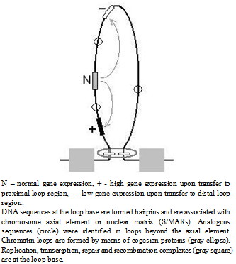
Figure 7. Relationship between gene activity and gene position in the chromatin loops
It is hypothesized that during the life cycle, from zygote to adult, “mutant” variants of the loop domain formation appear in the chromosomes with altered gene localization relative to the loop base and the potential expression of the corresponding genes (Fig. 7). The number of abnormal loops (MDMs) per generation depends on the life-cycle duration[49]. Thus, MDMs disrupt the formation and inheritance of a specific functional state of the genome rather than alter DNA structure[35], i.e., are epigenetic in their nature. The viability restoration in hybrids carrying chromosomes with independently appeared mutations suggests that both an increase and a decline in the gene activity are disadvantageous for the development. In hybrid heterozygotes, such opposite in activity mutant homologous genes within symmetrically mutant loops can compensate one another, providing normal development. Since the formation of a “mutant” loop involves on average two standard loops, the number of genes with altered activity can exceed more than 17. Moreover, the viability restoration is possible upon interaction of “mutant” but nonhomologous genes, whose products are involved in the same or connected developmental chains (an analogue of interallelic complementation). This is also evidenced by the results of the present study (significant viability restoration (P>0.99) in hybrids heterozygous for different chromosomes with MDMs mei-W68/+, c(3)G17/+ (0.71 ± 0.02 in comparison with 0.79) (Fig. 6).
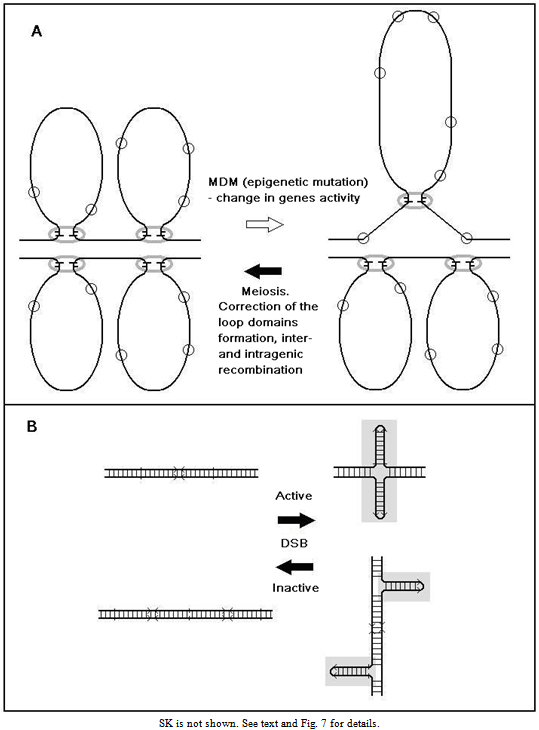
Figure 8. The directional correction of the loop domain formation in meiosis (A) is accompanied by the rearrangement of the secondary structure of S/MAR repeats (B) by means of double-strand DNA breaks (DSB)
Normally, nonstandard loop domains are probably corrected during meiosis (Fig. 8A). Their correction, mediated by stabilizing function of the genome of each species[50] seems to be based on the rearrangement of the secondary structure of S/MARs (palindrome hairpins], also accompanied by DNA breaks (Fig. 8B). Thus, the function of meiosis, in addition to recombination and chromosome segregation, includes directional correction of invariant development of organisms (zygotes), depending on the structural chromatin organization in chromosomes. This correction is very effective: approximately 20% MDMs per meiotic cycle are eliminated in a heterozygote with the normal chromosome[23]. This is much higher than could be compared to the restoration of the original genetic material by means of recombination, to say nothing of reverse mutations whose rate is orders of magnitude lower than that of direct mutations. The maximum probability of random reverse mutation of loop domains is (1/690)2 ≈ 0.0000021. Classical point mutagenesis is practically irreversible: (1/13767)2 ≈ 0.0000000053 (without taking into account specific base substitutions in DNA). Since most of genic mutations are deleterious, they are either eliminated, in case of being very disadvantageous or lethal, or are inherited but they can never return to their initial state. In the case of MDMs, we deal with the functional restoration of the loop domain structure standard for the species - the code of the species ontogeny. The restoration of the original functional state of the genes in meiosis, which we report, is a characteristic feature of “epimutations”.
Ontogeny can be viewed as unfolding a branched chain of gene activities. Each link of this chain relative to the subsequent links represents only a possibility for development, which is determined, apart from quality, by the number and the functional properties of critical products (regulators). Because of this, the MDM manifestation is probabilistic and increases with their accumulation. It may well be that in a genotype with altered activity of the gene cluster, genes of development exhibit altered sensitivity to the gradient and/or the concentration of the regulators is changed. In this case, due to the existence of a sensitivity threshold, defects in the temporal pattern of the gene expression (heterochronization) are inevitable. All this at some stage of development of organisms[51] should lead to changes in adaptation to the environment. Naturally, the influence of MDMs starts at the early developmental stages and continues throughout the development.

4. Conclusions

Thus, the relationship between survival and generation number upon MDMs accumulation suggests that MDMs interaction corresponds to their synergistic epistasis, that impair the formation of DNA breaks is more effective in accumulation of MDMs. The viability in progeny after meiosis in heterozygotes with chromosome with accumulated MDMs and normal chromosome, and in heterozygotes with independently accumulated MDMs chromosomes was shown to be partially restored (20% and 50% respectively). Our results support the hypothesis that MDMs have epigenetic nature. The potential polyvariant character of the chromatin loop formation and the existence of genes with the expression decreasing from the loop base, taken together with our results unambiguously support our conclusion on the nature of MDMs.
The sexual reproduction is important for generating the evolutionary potential of the species and its further evolution, but it’s most vital function is conferring stability to the species by means of meiosis. In essence, resolving this issue will provide insight in understanding evolution, which implies increasing the complexity of the structural organization of life upon the presence of nearly equal standard gene set in the majority of higher organisms.
The simple argument for epigenetic nature MDM may be obtained through using the method of comparative DNA loop by mapping a number of genes in wild chromosomes and chromosomes carrying mei-mutations.

ACKNOWLEDGEMENTS

The author is very grateful to Ivan P. Glushkov, T.M. Grishaeva and A.G. Imasheva for technical assistance. The study was supported by the Russian Foundation for Basic Research (grant nos. 05-04-49052, 08-04-01725a).

References

[1]  M. Kimura, T.Maruyama, “The mutation load with epistatic gene interactions in fitness,” Genetics, vol. 54, pp. 1337 – 1351, 1966.
[2]  J. Maynard Smith, “The evolution of sex,” Cambridge: Cambridge University Pres. 236 pp, 1978.
[3]  A.S. Kondrashov, “Classification of hypotheses on the advantage of amphimixis,” Journal of Heredity, vol. 84, pp. 372 – 387, 1993.
[4]  H.J. Muller, “Some genetic aspects of sex,” American Naturalist, vol. 66, pp. 118–138, 1932.
[5]  H.J. Muller, ”The relevance of mutation to mutational advance,” Mutation Research, vol. 1, pp. 2–9, 1964.
[6]  A.S. Kondrashov, “Deleterious mutations and evolution of sexual reproduction,” Naturе, vol. 336, pp. 435 – 440, 1988.
[7]  A.S. Kondrashov, “Sex and U,” Trends in Geneics, vol. 17, pp. 75 – 77, 2001.
[8]  F. Jacob, J.Monod, “Genetic regulatory mechanisms in the synthesis of proteins,” Journal of Molecular Biology, vol. 3, pp. 318-356, 1961.
[9]  T. Мukai, C.C. Cockerham, “Spontaneous mutation rates at enzyme loci in Drosophila melanogaster,” Proceeding of the National Academy of Sciences of the USA, vol. 74, pp. 2514 – 2517, 1977.
[10]  E.G. Pasyukova, E.S. Belyaeva, L.E. Ilyinskaya, V.A. Gvozdev, “Outcross-dependent transpositions of copia-like mobile genetic elements in chromosomes of an inbred Drosophila melanogaster stock,” Molecular and General Genetics, vol. 212, pp. 281 – 286, 1988.
[11]  P.D. Keightly, “Nature of deleterious mutations load in Drosophila,” Genetics, vol. 144, pp. 1993 – 1999, 1996.
[12]  H.J. Muller, ”The measurement of gene mutation rate in Drosophila, its high variability, and its dependence upon temperature,” Genetics, vol. 13, pp. 279-357, 1928.
[13]  V.L. Chubykin, “Survival of Drosophila melanogaster progeny after prolonged suppression of pairing and recombination in autosome 3,” Russian Journal of Geneics, vol. 40, pp. 1223 – 1228, 2004.
[14]  T. Mukai, “The genetic structure of natural populations of Drosophila melanogaster. I. Spontaneous mutation rate of polygenes controlling viability,” Genetics, vol. 50, pp. 1 – 19, 1964.
[15]  O. Ohnishi, “Spontaneous and ethylmethane-sulfonate-induced mutations controlling viability in Drosophila melanogaster. II. Homozygous effects to polygenic mutations,” Genetics, vol. 87, pp. 529 – 545, 1977.
[16]  A. Cabаllero, E. Cuсi, C. Garсia, A. Garсiа-Doradа, “Accumulation of deleterious mutations: additional Drosophila melanogaster estimates and a simulation of the effects of selection,” Evoution: Inernational of Organic Evoution, vol. 56, pp. 1150-1159, 2002.
[17]  T. Mukai, S.T. Chigusa, L.E. Mettler, J.F. Crow, “Mutation rate and dominance of genes affecting viability in Drosophila melanogaster,” Genetics, vol. 72, pp. 335 – 355, 1972.
[18]  D.L. Lindsley, E.H. Grell, “Genetic variations of Drosophila melanogaster,” Carnegie Institution of Washington Pubication, № 627, 472 pp, 1968.
[19]  M.S. Gowen, J.W. Gowen, “Complete linkage in Drosophila melanogaster,” American Nauralist, vol. 56, pp. 286 – 288, 1922.
[20]  B.S. Baker, A.T.C. Carpenter, P. Ripol, “The utilization during mitotic cell division of loci controlling meiotic recombination and disjunction in Drosophila melanogaster,” Genetics, vol. 90, pp. 531-578, 1978.
[21]  K.S. McKim, A. Hagashi-Hagihara, ”mei-W68 in Drosophila melanogaster encodes a spo11 homolog: mechanism for initiating meiotic recombination is conserved,” Genes and Development, vol. 12, pp. 2932-2942, 1998.
[22]  B.S Baker, A.T.C. Carpenter, “Genetic analysis of sex chromosomal meiotic mutants in Drosophila melanogaster,” Genetics, vol. 71, pp. 255-286, 1972.
[23]  V.L. Chubykin, “Deleterious mutations in various Drosophila melanogaster strains carrying meiotic mutation c(3)G17,” Russian Journal of Genetics, vol. 44, pp. 1054 – 1060, 2008.
[24]  P.A. Smith, R.C. King, “Genetic control of synaptonemal complex in Drosophila melanogaster,” Genetics, vol. 68, pp. 335 – 351, 1968.
[25]  Yu.F. Bogdanov, S.Ya. Dadashev, T.M. Grishaeva, “Gene CG17604 of Drosophila melanogaster in silico may be the c(3)G17,” Drosophila Information Service, vol. 84, pp. 84-88, 2001.
[26]  S.K. Sen, S.K. Hazra, G.A.S. Iyengar, R.S. Banerjee, “Comparison of intragene and intergene recombination in the meiotic mutant c(3)G17 of Drosophila melanogaster,” Genetica, vol. 55, pp. 47 – 50, 1981.
[27]  V.L. Chubykin, “Structural Characteristics of the Chromocenter in Ovary Cells with the c(3)G17 and NOD Mutations of Drosophila melanogaster,” Russian Journal of Genetics, vol. 37, pp. 1032 – 1040, 2001.
[28]  A. Garсia-Doradа, A. Caballero, “On the average coefficient of dominance of deleterious spontaneous mutations,” Genetics, vol.155, pp. 1991-2001, 2000.
[29]  V.L. Chubykin, L.V. Omelyanchuk, “Mutual positions of nonhomologous chromosomes inferred from the data on interchromosomal exchanges in Drosophila melanogaster,” Genetika (Moscow), vol. 25, pp. 292 – 300, 1989.
[30]  A. Chovnick, “Gene conversion and transfer of genetic information within the invert region of inversion heterozygotes,” Genetics, vol. 75, pp. 123-131, 1973.
[31]  W.J. Gong, K.S. McKim, R.S. Hawley, “All paired up with no place to g: pairing, synapsis, and DSB formation in a balancer heterozygote,” PLoS Genetics, Nov; 1(5):e67. Epub 2005 Nov 18.
[32]  S.W. Schaeffer, W.W. Andersen, “Mechanisms of genetic exchange within the chromosomal inversions of Drosophila pseudobscura,” Genetics, vol. 171, pp. 1729 – 1739, 2005.
[33]  H. Liu, J.K. Jang, N. Kato, K.S. McKim, “ mei-P22 encodes a chromosome associated protein required for the initiation of meiotic recombination in Drosophila melanogaster,” Genetics, vol. 162, pp. 245 – 258, 2002.
[34]  A.D. Gruzdev, “DNA topology in heterochromatin (a hypothesis),” Journal of Theoretical Biology, vol. 207, pp. 255-264, 2000.
[35]  L.I. Korochkin, “What Is Epigenetics,” Russian Journal of Genetics, vol. 42, pp. 958-965, 2006.
[36]  P.R. Cook, “The organization of replication and transcription,” Science, vol. 284, pp. 1790 – 1795, 1999.
[37]  H.H.Q. Heng, W.J. Chamberlain, X-M. Shi, B. Spyropoulos, L-C. Tsui, P.B. Moens, “Regulation of meiotic chromatin loop size by chromosomal position,” Proceeding of the National Academy of Sciences of the USA, vol. 93, pp. 2795 – 2800, 1996.
[38]  G. Micheli, A.R.C. Luzzatto, M.T. Carri, A. Capoa, P. Pelliccia, “Chromosome length and loop size during early embryonic development of Xenopus laevis,” Chromosoma, vol. 102, pp. 478-483, 1993.
[39]  H.H.Q. Heng, S. Goetze, C.J. Ye, G. Li, J.B. Stevens, S.W. Bremer, J. Bode, S.M. Wykes, S.A. Krawetz, “Chromatin loops are selectively anchored using scaffold/matrix attachment regions,” Journal of Cell Science, vol. 117, pp. 999 – 1008, 2004.
[40]  A.T. Akhmedov, C. Frei, M. Tsai-Pflugfelder, B. Kemper, S.M. Gasser, R. Jessberger, “Structural maintenance of chromosomes protein C-terminal domains bind preferentially to DNA with secondary structure,” Journal of Biological Chemistry, vol. 273, pp. 24088 – 24094, 1998.
[41]  J.R. Davie, “The nuclear matrix and the regulation of chromatin organization and function.” International Review of Cytology, vol. 19, pp. 191 – 250, 1995.
[42]  M.G. Gerdes, K.C. Carter, P.T. Moen, J.B. Lawrence, “Dynamic changes in the higher level chromatin organization of specific sequences revealed by in situ hybridization to nuclear halos,” Journal of Cell Biology, vol. 126, pp. 289 – 304, 1994.
[43]  S.V. Razin, “The nuclear matrix and spatial organization of chromosomal DNA domains,” Austin: Landes, 212 pp, 1997.
[44]  B.D. Hendrich, H.F. Willard, “Epigenetic regulation of the gene expression: the effect of altered chromosome structure of yeast to mammals,” Human Molecular Genetics, vol. 4, pp. 1765 – 1777, 1995.
[45]  S.V. Razin, “Spatial organization of the eukaryotic genome and the action of epigenetic mechanisms,” Russian Journal of Genetics, vol. 42, pp.1353 – 1361, 2006.
[46]  J.E. Krеbs, C.L. Peterson, “Understanding “active” chromatin: a historical perspective of chromatin remodeling,” Critical Reviews in Eukaryotic Gene Expression, vol. 10, pp. 1 – 12, 2000.
[47]  N.M. Cai, P. Shen, “Effects of cis arrangement of chromatin insulators on enhancer-blocking activity,” Science, vol. 291, pp. 493 – 495, 2001.
[48]  J.S. Heslop-Harrison, “Plannings for remodeling: nuclear architecture, chromatin and chomosomes,” Trends in Plant Science, vol. 8, pp. 95 – 198, 2003.
[49]  P.D. Keightly, A. Eyre-Walker, “Deleterious mutation and the evolution of sex,” Science, vol. 290, pp. 331 – 333, 2000.
[50]  J. Loidl, H. Scherthan, J.K. Den Dunnen, F. Klein, “Morphology of a human-derived YAC in yeast meiosis,” Chromosoma, vol. 104, pp. 183 – 188, 1995.
[51]  J. Green,”Morphogen gradients, positional information and Xenopus: interplay of theory and experiment ,“ Developmental Dynamics, vol. 25, pp. 392 – 408, 2002.