Urinova Xulkar Shokirovna
Teacher of Navoi State University of Mining and Technology, Uzbekistan
Correspondence to: Urinova Xulkar Shokirovna, Teacher of Navoi State University of Mining and Technology, Uzbekistan.
| Email: |  |
Copyright © 2023 The Author(s). Published by Scientific & Academic Publishing.
This work is licensed under the Creative Commons Attribution International License (CC BY).
http://creativecommons.org/licenses/by/4.0/

Abstract
The scientific article examines the fact that the third largest class of the Fabaceae family, Indigofera, contains about 800 species, and this species grows naturally at altitudes up to 1650 meters above sea level. There are more than 600 species in Africa, about 200 species in Asia, 80 species in America and 50-60 species in Australia. In our research, we aimed to study the bioecological characteristics of Indigofera tinctoria L. grown under introduction conditions in Kyzylkum.
Keywords:
Indigofera, Natural state, Indigofera tinctoria L, Bioecological properties, "Indigo" dye, Vegetative organ, Biochemical and physiological processes
Cite this paper: Urinova Xulkar Shokirovna, Anatomical Structure of Leaf and Stem of Indigofera Tinctoria, American Journal of Polymer Science, Vol. 12 No. 1, 2023, pp. 7-16. doi: 10.5923/j.ajps.20231201.02.
1. Inctoduction
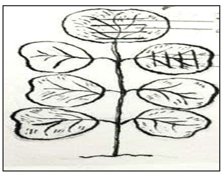
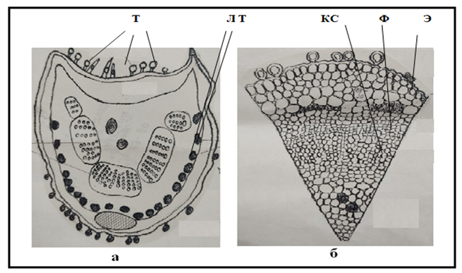
I. tinctoria is a plant of the tropical region, from its leaves, is obtained the famous air colour "Indigo", the king of dyes. The use of the natural dye "Indigo", a fixed air colour obtained from plants, spans thousands of years. But growing this plant and getting dye from it remains a secret in countries like India, Japan and China. In the scientific literature, there is insufficient information on the bio ecology, morphology, anatomical structure of vegetative organs, water regime, and agrotechnical of I. tinctoria in Kyzylkum conditions. Therefore, it is important to determine the diagnostic signs indicating the presence of dyeing properties by studying the bio ecological characteristics of the species, and the anatomical structure of its vegetative organs.For many years, India, China, and Africa have been using this plant to obtain dyes, but morpho-anatomical studies have not been carried out on which vegetative organ of the plant grown in our Kyzylkum conditions has the ability to give dyes. Our goal is to show the dye-yielding feature of the vegetative organs of the plant based on the diagnostic signs of its anatomical structure.The anatomical structure of the leaf: Biochemical and physiological processes such as transpiration, photosynthesis and gas exchange take place in the leaf, which is considered a vegetative organ of plants.The leaf of I. tinctoria belongs to the group of compound leaves, that is, there are 3-5-7 leaflets in one leaf band (Fig. 1, Fig. 2, Fig. 5).The structure of the leaf blade is kidney-shaped, located in a small 2-3 mm leaf band. The upper leaf is larger than the lower leaves. The upper part of the leaf is covered with small hairs and is dark green in color.In the cross-section of the leaf, the leaf has a long ribbon-like structure. The flesh of the leaf (mesophyll) is composed of 2 rows of columnar cells in the upper part of the leaf, and 1 row below. In their middle layer there are long narrow cloud-like cells that stick to each other.In the mesophyll of the leaf, that is, in the columnar cells, it can be observed that there is a large amount of black substances.Even in the small leaves of the leaf blade, when viewed in section, the same similarity can be seen as in the large leaf.In the leaf section, the mesophyll of the plant differs in thickness and thinness.When the outer part of the leaf, that is, the epidermis, was removed, it was observed that the epidermal cells in the upper part of the leaf were larger than those in the lower part.Leafhoppers are found both above and below the leaf. Depending on their location, epidermal cells can be classified into 2 types. That is, it has a (hemiparasite and parasite) structure. It is named so because 3-4-5-6 side cells surround the mouth.In the bark part of the leaf, that is, when the lower and upper layers are removed, hairs similar to the letter (T) can be seen under the microscope, and a large number of round hairs can be seen in the cross section (Fig.3, Fig.4).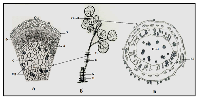 | Figure 1. a- anatomical structure of I. tinctoria plant branch, b- schematic view of leaf band, v- anatomical structure of thin branch. Conditional symbols: F-phloem, S-stem part, QD-black spot (painting pigment), E-phloem, L-libriform, KL-collenchyma |
 | Figure 2. Stem and leaves of I. tinctoria, view of fruiting branch |
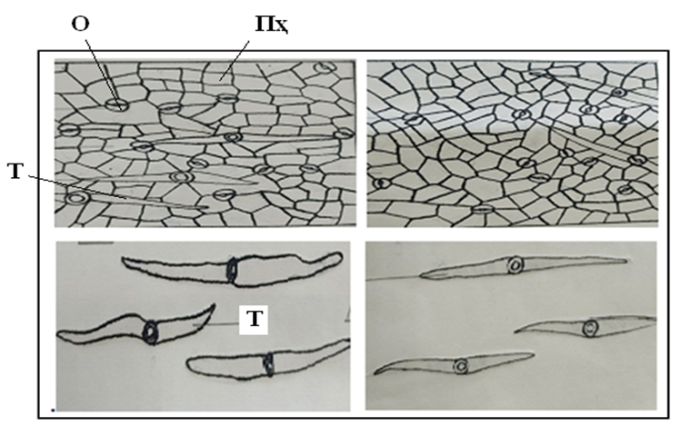 | Figure 3. Appearance of leaf sheath and hairs: Conditional characters: O-mouth, Ph-parenchyma cells, T-trichome |
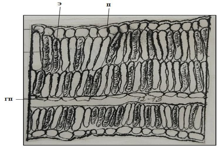 | Figure 4. Anatomical structure of the epidermis of a smaller leaf blade.Conventional symbols: E-epidermis, P-parenchyma, GP- porous parenchyma |
 | Figure 5. Compound leaf (its cut areas) |
2. In Conclusions
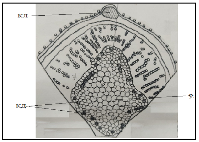
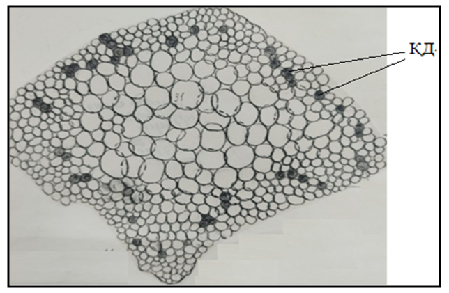
The leaves of I. tinctoria were found to contain a large amount of black pigment. It can be observed in all large and small leaves. Indigo dye can be obtained from the leaves of I. tinctoria more than other organs of the plant.The structure of a thin branch with compound leaves. Its microscopic structure is as follows: when a branch is cut, it is round in shape, covered by a row of epidermis, that is, bark cells, below it there is 4-6 rows of small bark parenchyma, and then the phloem layer is composed of small cells that surround the woody part. A large number of conductive grooves can be seen in the woody part of the stem. Radial rays are clearly visible between the tubes. The core part of the branch has a much larger size, and the parenchyma cells in it are large and small, thin-walled. A large number of dark-spotted cells can be observed around the stem cells.Anatomical structure of the large band that holds the compound leaf plates. The upper part of the leaf band consists of the following. In the section of the leaf band, it has a crescent-shaped structure. The middle part is bulging in the form of a circle. 1 set of collenchyma can be seen under the bark cell in the raised part. 1 row of bark cells is covered with thick hairs. Between the cells of the bark can be seen clusters of lobules, followed by a thick layer of phloem cells and a series of conducting tubules. The parenchyma cells in the core of the stem are small, and there are also black parenchyma cells.The structure of a small band of leaf petals. The following view can be observed in the cross section. The cross-section is oval-shaped, and it is surrounded by the 1st row of cortical cells from the upper side. They have a lot of hair. Under the epidermis, the parenchyma of the bark is located in the form of a ring. Lube fibers in bunches, in large quantities. A wooden layer can be observed under it. The size of the transfer tubes is smaller. The core parenchyma containing dark staining material can be seen in many of the parenchyma cells surrounding the small and thin-walled core cells with parenchymal tissue in the core of the small band. Therefore, it was observed that in the small leaf band, as well as in the large leaf band, the core tissue consists of a large number of black spotted cells (Fig.6, Fig.7).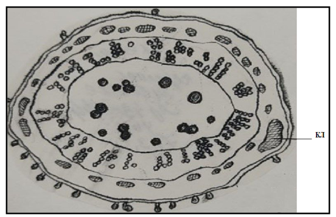 | Figure 6. The structure of the leaf band. Conditional signs: KL-collenchyma |
 | Figure 7. A view of a thicker section of a leaf band. Conventional symbols: T-trichome, LT-lub fibers, X-xylem, F-phloem, E-epidermis |
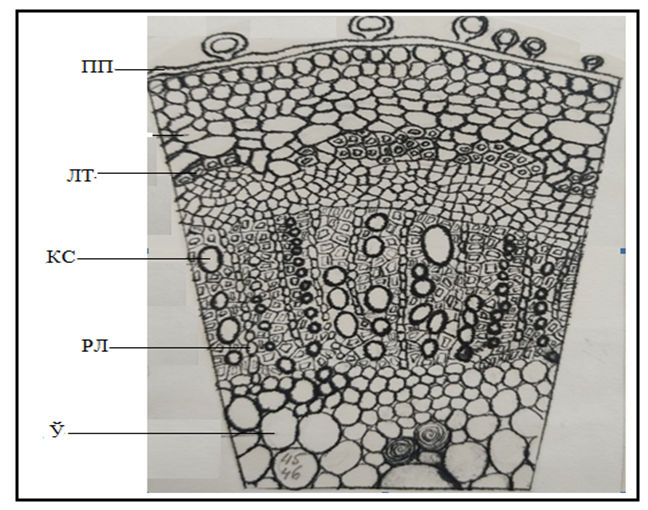
Anatomical structure of the stemAnatomical structure of the stem of I. tinctoria. When a small branch from a fixed plant branch is cut from the lower part, its shape has the shape of a rounded circle. Its upper part is surrounded by a thin row of bark cells, in its lower part 5-6 rows (black), bark parenchyma, can be seen. Lube fibers can be seen between its cells. The wall of this cell differs slightly from the wall of other cells by its thickness. In its lower part, the wooden part surrounding the stem in the form of a ring is noticeable. A large number of small and large conductive tube cells can be observed in the woody part. Tubes can be seen as elongated cells arranged in a chain, radial ray cells. The core parenchyma is located in the middle of the woody part. The wall of its cells is thin, round and oval in shape (Fig. 8).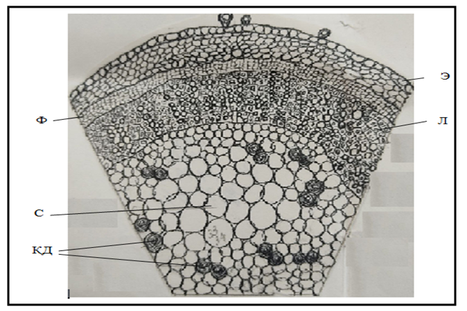 | Figure 8. Anatomical structure of the branch (thin variety) |
It did not find black cells in the lower part of the stem, in the cells.When the upper part of the stem is cut, it can be seen that the lignification is thinner compared to the lower part of the stem. The cross-section of the branch is surrounded by 1 layer of small bark cells from the upper side, in which a large number of hairs can be seen. Under the bark (black), that is, it forms bark parenchyma. Below it is a thin phloem, after which the woody part begins. In this part, there are a large number of large and small round-shaped conducting tubes. Between them can be seen radial cells (radial light cells). The core of the stem is covered with thin-walled parenchyma cells. The stem cells are surrounded by a large number of dark colored cells (Fig. 9).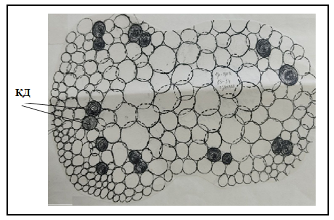 | Figure 9. The core part of the rod section. Conditional sign: QD-black spot (pigment) |
In conclusion, black cells that are not found in the lower part of the stem are found in the upper part of the stem.Anatomical structure of a short stem with fruit. When the cross-section of the fruiting stem was cut, it was found that the stem had a high woody quality. The upper part of the stem is covered by a bark cell, on which there are hairs. Below it, you can see 5-6 rows of (black) bark parenchyma, between which there are bundles of lub fibers, and in its lower part, a thin-walled phloem layer. After that, the people have a layer of wood in shape. The conducting pipes in it are located in a columnar shape (of different sizes). The wall is thick. There are 2-3 rows of radial rays between them. The core of the stem consists of thin-walled parenchyma cells. Dark spots can be seen in these parenchyma tissues (Fig. 10, Fig. 11, Fig. 12, Fig. 13).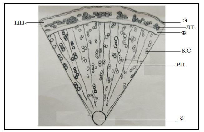 | Figure 10. Cross-sectional view of the lower part of the stem. Conventional symbols: E-epidermis, LF-lub fibers, Ph-phloem, X-xylem, RR-radial rays, C-core, BP-bark parenchyma |
 | Figure 11. Cross-sectional view of the tip of the stem. Conditional symbols: KL-xylem, BS-black spot (pigment), C-core |
 | Figure 12. A view of the core of a stem cross-section. Conditional signs: QD-black spot (pigment) |
 | Figure 13. Cross-sectional view of a stem with fruit |
The number of such cells is less than in the upper part of the stem.Anatomical view of the smaller root of I. tinctoria. When observing the cross-section of the end of the root, its shape is round, above it is covered with 1 layer of bark, below it is bark parenchyma, and in between there are bunches of lube fibers.The wood layer occupies the central part, it has a large number of conducting tubes, and consists of large and small circular tubes. Between the tubes there are long radial ray cells. Tubes in the central cylinder deliver nutrients from the root to the stem tubes, while tubes in the stem deliver nutrients to the plant's branches and leaves. This type of plant is supplied with food. In the root section, we did not observe black spots in the cells (Fig. 14).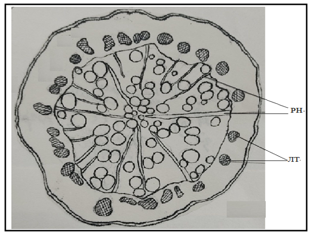 | Figure 14. Cross section of the root |
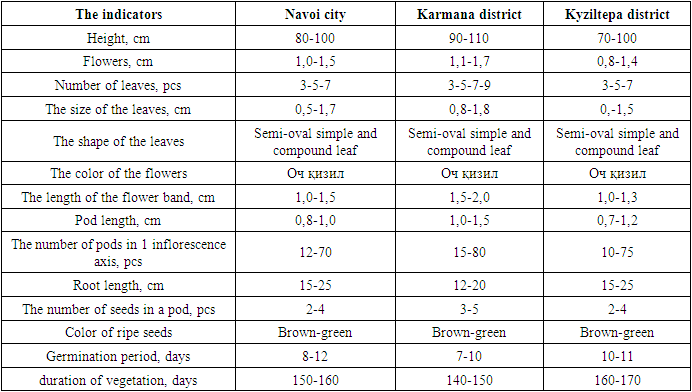
The growth and development of I. tinctoria were studied in Kyzylkum conditions (Navoi city, Karmana district, Kyziltepa district) and some biomorphological indicators were comparatively analyzed (Table 1).Table 1. Comparative analysis of vegetative and generative organs of I. Tinctoria
 |
| |
|
In our research, the flowering biology of I. tinctoria in Kyzylkum conditions was studied and scientifically analyzed. The seasonal flowering period of I. tinctoria in Kyzylkum conditions was observed in Navoi city, Karmana district and Qiziltepa district in 2017-2019 (Table 2). The flowering phase of I. tinctoria started in the third month of the growing season. Budding of I. tinctoria flowers lasted from the second decade of July to the second decade of August.Table 2. Seasonal flowering of I. tinctori a (2017-2019)
 |
| |
|
3. Summary
It can be said that I. tinctoria is a dye plant by looking at its anatomical structure. The coloring substance is found in the leaf of the plant (more), in the leaf bands, in smaller quantities, in the upper part of the stem, more than in the lower part of the stem. In the root part of I. tinctoria, it was not found at all.
References
| [1] | Ergashev A. et al. Abiotechnology of Indigofera tinctoria L. on the saline land of Aral Sea Basin and producing of the natural plant indigo pigment for the industry // Journal of Chemistry and Chemical Engineering. – 2014. – Т. 8. – №. 7. |
| [2] | Ахмедова, Н. М., Турсунова, И. Н., Урунова, Х. Ш., & Уринова, Х. Ш. (2018). Художественно-эстетический анализ миниатюры Бехзада на внеаудиторных занятиях в педагогических высших учебных заведениях. Проблемы современной науки и образования, (3 (123)), 69-71. |
| [3] | Urinova X. Sh. INTER, FIBER LENGTH IN. "An International Multidisciplinary Research Journal." An International Multidisciplinary Research Journal 41.43 (2017). |
| [4] | Rajabovna, Shomurodova Z. "Peculiarities of Translating Technical Texts from Uzbek into English." European Journal of Research Development and Sustainability, vol. 3, no. 2, 2022, pp. 15-17. |
| [5] | M.N Mirzaeva Technology forming professional competence of students of technical university in the lessons of foreign languages // Научные горизонты, 2019. |
| [6] | Mirzaeva M.N. Formation of the concept and technology of intercultural competition among students in universities. International scientific journal "Scientific Horizons" No.5 / 2018- P. 81-84p. |
| [7] | Bazarova U.M. The role of spiritual and moral education of students of technical university in the lessons of foreign languages // Theoretical & Applied Science. – 2019. – №. 11. – С. 614-616. |
| [8] | Bazarova U.M. The state of the problem of moral and aesthetic education of students by means of a foreign language at the present stage // International scientific journal" Scientific Horizons" no. – Т. 1. |
| [9] | Bazarova U.M. Improvement of mechanisms of moral and aesthetic education of students in foreign language lessons of a technical university // Asian Journal of Research in Social Sciences and Humanities. – 2021. – Т. 11. – №. 11. – С. 7-9. |
| [10] | Mansurova Dilfuza Zokir kizi, Abduganiyev Firdavs Sherzod ugli, & Bazarova Umida Mamurjanovna. (2022). Features of the classification of types of geotechnological terminology in the English language. Youth, Science, Education: Topical Issues, Achievements and Innovations, Prague, Czech. https://doi.org/10.5281/zenodo.6643026. |

 Abstract
 Reference
 Full-Text PDF
 Full-text HTML