American Journal of Polymer Science

p-ISSN: 2163-1344    e-ISSN: 2163-1352

2019;  9(1): 1-9

doi:10.5923/j.ajps.20190901.01


Nanostructures Obtaining on the Basis of Bombyx Mori Chitosan Hydroxyapatite

Vokhidova N. R., Ergashev K. H., Yugay S. M., Rashidova S. Sh.

Institute of Polymer Chemistry and Physics of Academy of Sciences of Uzbekistan, Tashkent, Uzbekistan

Correspondence to: Vokhidova N. R., Institute of Polymer Chemistry and Physics of Academy of Sciences of Uzbekistan, Tashkent, Uzbekistan.

Email:

Copyright © 2019 The Author(s). Published by Scientific & Academic Publishing.

This work is licensed under the Creative Commons Attribution International License (CC BY).
http://creativecommons.org/licenses/by/4.0/

Abstract

Composites of Bombyx mori chitosan with hydroxyapatite (Ca10(PO4)6(OH)2) have been obtained. Obtained chitosan - hydroxyapatite composites were studied by elemental analysis, conductometric titration and XRD, FTIR, UV and AFM methods. It was shown that interaction takes place between functional groups of chitosan and hydroxyapatite by hydrogen bonds. It was revealed that nanostructure composites with minimal sizes from 2 to 15 nm are forming at the selected synthesis conditions. Methods of composites regulation have been elaborated. The obtained composites are of an interest in production of biomaterials for traumatology, dentistry and veterinary.

Keywords: Bombyx mori chitosan, Hydroxyapatite, Nanocomposite

Cite this paper: Vokhidova N. R., Ergashev K. H., Yugay S. M., Rashidova S. Sh., Nanostructures Obtaining on the Basis of Bombyx Mori Chitosan Hydroxyapatite, American Journal of Polymer Science, Vol. 9 No. 1, 2019, pp. 1-9. doi: 10.5923/j.ajps.20190901.01.

1. Introduction

Biopolymer chitosan (CS) and its derivatives are of a great interest for practical application due to their valuable properties such as biocompatibility, biodegradation, bactericide and others. The special attention is devoted to composites on the base of biopolymer including CS and hydroxyapatite (HA) with biological active properties, represented a new class of preparations for osteoporoses therapy and substitution of human bone tissue [1-6]. CS scaffolds alone cannot imitate all properties of a natural bone. Composite materials with CS can mimic all properties of a bone. Calcium phosphate materials are osteoconductive and mimic the inorganic portion of natural bone, while CS/HA composite materials were proved to be promising in imitation as the organic and inorganic portion of a natural bone. Several studies have been conducted with CS/HA composite materials for bone tissue engineering [7-9].
Polymers like СS have a higher degradation rate in compares on to bioceramics. Incorporation of HA into a СS polymer matrix has been shown to increase osteoconductivity and biodegradability with significant enhancement of mechanical strength [10]. Calcium phosphate compounds are of interest in the field of bone tissue engineering. HA is one of the most stable forms of calcium phosphate and it occurs in the bone as a major component (60 to 65%) [11]. Chitosan’s primary attractive features such as biocompatibility, biodegradability, flexibility, adhesiveness and anti-infectivity, make it as a feasible wound healing agent and the best polymeric matrix for HA ceramic [12].
Recently, an interesting bio-inspired route for the design of composite materials containing CS and nanosized HA has been reported [13]. The authors suggested that a biomimetic approach should enable both the architecture (the structure) and the chemistry (the composition) of synthetic biomaterials to be controlled.
The method consisted in of the formation of dicalciumphosphate dihydrate (DCPD) (CaHPO4*2H2O) in bulk CS acidic solution and their co-precipitation by gradually increasing the pH of the reaction medium by the dropwise addition of NaOH solution. It should be noted that all of the above mentioned approaches lead to a particular type of CS/HA composite material with a distinctive structure and properties. By a similar way, Peсa et al. [14] dispersed a commercial DCPD powder in a CS solution in diluted citric acid. The resulting viscous slurry was transformed at room temperature into a CS/Ca-deficient apatite/composite being poured into NaOH solution. DCPD is a well-known precursor of apatite and in different aqueous solutions can be hydrolyzed into octacalcium phosphate or apatite. Literature [1, 15-19] reported the FTIR spectra, X-ray powder diffraction (XRD) of apatite, CS/HA and their respective composites. Ca2+-ions appear on the terminated surface of HA crystals, which have coordination number of seven and are strictly held in the structure. Therefore, there is a possibility to form coordination bonds between the -NH2 of CS and Ca2+ of HA [5, 21, 22].
The aim of this investigation is to investigate the composition and structure of Bombyx mori CS composites with HA.

2. Materials and Methods

For obtaining of HA composites of Bombyx mori CS with molecular weight 280000 and degree of deacylation 86%, CaCl2 and KH2PO4 of “chemically pure” type have been used. Obtained samples were investigated by the elemental analysis.
Samples were dried until constant mass using a freeze drier ALPHA 1-2 LD plus (Germany).
The molecular weight of the polymer was measured by viscometry [23]. The degree of chitosan deacetylation and content of free (non-bonded) amino groups of CS in CS-HA was determined by conductometric titration. Final, total nitrogen content in chitosan was estimated by the Dumas method, using sample combustion in a quartz tube under carbon dioxide [24].
UV spectra of cobalt acetate and chitosan with cobalt ware solutions were recorded on a Specord 210 UV-Vis spectrophotometer in the region 190-1000 nm with the use of 1 cm quartz cuvettes. The scanning rate was 5 nm/s.
FTIR spectra were obtained with a SHIMADZU FTIR spectrometer. Samples were prepared in the form of KBr discs. Spectra were recorded over the range 4000-400 cm-1 at 4 cm-1 resolution.
XRD investigation was carried out using a Multi Plot in the following conditions: Condition X-ray Tube: Cu (1.54060 A) Voltage: 30.0 kV Current: 30.0 mA ScanRange: 4.0000<->80.0000 deg Step Size: 0.0200 deg Count Time: 0.30 sec Slit DS: 1.00 deg SS: 1.00 deg RS:0.30 mm.
Morphology of nanostructured film systems were studied on an atomic-force microscope AFM Agilent 5500 (USA) at room temperature. Silicon cantilevers with rigidity 9.5 N/m with frequency 145 kHz were used. Maximum field of scanning at AFM by Х, Y was 15x15μm2, by Z – 1 μm.

3. Results

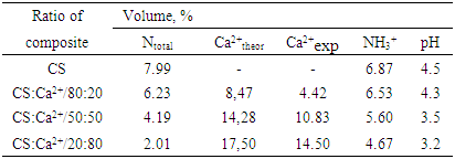
Compositions were obtained by addition in 2% solution of CS in acetic acid water solution of CaCl2 and KH2PO4 at a ratio Ca/P = 1.67. Ratio of chitosan and Ca2+-ions (CS:Ca2+) varied in intervals of 20:80; 50:50 and 80:20 (Table 1).
Table 1. Influence of synthesis conditions on the composition of Bombyx mori chitosan composites with hydroxyapatite
     
Results of the elemental analysis have shown that in increasing of content of Ca2+-ions in the reaction medium from 20 to 80 mols NH2 (CS)/Ca2+, their content in composites increased from 4.42 to 14.5%, correspondingly. With increasing of calcium content in composite, pH of the reaction mixture decreases to 3,2 that proved the hydrochloric acid (HCl) formation in the reaction:
CaCl2(aq) + KH2PO4 (aq) + 2H2O → CaHPO4 • 2H2O(S) + HCl(aq) + KCl(aq)
Decreasing the content of free amino groups of CS is possibly related to the chitosan amount decreasing in the reaction mixture.
CS-НА granules were obtained according to Pogorielov M.V., et al. 2014 [4]. As granules were obtained in alkali medium, they were washed in bi-distilled water until pH=7.
At that the solubility of chitosan composites with HA has decreased. Comparative UV-spectroscopy investigation of CS and its composites with ions of Ca2+ has been carried out. UV-spectrum of CS solution shown that absorption bands of acetamide and amino groups appeared at λmax=235 and 290 nm, accordingly (Figure 1, Curve 1).
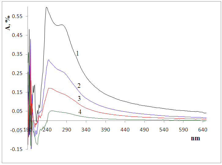
Figure 1. UV spectra of Bombyx mori CS (1) and mixtures CS:Са2+/80:20 (2); 50:50 (3) and 20:80 (4)
According to literature, absorption bands of Ca2+-ions appeared at λmax=280 nm [3], i.e. bands of absorption of chitosan amino groups and Ca2+-ions were practically coincident.
At increasing of Ca2+-ions in the composites, the intensity of absorption has decreased and bands of absorption blended in the range of 235-250 nm. That is connected with Ca2+-ions introduction into macromolecules of chitosan (Figure 1, Curves 2, 3, 4).
Structure of composites also was investigated by FTIR. In IR-spectrum of chitosan the wile band in range 3368-3289 cm-1 attributed to valent vibrations of -OH and -NH2 groups, and also vibrations of -CH2 groups at 2921-2877cm-1 have been observed (Figure 2).
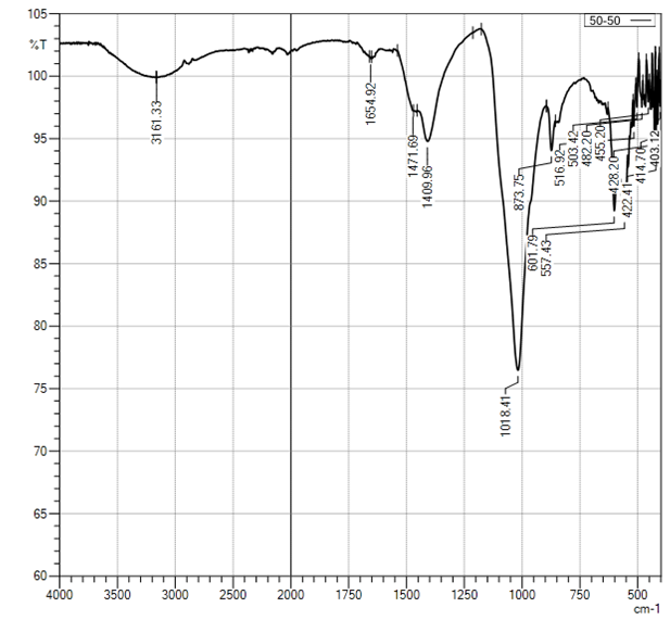
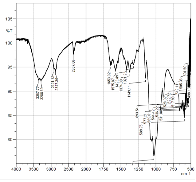
Figure 2. Curve of FTIR of Bombyx mori CS sample
In the ranges of 1653-1575 cm-1 and 1423-1321 cm-1, the deformation vibrations of -NH2 and -OH; -CH groups; at 1150-1070 cm-1 valent vibrations of other bands –C-O-C- were observed. Structures of bands in the range of 1700-920 cm-1 for chitosan and chitosan -Ca2+ samples were different in a great degree.
It is necessary to note that IR-spectra for all obtained samples of chitosan-Ca2+ were identical and characterized by absorption bands at 1150-920 and 561-602 cm-1 attributed to asymmetric valence and deformation vibrations of P-O bonds in PO43- (Figure 3). It should be noted that, in IR spectra of sample CS-Ca2+/80:20 very intensive absorption band of A-type carbonate at 1425 cm-1 and B-type carbonate at 879.54 cm-1 appeared.
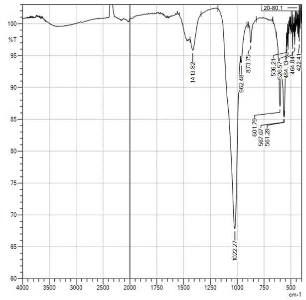
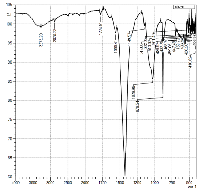
Figure 3. Curve of FTIR of sample CS-Ca2+/80:20
In spectra of samples with different CS/Ca2+ ratios, the bands of absorption -OH- (chitosan) and -PO43- groups have been overlapped at 1027 and 1120 cm-1. This has led to increasing of their intensity for 2, 7, 4, and 5 times.
In the IR-spectra of CS-Ca2+/50:50 composite an intensive absorption band of B-type carbonate appeared, in contrast to spectra of CS-Ca2+/80:20.
Figure 4. Curve of FTIR of sample CS-Ca2+/50:50
In the spectra of CS-Ca2+/20:80 the composite absorption bands relevant for CS appeared with low intensity; probably, this is related to low amount of CS in the composite (Figure 5). Nevertheless, an intensive absorption band of A-type carbonate, and a low intensive absorption band of B-type carbonate were detected.
Figure 5. Curve of FTIR of sampleCS-Ca2+/20:80
Also, it is necessary to note that in samples CS-Ca2+/50:50 and CS:Ca2+/20:80 spectra characteristic bands of functional groups of chitin and chitosan are presented as fuzzy edged small shoulder. Obviously, it is attributed to increasing content of PO43--ions.
It is known that chitosan and its derivatives have ability to form chelated complexes with ions of some metals. Formation of chelated compounds and bonds between CS and Ca2+-ions can promote deprotonation of amino-groups of chitosan. But, taking into account low ability of Ca2+-ions to form complex with chitosan, it is may be suppose that in experimental synthesis conditions the compositional materials CS-Ca3PO4 have been obtained.
A comparative XRD analysis of CS and its compositions with calcium ions has been carried out. Characteristic peaks of CS appeared at 2Ө 10 and 20°. Natalia Davidenko et al. 2010 [10] found intensive peaks of apatite crystals at 2Ө 25, 27, 28, 31.5 32, 33, 39 and 20° (Figure 6).
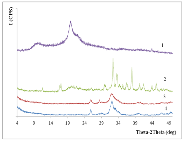
Figure 6. X-ray diffraction of samples Bombyx mori CS (1), CS:Са2+ /80:20 (2), 50:50 (3), and 20:80 (4)
Thus, in X-ray diffraction results of hydroxyapatite, chitosan not maintained the characteristic peaks of initial chitosan and appeared as peaks at 2Ө 26, 29 and 33º. Also, increasing of apatite content in the composites in 20:50:80 moles leads to appearance of new peaks at 30-50°, where the appropriate peaks intensity increases at 2Ө 31.5-34 and 37°. By increasing the content of apatite in the polymer (20→80 mole), solubility of composites decreased. Probably, this is related to calcium apatite introduction into CS macromolecule followed by increasing of composites crystallinity level [26].
Morphology of obtained hydroxyapatite samples of chitosan has been investigated by microscopic method (Figures 7-8-9).
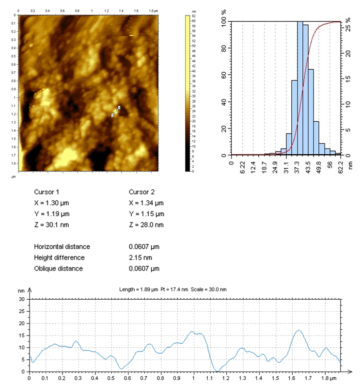
Figure 7. AFM image of CS:Са2+/20:80
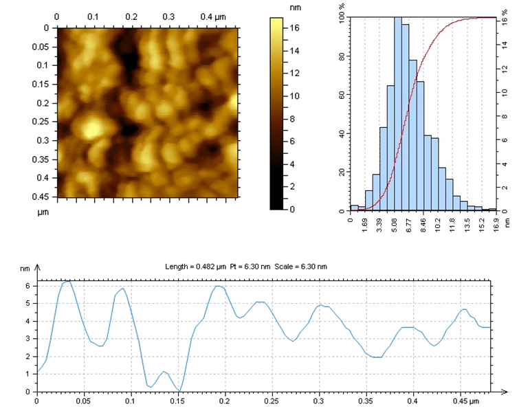
Figure 8. AFM-image of CS:Са2+/50:50
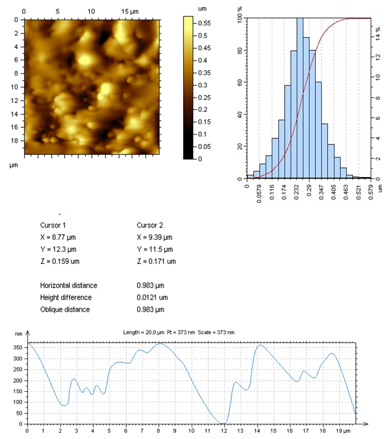
Figure 9. AFM-image of CS:Са2+/80:20
Obtained results shown that spherical nanoparticles of 25-60 nm were formed in the system at the ratio of CS:Са2+/20:80. Overall size was 40 nm and arranged 27% from general content of nanostructures. AFM-images proved that the nanoparticles were practically uniformly distributed on the polymer matrix. According to surface roughness (of the film) we can argue that at the selected synthesis conditions polydisperse nanoparticles were formed with narrow distribution along the whole surface of polymer matrix.
AFM results shown that nanoparticle sizes restricted to 2-15 nm at the ratio CS:Са2+/50:50 in comparison to CS:Са2+/20:80. Nanoparticles of an average size of 5.5 nm were 16% of total amount of particles. According to roughness of films, it was seen that the particles were distributed uniformly along the whole surface.
Obtained results show that spherical and no spherical nanoparticles in size range of 58-500 nm formed at ratio CS:Са2+/80:20 in the system. Nanoparticles with an average size of 260 nm reached 16% of total content of nanoparticles. AFM results revealed a big dispersion of nanoparticles by size and distribution along the polymer matrix. According to roughness of the surface (film), we can argue that at selected synthesis conditions polydisperse nanoparticles formed and uniformly distributed along the whole surface of the polymer matrix.
Thus, it is seen from experimental data that in the sample CS:Са2+/50:50 nanoparticles had the minimal size and were the most uniform, which is important factor in the compatibility of calcium ions with living organism. By the roughness we also evaluated the activity of hydroxyapatite chitosan (CS:Са2+/50:50), the sample had a maximal surface directly related to the biological activity of nanoparticles.
Interaction of chitosan to with calcium ions
There are different assumptions on mechanism of chitosan interaction to with hydroxyapatite, namely, with calcium ions. It was established that calcium ions interact with amino groups of CS and form coordination bonds [2, 5]. However, Davidenko N.et al. [1] showed that during the interaction of calcium ions with electron donors, functional groups of CS, they absorb by the macromolecule. Adsorption mechanism of metal ions by chitosan depends on several factors such as pH of solution, molecular mass characteristics of CS, molecular mass (ММ) and deacetylation level (DAL), particle size, porosity etc. [27].
Investigation results showed that interaction of CS with hydroxyapatite occurs in sharp decreasing of рН (Table 1). It is known that CS in diluted solutions of organic and mineral acids remains as a salt, which obstruct the interaction of amino groups with metal cations:
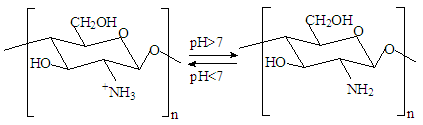
In the selected conditions for obtaining the composites of CS with Са2+, рН of reaction system рН<7 prevents interaction of Са2+ with NH3+. The results of conductometric titration, UV and FTIR investigations shown that interaction of chitosan macromolecule with hydroxyapatite molecule occurs due to hydrogen bonds of CS and НАfunctional groups.

4. Conclusions

Composites of Bombyx mori chitosan with hydroxyapatite with content of Ca2+-ions till 17.5% have been obtained. It was found that, at the selected synthesis conditions, nanostructure composites with minimal sizes from 2 to 15 nm obtained.
It was established that the interaction occurs between the functional groups of chitosan and hydroxyapatite by hydrogen bonds. It was shown that the introduction of hydroxyapatite into chitosan macromolecule leads to formation of insoluble composites. Low soluble composites CS-Ca2+ were obtained in increasing of Ca2+-ions content in the system. They are of interest of the biomaterials obtaining for traumatology, dentistry and veterinary.

References

[1]  Davidenko N., Carrodeguas R.G., Peniche C., Solis Ya., Cameron Ruth E. Chitosan/apatite composite beads prepared by in situ generation of apatite or Si-apatite nanocrystals. Acta Biomaterialia 2010. 6:рр.466–476. [doi: 10.1016/j.actbio.2009.07.029]
[2]  Rozita A.R., Rohana A., Abu В.,Sam’an M.M. Synthesis and Characterization of Pure Nanoporous Hydroxyapatite. Journal of Physical Science, 2011 Vol. 22(1): pp.25–37. [doi:http://web.usm.my/jps/22-1-11/22.1.2.pdf]
[3]  Luciano P., Magdalena K. Properties and structure of microcrystalline chitosan and Hydroxyapatite composites. Journal of Biomaterials and Nanobiotechnology, 2014. 5: pp. 128-138. [doi:https://file.scirp.org/pdf/JBNB_2014042515442779.pdf]
[4]  Pogorielov M.V., GusakYe.V., Babich I.M., Kalinkevich O.V., Kalinkevich A.N., Somokhvalov I.I., Danilchenko S.N., Skliar A.M. Trace elements sorption by the chitosan-based materials. J. Clin. Exp. Med. Res., 2014. 2(1): pp.88–99.
[5]  Jayachandran V., Se-Kwon Kim. Chitosan Composites for Bone Tissue Engineering–An Overview// Mar. Drugs 2010.8: pp.2252-2266. [doi:10.3390/md8082252]
[6]  Danilchenko S.N., Kalinkevich O.V., Pogorelov M.V., Sklyar A.M., Kalinichenko T.G., Kalinkevich A.N., Starikov V.V., and etc. Experimental substantiation of the use of composite materials based on chitosan and calcium phosphates for the replacement of bone defects. Orthopedics, traumatology and prosthetics. 2009. Vol.1. pp. 66–72. (in Russian)
[7]  Di Martino A., Sittinger M., Risbud M.V. Chitosan: A versatile biopolymer for orthopaedic tissue-engineering. Biomaterials 2005. 26:pp.5983–5990. [doi: 10.1016/j.biomaterials.2005.03.016]
[8]  Hu Q., Li B., Wang M., Shen J. Preparation and characterization of biodegradable chitosan/hydroxyapatite nanocomposite rods via in sit hybridization: a potential material as internal fixation of bone fracture. Biomaterials 2004. 25:pp.779–785. [doi:https://www.ncbi.nlm.nih.gov/pubmed/14609666]
[9]  Teng S., Lee E., Yoon B., Shin D., Kim H., Oh J. Chitosan/nanohydroxyapatite composite membranes via dynamic filtration for guided bone regeneration. J. Biomed. Mater. Res. Part A 2009. 88: pp.569–580. [doi:10.12691/ajmse-5-1-2]
[10]  Yamaguchi I., Tokuchi K., Fukuzaki H., Koyama Y., Takakuda K., Monma H., et al. Preparation and microstructure analysis of chitosan/hydroxyapatite nanocomposites. J of Biomedical Materials Research, 2001. 55:pp.20–27. [doi:10.1186/1423-0127-16-65]
[11]  Kim S.K., Mendis E. Bioactive compounds from marine processing byproducts-A review. Food Res. Int. 2006. 39: pp. 383–393. [doi:https://doi.org/10.1016/j.foodres.2005.10.010]
[12]  Rusu V. M., Ng C. H., Wilke M., Tiersch B., Fratzl P., Peter, M. G. Size-controlled hydroxyapatite nanoparticles as self-organized organic–inorganic composite materials. Biomaterials 2005. 26: pp. 5414–5426. [doi: 10.1016/j.biomaterials.2005.01.051]
[13]  Pena J., Izquierdo-Barba I., Martinez A., Vallet-Regi M. New method to obtain chitosan/apatite materials at room temperature. Solid State Sci. 2006. 8(5): pp.513-519.
[14]  Monica V.Z., Richard A.S., Benjamin T.R., Jessica A.J., Jared O.C., Warren O.H., Joel D.B. Physical properties and in vitro evaluation of collagen–chitosan–calcium phosphate microparticle-based scaffolds for bone tissue regeneration. Journal of Biomaterials Applications 2012. 28(4):pp.566–579. [doi:https://doi.org/10.1177/0885328212465662]
[15]  Wilson R.M, Elliott J.C., Dowker SEP, Rodriguez-Lorenzo L.M. Rietveld Refinements and spectroscopic studies of the structure of Ca-deficient apatite. Biomaterials 2005; 26: pp.1317–27. [doi:10.1016/j.biomaterials.2004.04.038]
[16]  Bachurin A.V. Analysis of osseointegration of titanium implants with additional stimulation of their surface osteoinductive effect. Bulletin of Volgograd State University. Series 9. Issue. 11. 2013. pp. 58-61. (in Russian)
[17]  Starikov V.V., Rudchenko S.O. Optimization of the properties of a composite based on hydroxyapatite and chitosan by varying its composition and heat treatment regimes. Bulletin of Kazan National University, No. 915, Series "Physics", Issue. 2010.14:pp.35-39. (in Russian)
[18]  Haiguang Z., Lie M., Changyou G. Jiacong Sh., Fabrication and properties of mineralized collagen-chitosan/hydroxyapatite scaffolds// Polym. Adv. Technol. 2008; 19: pp.1590–1596.[doi:10.1002/pat.1174]
[19]  Xianmiao C., Yubao L., Yi Z., Li Z., Jidong L., Huanan W. Properties and in vitrobiological evaluation of nano-hydroxyapatite/chitosan membranes for bone guided regeneration. Mater. Sci. Eng. C 2009. 29:pp.29–35. [doi: 10.3390/md8082252]
[20]  Kikuchi M., Ikoma T., Itoh S., Matsumoto H., Koyama, Y., Takakuda K., Shinomiya K., Tanaka J. Biomimetic synthesis of bone like nanocomposites using the self-organization mechanism of hydroxyapatite and collagen. Compos. Sci. Technol. 2004. 64:pp.819–825. [doi: 14580909]
[21]  Ergashev К.H., Vokhidova N.R., Rashidova S.Sh."On the preparation of the compositions of chitosan Bombyx mori with calcium phosphate", Rep. conf. Urgench, 20-21 April 2017. 2b:pp.22-23. (in Russian)
[22]  Pavlov G.M., Selyunin S.G. Polym. Sci.1925. 8(8). pp.1986.
[23]  Klimova V.A. Main Micromethods of Organic Compound Analysis. Chemiya, Moscov. 1980. (in Russian)
[24]  Pogorielov M.V., GusakYe.V., Babich I.M., Kalinkevich O.V., Kalinkevich A.N., Somokhvalov I.I., Danilchenko S.N., Skliar A.M. Trake elements sorption by the chitosan-based materials. J. Clin. Exp. Med. Res. 2014. 2(1):pp88-99.
[25]  Tager A.A. Physicochemistry of polymers. (4th edition.). / Edited by Askadsky A.A. М: Nauchniy mir. 2007. pp. 576. (in Russian).
[26]  Vokhidova N.R. Synthesis, properties and application of polymer metal complexes of chitosan Bombyx mori and the chitosan stabilized nanoparticles of d-metals. Abstract of the doctoral dissertation. Tashkent. 2016. P.190.
[27]  www.pharmencyclopedia.com.ua/article/7074/kalciyu-xlorid