American Journal of Organic Chemistry

p-ISSN: 2163-1271    e-ISSN: 2163-1301

2016;  6(3): 86-92

doi:10.5923/j.ajoc.20160603.02


Assembled Nanoparticle Films with Crown Ether Derivatives as Sensors for Metal Ions

Kevin W. Kittredge1, Justin T. Malinowski1, Alison M. Washington1, Robert W. Dayand2, Michael C. Leopold2

1Department of Chemistry, Virginia Wesleyan College, Wesleyan Dr., Norfolk, VA, USA

2Department of Chemistry, University of Richmond, Richmond, VA, USA

Correspondence to: Kevin W. Kittredge, Department of Chemistry, Virginia Wesleyan College, Wesleyan Dr., Norfolk, VA, USA.

Email:

Copyright © 2016 Scientific & Academic Publishing. All Rights Reserved.

This work is licensed under the Creative Commons Attribution International License (CC BY).
http://creativecommons.org/licenses/by/4.0/

Abstract

This work represents our ongoing studies of potentially novel sensing materials that utilize the ionophoric capacity of crown ethers attached to metallic nanoparticles by aromatic linkers and assembled as films. These films were better able to detect changes in potassium ion concentration than those previously reported using alkyl linkers. These nanomaterials are small enough to be considered for invivo and remote sensing applications as well as portable measurement devices.

Keywords: Monolayer protected clusters films, Crown ether, Metal ion sensors, Nanoparticles

Cite this paper: Kevin W. Kittredge, Justin T. Malinowski, Alison M. Washington, Robert W. Dayand, Michael C. Leopold, Assembled Nanoparticle Films with Crown Ether Derivatives as Sensors for Metal Ions, American Journal of Organic Chemistry, Vol. 6 No. 3, 2016, pp. 86-92. doi: 10.5923/j.ajoc.20160603.02.

1. Introduction

Each year in the United States, there are more than 200 million clinical tests performed for metal cations in physiological systems [1]. Potassium and sodium play critical roles in many of the body’s important functions including nerve conduction, muscle activity, acid-base equilibria, and cardiac regulation. Insufficient levels of potassium, a condition known as hypokalemia, can cause muscle weakness, paralysis, and eventually cardiac arrest. Hyperkalemia or elevated levels of potassium can lead to mental confusion, respiratory muscle failure, and cardiac abnormalities. Even small changes in potassium concentration can signal the onset of some of these conditions [2]. Considering potassium’s important role in vital physiological functions, the measurement of potassium concentration is an excellent diagnostic tool for disorders and it is crucial that clinicians can effectively monitor potassium levels, especially during emergencies.
Currently, the majority of these measurements are performed using potentiometry with ion selective electrodes (ISEs) that are capable of selectively detecting individual target analytes in a complex matrix such as blood or urine [3].
While this method of analysis has proven effective for years, several disadvantages persist. The ISEs themselves have a limited lifetime and are of substantial cost. ISEs are highly susceptible to fouling, resulting in diminished performance, especially at lower concentration levels where they lack adequate sensitivity [4]. More importantly, the manner in which potassium is currently measured is time consuming at a moment when more immediate information would be more beneficial for expedited diagnosis and treatment of patients.
The booming research efforts into nanotechnology have afforded a plethora of new materials with unique properties [5]. One of these materials is metallic nanoparticles or colloidal metal whose size and properties are intermediate between molecular and bulk metal [6]. Of particular interest has been the monolayer protected cluster nanoparticle (MPC) [7], which consists of a metallic core with a peripheral layer of stabilizing molecules, commonly alkanethiolates (Figure 1) [8]. The protective layer of molecules prevents coagulation of the metallic cores back to bulk metal and allows them to be easily handled, dissolved in solution, and recollected from solution [7]. MPCs can be easily modified with additional ligands through facile place-exchange reactions where a functionalized ligand (i.e. fluorophore, redox molecule, or simple functional group) can be incorporated into the peripheral layer of the material (Figure 1A→1B) [9]. Their amenity to functionalization, along with their large surface to volume ratio, makes these nanoparticles an attractive material for potential sensing applications.
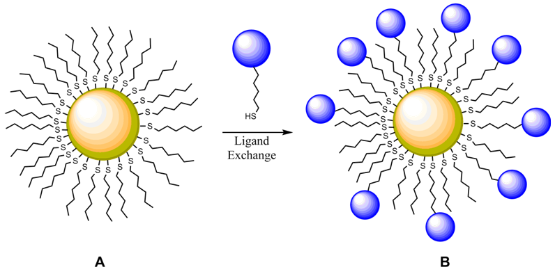
Figure 1. Monolayer protected cluster (MPC) nanoparticle undergoing ligand exchange
A nanoparticle’s geometry, size, type of protecting ligand, polarity of its medium, and any change in its proximate surroundings affect its surface plasmonband (SPB), which can be monitored by spectroscopy in the visible spectrum of light [10, 11]. Therefore, if ananalyte of interest (i.e. potassium) can somehow induce a noticeable change in a nanoparticle’s SPB, then this process can be exploited as a method of analyte detection. Chen and co-workers were successful in modifying citrate stabilized MPCs with ionophoric crown-ether (CE) moieties [10, 11]. CE molecules possess a cavity that is size selective for a specific metal ion [12]. Chen incorporated 15-crown-5 groups (Figure 2) in MPCs to create nanoparticles in solution that were self-selective for K+, sandwiching the metal ion between two CE moieties from separate nanoparticles, and for Na+, where a single ion complexes with a single crown. A solution of these modified nanoparticles displays a colorimetric shift from red (Na+complexes) to blue as CE groups “sandwich” K+ and consequently aggregate the MPC cores. Chen successfully applied this system to K+ detection in a high salt matrix (simulated serum) [10] and in human urine [11]. The visible color change observed is a function of the core proximity, but closer inspection of Chen’s results, reveals that the MPCs, upon exposure to K+, are actually aggregated and precipitating out of solution, meaning the test is not reversible and not practical for any type of in vivo potassium measurements.
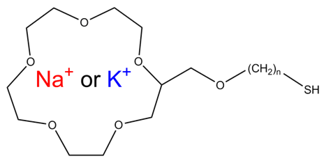
Figure 2. Structure of alkanethiol modified 15-crown-6 ether
Previously we have reported that MPCs incorporated into thin films in non-aqueous environments can be used as a potassium specific detection method [13, 14]. The film geometry offers many advantages for the aforementioned sensing applications, rendering precipitation a non-issue. Earlier research has indicated that MPC assembled films are extremely flexible and reactive to their local environment, exhibiting increases and decreases in inter-core spacing as the film swells with solvent and is subsequently exposed to air, respectively. MPC films are already widely investigated for their chemiresistive sensing capabilities of liquids and chemical vapor by observing differences in the film’s spectroscopic signature, mass, and conductivity upon exposure to a variety of metal ions [13-20].
As for film assembly itself, we and others have reported how networking functionalized MPCs into films affects sensing capabilities, including the use of dithiol [19, 20], estercoupled [14], and metalcarboxylate interparticle linkages [21, 22]. Ionophoric crown ether moieties were attached to the monolayer protected clusters (CE-MPCs) and used as an integral part of a novel linking mechanism. CE modified MPCs were linked together and grown on a substrate utilizing CEmetal ion-CE sandwiches – an interaction shown to be effective enough to grow substantially thick films. The films networked in this manner are sufficiently electronically connected as evidenced by voltammetric measurements [22].
Other CE- and non-CE modified MPC films have been studied extensively as chemiresistive sensors [23-27]. These films are reported to be extremely flexible materials which could expand or contract in the presence of certain solvents. Changes in spacing between the film's layers resulted in changes in their conductivity, mass, and surface plasmon band emission [27, 28].
It is clear that the SPB can be used to diagnoseinterparticle spacing within nanoparticle solutions and films [15]. However, film structures are severely constrained compared to solution systems so the response to metal ions was found to be much more subtle. In this paper, CE moieties have been modified with a conjugated aromatic section (Figure 3) to create a more rigid structure, than compared to simple alkyl linkers. This type of linker may facilitate electronic communication between the CE-metal ion complex and the metallic nanoparticle, most likely by a through-space mechanism [29]. This work shows that CE-MPC films may serve as selective, metal ion sensitive materials.
Figure 3. Structure of biphenyl thiol modified 15-crown-6 ether monolayer protected cluster (CE-MPC)

2. Experimental

NaH (Acros, 80% dispersion in mineral oil), THF (Acros, 99.9% anhydrous), 2(hydroxymethyl)15-crown-5 ether (Acros), 4,4’-Bis(bromomethyl)biphenyl (TCI America), tetrabutylammonium fluoride (Acros, 1M in THF containing 5% water), hexamethyldisilathiane (Aldrich), tetrachloroauric(III) acid (Acros), tetranoctylammonium bromide (Avocado), sodium borohydride (Fisher), 1-hexanethiol (Acros), toluene (Aldrich), CH2Cl2 (Acros, extra dry, water <30 ppm, stabilized), and (3mercaptopropyl)-trimethoxysilane (Aldrich) were used as received without further purification. All reactions were run under argon atmosphere. Flash column chromatography was performed with Fisher 60-200 mesh silica gel. All glassware was dried before use at 160°C. 1H and 13C nuclear magnetic resonance spectra were record on aJeol 400 MHz spectrometer in CDCl3 or CD2Cl2.High Resolution mass spectral data (HRMS) were recorded on an Agilent Technologies 6210 LC-TOF by use ofelectron spray ionization (ESI). UV-Vis spectra were recorded on a Perkin Elmer Lambda 35 spectrometer.
The synthesis of 2-(4'-Mercaptomethyl-biphenyl-4-methoxymethyl)-15-crown-5 ether (2) is described below and outlined in Scheme 1.
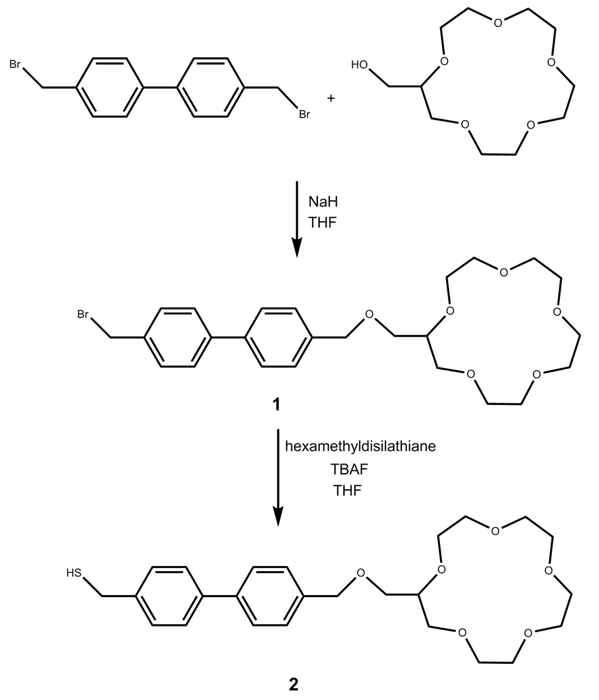
Scheme 1. Synthesis of 2-(4'-Mercaptomethyl-biphenyl-4 methoxymethyl)-15-crown-5 ether (2)
2-(4'-Bromomethyl-biphenyl-4-methoxymethyl)-15-crown-5 ether (1).
2(hydroxymethyl)15-crown-5 ether (1.0 g, 4 mmol) was dissolved in THF (15 mL) and stirred at room temperature. NaH (0.22 g, 9.2 mmol) was added, and stirred until fizzing stopped. 4,4’Bis(bromomethyl)biphenyl (5.0 g, 14.7 mmol) was added to THF (10 mL) in a separate flask. The crown ether solution was then added to the biphenyl solution via an addition funnel dropwise at 0°C. The reaction was then allowed to warm to room temperature and stirred for 48 hours. The reaction was then quenched with isopropanol (25 mL) and distilled water (15 mL). All solvents were removed in vacuo yielding a white/yellow crude product. The product was then dissolved in CH2Cl2 and loaded onto a short column. This was washed with EtOAc (300 mL), CH2Cl2 (300 mL), and methanol (150 mL). The methanol fraction containing the product was dried and dissolved in CH2Cl2, and the white insoluble material was filtered out leaving a pale yellow solution. Solvent was removed in vacuo yielding a pale yellow oil. 1H NMR (400 MHz, CDCl3) δ 3.4-4.0 (m, 21 H), 4.45-4.75 (d, 4 H), 7.3-7.65 (d, 8 H). 13C NMR (100 MHz, CDCl3) δ 22.2, 33.3, 69.4, 69.7, 70.6, 73.5, 127.2, 128.4, 137.1, 140.3. HRMS (calcd for C25H33BrO6) 508.1461, found 508.1440.
2-(4'-Mercaptomethyl-biphenyl-4-methoxymethyl)-15-crown-5 ether (2). Bromide 1 (0.10 g, 0.20 mmol) was dissolved with stirring in CH2Cl2 (5 mL) and cooled to 0°C. Hexamethyldisilathiane (0.043 g, 0.24 mmol) was then added along with tetrabutylammonium fluoride solution (0.222 g, 0.22 mmol). This solution was allowed to warm to room temperature while being stirred overnight. The product was then diluted with CH2Cl2 (10 mL), washed eight times with saturated ammonium chloride solution (5 mL), followed by two washes with DI water (5 mL), and then dried with MgSO4. The CH2Cl2 was then removed in vacuo yielding a viscous yellow oil. 1H NMR (400 MHz, CDCl3) δ 3.5-3.85 (m, 22 H), 4.5-4.75 (m, 4 H), 7.3-7.6 (d, 8 H). 13C NMR (100 MHz, CDCl3) δ 33.2, 68.6, 69.1, 69.6, 70.6, 73.3, 127.1, 127.2, 127.6, 128.4, 128.9, 129.6, 137.1, 160.9. HRMS (calcd for C25H34O6S) 462.2076, found 462.2078.
Synthesis of monolayer protected clusters (MPC). Tetra-n-octylammonium bromide (1.1 g, 2.01 mmol) was weighed into a 250 mL flask, followed by the addition of toluene (30 mL). Tetrachloroauric (III) acid (0.27 g, 0.787 mmol) was added to the flask along with distilled water (25 mL). This biphasic solution was stirred vigorously for 30 minutes. The water layer was discarded and hexanethiol (0.186 g, 1.57 mmol) was added. This solution was stirred until it turned clear (20 minutes). It was chilled to 0°C and a cold sodium borohydride (0.38 g, 10.3 mmol) solution in water (25 mL) was added. The solution immediately turned black and was stirred at 0°C for 24 h. The water layer was discarded and the toluene layer was dried in vacuo. The black solid was suspended in acetonitrile (200 mL) and stirred overnight. This solution was filtered through a medium porosity frit and washed with acetonitrile (200 mL), yielding a black solid. 1H NMR (400 MHz, CDCl3) δ 0.7-0.95 (s), 1.2-1.45 (d), 1.5-1.55 (s) 1.6-1.8 (d).
Ligand exchange. MPC 1 (0.140 g) was added to a 200 mL flask. CH2Cl2 (50 mL) was then added to the flask along with the thiol ligand 2 (0.400 g). The flask was sealed, covered with aluminum foil to block light, and stirred for 72 hours. The solvent was then removed in vacuo. The black solid was recrystallized with acetonitrile and collected by filtration. 1H NMR (400 MHz, CDCl3) δ 0.7-0.95 (s), 1.2-1.5 (s), 1.5-1.9 (s), 3.4-3.8 (s), 4.4-4.7 (s), 7.2-7.6 (d).
CE-MPC film assembly. Films were prepared by a variation of the protocol outlined by Pompano et al [19]. Quartz slides (2.5 cm2) were washed with Piranha solution (2:1 sulfuric acid:hydrogen peroxide), rinsed liberally with distilled water, and dried. A 5 mM solution of (3-mercaptopropyl)-trimethoxysilane in isopropanol was prepared. This was heated to 50°C. The slides were immersed in the solution for 45 minutes, and washed with isopropanol. They were then cured at 100°C for several hours. This silanization process was then repeated to ensure adequate coverage of silane on the glass would serve as an anchoring layer. Slides were then dipped into a solution of 10 mg CE-MPC in 10 mL CH2Cl2 for 45 minutes. They were then rinsed with acetonitrile, dried in a stream of nitrogen, and immersed in an aqueous 0.1 M sodium acetate solution for 15 minutes. After rinsing with water, the process was repeated 15-20 times until films reached a desired thicknessas monitored by UV-Vis spectroscopy.
Testing CE-MPC film for metal ion sensing. Several solutions of potassium nitrate in water (0.1 mM, 0.5 mM, 1 mM, 5 mM, 10 mM, 20 mM, 30 mM, 40 mM, and 50 mM) were prepared. The metal sensing ability of the multilayer films was measured by taking baseline spectra of the film both dry in a cuvette and wet (distilled water). The slide was then placed in a cuvette filled with the desired potassium nitrate solution and the absorbance spectrum was measured (Figures 6 & 7).

3. Results and Discussion

Film Growth and Structure
For these films to be useful as metal ion sensors the linking and sensing mechanisms must be separate. Also, the films must be quite flexible within their networks so any solvent and ionophores may permeate through the film. Film growth dynamics were monitored spectroscopically, as previously described [12-15, 24]. Films were assembled in dichloromethane, which completely dissolved the CE-MPCs. The biphenyl pendant chain on the crown ether was selected to serve two purposes. First, the two aromatic rings in the biphenyl system would provide a more structurally rigid scaffold upon which the layered film would be constructed, thereby reducing the density of the film upon the addition of subsequent layers. Second, the extended conjugated system would facilitate electronic communication between the coordinated metal in the crown ether and the gold nanoparticle. Our earlier work showed that while electronic communication was occurring, it was seen in only the surface plasmon emission band. This gave very small changes in absorption intensity in the UV-Vis spectrum as a response to metal ion coordination. Figure 4 shows the CEmetal ion – CE bridges that act as the linking mechanism between neighboring nanoparticles. Figure 5 is the absorbance spectra of film growth using sodium ions as the linking metal coordinated in a sandwich configuration. The spectra are typical of MPC films and their growth with a decreasing absorbance at higher wavelengths and a surface plasmon band (SPB) near 540 nm that increases with each exposure to MPCs [19]. Interestingly, the biphenyl linkage produced another absorption in the UV-Vis spectrum at 260 nm. It follows that with each dip cycle, as the film becomes thicker with the immobilization of additional MPC possessing CE ligands. Also, as the film became thicker, a red shift in the absorbance maximum for the SPB was observed (Figure 5, inset), indicative of an increase in MPC cross-linking and a necessary decrease in interparticle spacing [12, 13, 20].
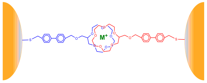
Figure 4. Structure of CE-MPC metal ion sensing film of biphenyl thiol modified 15 crown-6 ether "sandwich"
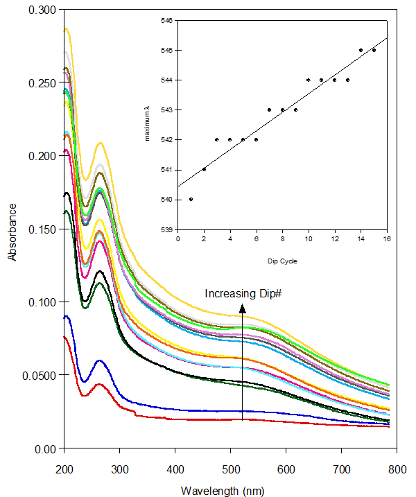
Figure 5. UV-Vis spectra after each dip cycle during the growth of the CE-MPC. Inset is the plot of maximum λ versus dip cycle. Increasing slope shows red shift as film layers thicken
Response to potassium ion in solution
It is hypothesized that these networked films of CE-MPCs should respond to metal ions in solution as they would in solution. Our CEMPCs films were assembled using sodium ion as the coordinated metal in the crown ether cavity. We have previously shown that these films are able to exchange sodium for other metal ions and undergo a small red or blue shift in the surface plasmon band [12, 13]. The films studied here showed a similar response to potassium ion, with a slight red shift in the SPB. Unfortunately, this shift was too small to be of practical use in ascertaining the concentration of potassium (Figure 6). However, a significant change in absorbance as well as a slight red shift of λmax for the absorbance is observed at lower wavelengths (Figure 7 and inset). This response to increasing concentrations of potassium, seen in Figure 7, is attributed to the interparticle conjugation of biphenyl linkers on neighboring nanoparticles. Because the absorbance increased with increasing potassium ion concentration, a calibration curve is able to be created (Figure 8).
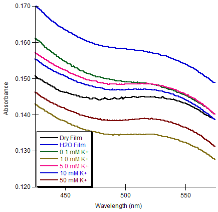
Figure 6. UV-Vis spectra showing response to potassium ion of a CE-MPC assembled film in the surface plasmon band region. A very slight red shift is shift is observed in the presence of increasing K+ metal ion concentration in the surface plasmon band maximum absorbance
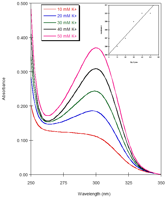
Figure 7. UV-Vis spectra showing response to potassium ion of a CE-MPC assembled film. Inset is the plot of maximum λ versus concentration of K+ ion and shows a very slight red shift as K+ metal ion concentration increases
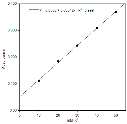
Figure 8. Plot of Absorbance versus potassium ion concentration for the CE-MPC films. λmax = 300 nm

4. Conclusions

The ability to construct a calibration curve demonstrates the utility of these films as diagnostic devices for potassium ion concentration. Future films will be designed containing rigid, aromatic (or polyaromatic) linkers whose purposes will be to strengthen the films structure and control the spacing between layers and help facilitate electronic communication between the metal cation captured in the crown ether and the metallic nanoparticles.

ACKNOWLEDGMENTS

The investigators gratefully acknowledge the National Science Foundation, CHE-0643709 (JTM & KWK) and CHE-1401593 (MCL) as well as Virginia’s Commonwealth Health Research Board (RWD) for support. KJD was supported by a fellowship from the Virginia Federation of Independent Colleges. Additional support for KWK was provided by a Virginia Wesleyan College Faculty Summer Development Grant.

References

[1]  Harris, D.C., Quantitative Chemical Analysis, 6th Edition, W.H. Freeman and Company, New York, 2003.
[2]  Evans, K.J.; Greenberg, A., Journal of Intensive Care Medicine 2005, 10, 272 and references therein.
[3]  N.W. Tietz; Fundamentals of Clinical Chemistry, Fifth Edition. W. B. Saunders Co., Philadelphia, PA, 2001.
[4]  G. Ramsay, Ed. Commercial Biosensors Applications to Clinical, Bioprocess, and Environmental Samples, John Wiley & Sons, Inc.: New York, 1998 and references therein.
[5]  a) “Nanotechnology for the Twenty-First Century: Toward a New Industrial Revolution,” report of the Interagency Working Group on Nano Science, Engineering and Technology, National Science Foundation, 1999. b) Wu, C. Prism 2004, 14, 1.
[6]  R. W. Murray, Chem. Rev. 2008, 108, 2688; and references therein.
[7]  Templeton A.C., Wuelfing, W.P.; Murray, R.W. Acc. Chem. Res. 2000, 33, 27; and references therein.
[8]  Grabar, K.C.; Freeman, R.G.; Hommer, M.B.; Natan, M.J. Anal. Chem. 1995, 67, 735.
[9]  Templeton, A.C.; Hostetler, M.J.; Warmoth, E.K.; Chen, S.; Hartshorn, C.; Krishnamurthy, V.M.; Forbes, M.D.E.; Murray, R.W. J. Am. Chem. Soc. 1998, 120, 4845.
[10]  Lin, S.Y.; Liu, S.W.; Lin, C.M.; Chen, C.H. Anal. Chem. 2002, 74(2), 330.
[11]  Lin, S-Y.; Chen, C.; Lin, M-C.; Hsu, H-F. Anal. Chem. 2005, 77, 4821.
[12]  Gokel, G.W.; Leevy, W.M.; Weber, M.E. Chem. Rev. B 2004, 104, 2723.
[13]  Pompano, R. R.; Russell, L. E.; Wortley, P. G.; Moatz, L. M.; Tognarelli, D. J.; Kittredge, K. W.; Leopold, M. C., Thin Solid Films 2006, 510, 311.
[14]  Russell, L. E.; Pompano, R. R.; Kittredge, K. W.; Leopold, M. C. J. Mat. Sci.2007, 42, 7100.
[15]  Zamborini, F.P.; Leopold, M.C.; Hicks, J.F.; Kulesza, P.J.; Malik, M.A.; Murray, R.W. J. Am. Chem. Soc. 2002, 124, 8958.
[16]  Tognarelli, D. J.; Miller, R.B; Pompano, R.R.; Loftus, A.F.; Sheibley, D.J.; Leopold, M. C. Langmuir 2005, 21, 11119.
[17]  Wohltjen, H.; Snow, A.W. Anal. Chem. 1998, 70, 2856.
[18]  Han, L.; Daniel, D.R.; Maye, M.M.; Zhong, C.-J. Anal. Chem. 2001, 73, 4441.
[19]  Grate, J.W.; Nelson, D.A.; Skaggs, R. Anal. Chem. 2003, 75, 1868.
[20]  Evans, S.D.; Johnson, S.R.; Cheng, Y.L.; Shen, T. J. Mater. Chem. 2000, 10, 183.
[21]  Loftus, A.; Reighard, K. P.; Kapourales, S. A.; Leopold, M.C. J. Am. Chem. Soc.2008, 130, 1649.
[22]  Sheibley, D.; Tognarelli, D.J.; Szymanik, R.; Leopold, M.C. J. Mater. Chem. 2005, 15, 491.
[23]  Kittredge, M. C.; Durst, T. S.; Kittredge, K. W. Thin Solid Films 2010, 518, 3949.
[24]  Nath, N.; Chilkoti, A.; Anal. Chem. 2002, 74, 504.
[25]  Zellers, E. T.; Cai, Q-Y. Anal. Chem. 2002, 74, 3533.
[26]  Krasteva, N.; Besnard, I.; Guse, B.; Bauer, R. E.; Mullen, K.; Yasuda, A.; Vossmeyer, T. Nano Lett. 2002, 2, 551.
[27]  Leopold, M. C.; Donkers, R. L.; Georganopoulou, D.; Fisher, M.; Zamborini, F. P.; Murray, R. W. Faraday Discuss. 2004, 125, 63.
[28]  Lowe, C. R.; Blyth, J.; Millington, R. B.; Mayes, A. G. Anal. Chem.2002, 74, 3649.
[29]  Kittredge, K. W.; Fox, M. A.; Whitesell, J. K. J. Phys. Chem. B 2001, 105, 10594.