Khabibulla Khayrullaevich Pulatov1, Abboskhuja Khikmatilla ugli Ubaydullaev2
1Associate Professor, Department of Human Anatomy and Operative Surgery with Topographic Anatomy, Tashkent State Medical University, Candidate of Medical Sciences, Uzbekistan
2Assistant, Department of Internal Medicine, Therapy in Family Medicine, Military Field Therapy, and Hematology, Termez Branch of Tashkent State Medical University, Uzbekistan
Correspondence to: Khabibulla Khayrullaevich Pulatov, Associate Professor, Department of Human Anatomy and Operative Surgery with Topographic Anatomy, Tashkent State Medical University, Candidate of Medical Sciences, Uzbekistan.
| Email: |  |
Copyright © 2026 The Author(s). Published by Scientific & Academic Publishing.
This work is licensed under the Creative Commons Attribution International License (CC BY).
http://creativecommons.org/licenses/by/4.0/

Abstract
Rats with diabetes mellitus that were fed pesticides were used to evaluate the morphological substrates of changes in the pancreas in specific cells and to study their characteristic features.The Ki67 marker is primarily manifested by its expression in the perinuclear region of any labile and stable cells and is considered a marker that determines proliferative activity. This is important for assessing the mitotic index specifically in the labile and stable cells of each organ.
Keywords:
Diabetes mellitus, Morphology, Pancreas, Pesticides
Cite this paper: Khabibulla Khayrullaevich Pulatov, Abboskhuja Khikmatilla ugli Ubaydullaev, Immunohistochemical Alterations in the Pancreas of Rats Under Chronic Pesticide Exposure and Experimental Diabetes Mellitus, American Journal of Medicine and Medical Sciences, Vol. 16 No. 6, 2026, pp. 2835-2840. doi: 10.5923/j.ajmms.20261606.15.
1. Introduction
Currently, diabetes mellitus is the third most prevalent disease globally and is considered a global medical and social problem for healthcare systems in all countries, affecting patients of all ages.According to WHO data [12], by 2030, 18-20% of the world's population will have this disease, with 80-90% of them having type 2 diabetes. In industrialized countries, the prevalence of diabetes is 5-6%, with a very high probability of incidence and increase primarily among the population over 40 years of age. The WHO projects that the number of patients with diabetes in developed countries will increase by 41% by 2027. Complications leading to a decreased quality of life, disability, and death in patients with diabetes include diseases affecting all internal organ systems. Despite extensive experience in treating diabetes, a majority of patients become disabled. Therefore, they represent a serious medical and social problem.
2. Materials and Methods
In this study, an experimental model of diabetes mellitus was induced in 180 white laboratory rats (at a dose of 0.11 mg/100 g of body mass). The white laboratory rats were divided into 3 groups. Group 1 consisted of 60 intact rats, serving as the control group. Group 2 was the experimental group, comprising 60 white laboratory rats in which an experimental model of diabetes mellitus was induced. Group 3 consisted of 60 rats with chronic pesticide poisoning under the conditions of experimental diabetes mellitus.
3. Results and Discussion
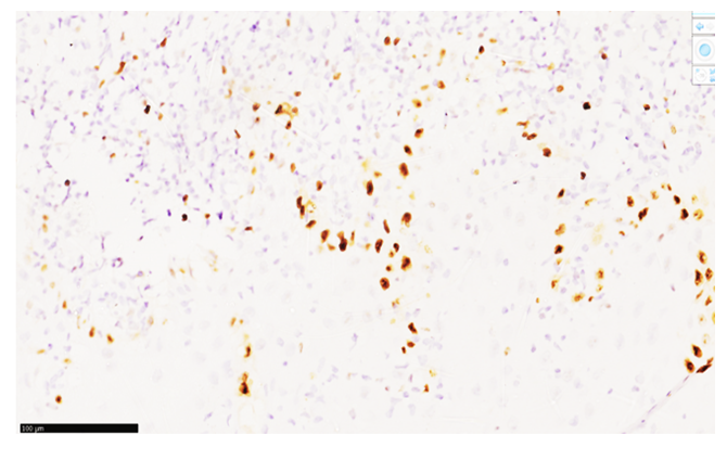
To evaluate the morphological substrates in specific cells and study their characteristics, we examined pancreatic changes in rats with diabetes mellitus that were fed with pesticides.The Ki-67 marker is considered a marker of proliferative activity, primarily manifested by its expression in the perinuclear region of any labile and stable cells. This is important for assessing the mitotic index in the labile and stable cells of each organ.Specifically, in our research, when determining the reaction of the Ki-67 marker in the pancreatic epithelium of rats with diabetes mellitus fed with pesticides, it was found that the cells' proliferative index showed high positive expression within the initial 3-month period. This is explained by the predominance of regeneration processes in the glandular epithelium damaged during the process, as well as the presence of active mitotic foci, mainly in mesenchymal cells.While a moderate level of expression was detected at the 3-month mark, a low level of positive expression in the acinar glands at the 6-month mark indicates an increase in the area occupied by connective tissue in the pancreas. This is due to the moderate positive expression of fibroblasts, histiocytes, and endothelial cells within the mesenchymal cells.This indicates that the process is progressing in a chronic manner. In the study, a moderate level of Ki-67 marker expression was observed in 33.5% of the rats, while 65.2% showed low-level expression and 1.3% had a negative reaction. It should be noted that not all cells expressing the Ki-67 marker were epithelial; 58.61% of the cells in the field of view were mesenchymal. When the Ki-67 marker was applied to all pancreatic epithelial cells to obtain an average indicator, the value was found to be 23.41±1.16% (Figure 1).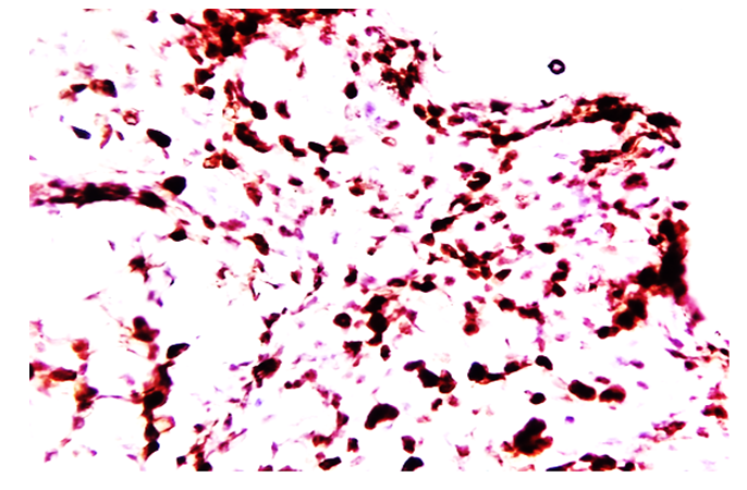 | Figure 1. High positive expression of the Ki67 marker in the pancreas of diabetic rats exposed to pesticides for 3 months. The sample was scanned using the QuPath-0.4.0.ink program to determine the level of expression. The expressing cells are dark brown. Stained with DAB chromogen. Magnification: 10x10 |
Thus, in immunohistochemical examinations of the pancreas of 3-month-old diabetic rats exposed to pesticides, the proliferative index of the Ki-67 marker in the pancreatic acinar epithelial cells was 23.41±1.16%, while in mesenchymal cells—histiocytes, fibroblasts, macrophages, and endothelial cells—it was found to be 18.46±1.01%.At the 6-month period, a low level of positive expression was detected, which indicates that the ongoing chronic negative effect of the process is causing damage and metabolic disruption in all glandular cells.In the study of 6-month-old diabetic rats receiving pesticides, moderate expression of the Ki-67 marker was observed in 16.5% of the rats, low expression in 82.6%, and a negative reaction in 0.9%. This means that not all cells expressing the Ki-67 marker were epithelial; 66.21% of the cells in the field of view were mesenchymal cells, and when the Ki-67 marker was applied to all pancreatic epithelial cells to obtain an average value, it was found to be 10.22±0.16%.Thus, in immunohistochemical examinations of the pancreas of 6-month-old diabetic rats exposed to pesticides, the proliferative index of the Ki-67 marker in pancreatic acinar epithelial cells was 10.22±0.16%, while proliferation in mesenchymal cells, such as histiocytes, fibroblasts, macrophages, and endothelial cells, was found to be 11.02±0.01%.At the 9-month period, a low positive expression of the Ki-67 marker was detected, indicating that the process is chronic and that the irritant effect is causing continuous damage and chronic metabolic disruption in all glandular cells.In the study of 9-month-old diabetic rats exposed to pesticides, moderate expression of the Ki-67 marker was observed in 8.91% of the rats, a low level in 90.2%, and a negative reaction in 0.89%. This indicates that in the studied rats, the expression of the Ki-67 marker in the pancreas is mainly due to mesenchymal cells; non-epithelial cells in the field of view accounted for 88.16% of the mesenchymal cells, and when the Ki-67 marker was applied to all pancreatic epithelial cells to obtain an average value, it was found to be 8.11±0.01%. Thus, during the immunohistochemical (IHC) examination of the pancreas of 9-month-old diabetic rats exposed to pesticides, the proliferative index of the Ki-67 marker corresponding to pancreatic acinar epithelial cells was 8.1±0.01%, while proliferation in mesenchymal cells—histiocytes, fibroblasts, macrophages, and endothelial cells—was found to be 10.01±0.01%.Changes in the Bcl-2 marker in the pancreas of rats with induced diabetes mellitus at 3, 6, and 9-month intervals.In the subsequent immunohistochemical study, the cells of the acinar system, the exocrine and endocrine systems, and the islets of Langerhans of the pancreas were examined. In particular, it inhibits the death (apoptosis) of pancreatic epithelial cells and most other cells.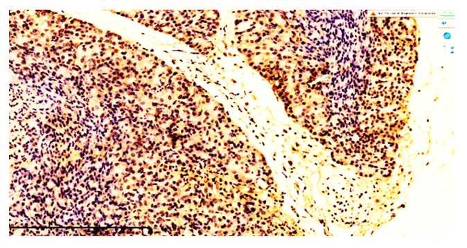 | Figure 2. Moderate positive expression of the pancreatic Bcl-2 marker in rats with diabetes mellitus that were administered pesticides for a 3-month period. Scanned and analyzed for expression levels using QuPath-0.4.0.ink. software. Expressed cells are dark brown. Stained with DAB chromogen. Magnification: 10x10 |
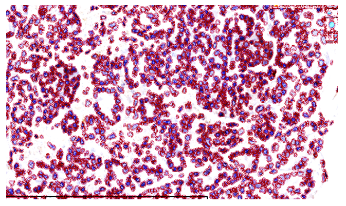 | Figure 3. Low positive expression of the Bcl-2 marker in the pancreas of diabetic rats exposed to pesticides for a 6-month period. Scanned and expression level determined using QuPath-0.4.0.ink. software. Expressed cells are dark brown. Stain: DAB chromogen. Magnification: 10x10 |
The results were evaluated as follows: absence of expression – 0 points, weak expression – 1 point, moderate expression – 2 points, strong expression – 3 points.In our study, over a 3-month period, rats with diabetes mellitus that consumed pesticides developed changes in the pancreas, manifesting as an increase in programmed cell death.The moderate positive expression of the Bcl-2 marker, in turn, is explained by the induction (enhancement of any stimulating factors) of cell apoptosis in rats with diabetes mellitus under the influence of pesticides. The positive expression of the Bcl-2 marker is explained by the intensification of the apoptosis process under the influence of pesticides in diabetes mellitus, and specifically by the abundance of labile cells in the pancreatic acini (labile cells normally have numerous mitotic foci, and epithelial cells rapidly regenerate). The uniform, diffuse positive expression of the Bcl-2 marker throughout all areas of the tissue, rather than being focal or confined to a specific area, is explained by the fact that pesticides stimulate the process of apoptosis in most tissue cells via the blood vessels in diabetes mellitus [8,10,11].During the chronic 3-month period of toxic poisoning from pesticides in diabetes mellitus, strong regenerative changes occurred in the pancreatic acinar epithelium, which is manifested by the binding of the APAF1 factor, an intracellular protein that blocks apoptosis, and the expression of the Bcl-2 marker. In this case, the premature death of the majority of apoptotic cells depends on the concentration of the stimulating factor; microscopically, cells that have died are identified by a 2-point result for Bcl-2 marker expression in monocellular and segmental patterns within the glandular epithelium. No changes are observed in the histotopographic structure of the pancreas; the trajectory of the glandular structures appears preserved. In paracellular glandular cells, the positive expression of the Ki-67 marker to replace lost glandular cells indicates that the processes of proliferation and apoptosis are occurring in parallel [8,10,11].During the subsequent 6-month period, it was determined that due to moderate regenerative changes in the pancreatic glandular epithelium of rats with diabetes mellitus under the influence of pesticides, the binding of the intracellular apoptosis-blocking protein APAF-1 factor and the expression of the Bcl-2 marker were at a moderately low level [8,10,11].In this case, the premature death of most apoptotic cells at the 6-month mark was reduced compared to the 3-month mark. This is explained by morphological adaptation, resulting in a decrease in the size of the glandular epithelial cells during their lifespan. Depending on the duration and concentration of the influencing factor, this is microscopically identified as foci of monocellular cell death within the glandular epithelium. Changes in the histotopographic structure of the pancreas were mainly manifested by an increase in clusters of cells that had decreased in size. The trajectory of the glandular structures is preserved, while in paracinar glandular cells, a low positive expression of the Ki-6 marker indicates the proliferation of a small number of glandular epithelial cells replacing lost ones. This, as mentioned above, is explained by the duration of the influencing factors.Thus, at the 6-month mark, the low positive expression of the Bcl-2 marker is defined as a weak expression score of 1 point, based on the number of cells showing positive expression through light golden-yellow staining, primarily in the cytoplasm of acinar glandular epithelial cells.During the subsequent 9-month period, it was found that due to moderately low-grade regenerative changes in the pancreatic epithelium of rats with diabetes mellitus exposed to pesticides, the binding of the intracellular apoptosis-blocking protein APAF-1 factor and the expression of the Bcl-2 marker were at a low level. In this case, the decrease in the rate of premature death of apoptotic cells at the 9-month mark, compared to the 3 and 6-month marks, is mainly explained by morphological adaptation, a reduction in the size of the glandular epithelial cells during their lifespan, and a decline in morphofunctional aspects. Depending on the chronic 9-month-long effect of the influencing factor, microscopic focal areas of monocellular cell death are identified in the glandular epithelium. Changes in the histotopographic structure of the pancreas were mainly manifested by an increase in the number of size-reduced acinar glandular epithelial cell clusters. The trajectory of the glandular structures is preserved, peri-acinar blood vessels appear anemic, and paracinar glandular cells have become smaller and uniform. A small number of cells with positive expression, appearing as a light golden color and masking the Bcl-2 marker, are detected in the cytoplasm. Specifically, at this 9-month point, the low proliferation rate confirms a direct correlation with the low positive expression of the Ki-6 marker in the glandular epithelial cells. This, as mentioned above, is explained by the duration of the influencing factors and the morphologically occurring adaptation process [8,10,11].Therefore, at the 9-month mark in the experimental conditions, in rats with diabetes mellitus that consumed pesticides, the continuous low positive expression of the Bcl-2 marker in the pancreas is microscopically defined as a weak expression score of 1 point. This is based on the number of cells showing positive expression through a small number of light golden-yellow stains, primarily in the cytoplasm of acinar glandular epithelial cells.The immunohistochemical investigation of our next group involves evaluating the specific morphological changes in the pancreas of rats with diabetes mellitus that were fed pesticides for periods of 3, 6, and 9 months.Specifically, in the pancreas of rats with diabetes mellitus fed pesticides for 3 months, a high level of Ki-67 marker expression was detected. This indicates that the direct and indirect effects of pesticides in diabetes mellitus disrupt cellular metabolism not only in the pancreas but in all organs.This means that a high degree of positive expression was detected in the acinar glands at the 3-month mark, indicating that the proliferation of connective tissue in the pancreas is ongoing, driven by moderate positive expression in mesenchymal cells such as fibroblasts, histiocytes, and endothelial cells.In the study, a high degree of Ki-67 marker expression was identified in a total of 63.8% of the rats studied, and it was found that mesenchymal cells constituted 23.8% of the identified cells. Therefore, of the total 40% positive expression rate, the breakdown is as follows: in the rats studied, 63.8% showed a high-level reaction, 30.16% a medium level, 4.2% a low level, and 1.84% a negative reaction.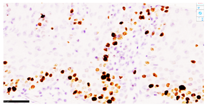 | Figure 4. Low positive expression of the Ki67 pancreatic marker in rats under the influence of pesticides in 9-month-old diabetes mellitus. It was scanned in the QuPath-0.4.0.ink. program and the degree of expression was determined. The expressed cells are dark brown. The dye Dab is chromogenic. Size 10x10 |
 | Figure 5. Low positive expression of the pancreatic Bcl-2 marker in rats with diabetes mellitus under the influence of pesticides for a 9-month period. Scanned and expression level determined using the QuPath-0.4.0.ink software. Expressed cells are dark brown. Stained with DAB chromogen. Magnification: 10x10 |
Thus, in immunohistochemical examinations of the pancreas of 3-month-old rats with diabetes mellitus that were fed pesticides, the proliferative index of the Ki-67 marker in the pancreatic acinar epithelial cells was 40.18±1.24%, while in mesenchymal cells—histiocytes, fibroblasts, macrophages, and endothelial cells—it was found to be 19.82±1.76%. This indicates an increased proliferative capacity of epithelial cells under the direct influence of pesticides on the pancreas in diabetes mellitus.In the subsequent 6-month period, a moderate positive expression of the Ki-67 marker was detected in the pancreas of rats with experimental diabetes mellitus that were fed pesticides. The process was characterized by a chronic negative impact, manifesting as damage to all glandular cells and a disruption of metabolic processes.In our 6-month study on the effects of pesticides in diabetes mellitus, a moderate expression of the Ki-67 marker was observed in 76.12% of the rats, a low-level positive expression was observed in 22.16%, and a negative reaction was seen in 1.72%. As we have consistently noted, among the cells expressing the Ki-67 marker, not only epithelial cells were present, but also mesenchymal cells, which constituted 21.02% of all cells in the field of view. It was found that in areas where proliferation occurred, there was an increase in fibroblasts replacing dead epithelial cells. Among the cells expressing the Ki-67 marker, the average proportion of epithelial cells was found to be 19.65±0.05%.Therefore, in immunohistochemical examinations of the pancreas of 6-month-old rats with experimentally induced diabetes mellitus that were fed pesticides, the proliferative index of the Ki-67 marker in the pancreatic acinar epithelial cells was 19.65±0.05%, while proliferation in mesenchymal cells—histiocytes, fibroblasts, macrophages, and endothelial cells—was found to be 12.11±0.01%. One noteworthy aspect is that the results of the immunohistochemical examinations confirm the findings from morphological studies of hematoxylin and eosin-stained microsamples. Specifically, due to the effect of pesticides on the pancreas in diabetes mellitus over time, a volumetric reduction and a decline in the morphofunctional indicators of the pancreas were observed during the rats' lifetime.In rats with 9 months of diabetes mellitus that were fed pesticides, a low-level positive expression of the Ki-67 marker was detected in the pancreas. This indicates that in this process, the morphological changes under the influence of pesticides in chronic diabetes mellitus primarily proceed as a decrease in pancreatic function and a chronic disruption of metabolism.In the study of rats with 9-month-long diabetes mellitus under the influence of pesticides, a moderate expression of the Ki67 marker was observed in a total of 60.18% of the rats. Of these, 51.16% were mainly mesenchymal cells, and 9.02±0.01% were acinar gland epithelial cells. A negative reaction was detected in 39.82% of the total cells in the field of view, while moderate positive reactions were detected in 60.18%. This is explained by the fact that in the rats studied, the expression of the Ki67 marker in the pancreas, under the influence of pesticides in persistent diabetes mellitus, occurs predominantly due to a large number of mesenchymal cells. It should also be noted that this process indicates a high probability of tumorigenesis over time. When the Ki-67 marker was applied to the total pancreatic epithelial cells, the average value was found to be 9.02 ± 0.01%.Thus, in the IHC examination of the pancreas of 9-month-old rats with diabetes that were fed pesticides, the proliferative index of the Ki-67 marker corresponding to pancreatic acinar epithelial cells was 9.02 ± 0.01%, while proliferation in mesenchymal cells—such as histiocytes, fibroblasts, macrophages, and endothelial cells—was found to be 51.16±0.01%.The results were evaluated as follows: no expression – 0 points, weak expression – 1 point, moderate expression – 2 points, strong expression – 3 points.In our study, it was established that in 3-month-old rats with diabetes exposed to pesticides, changes developed in the acinar glands of the pancreas, manifesting as a slowing of programmed cell death. The moderate positive expression of the Bcl-2 marker, in turn, is explained by its induction of cell apoptosis (slowing down any stimulating factors) in the rats given pesticides [1,2,3,4].The positive expression of the Bcl-2 marker, being uniformly diffuse across all areas of the tissue rather than focal or localized, is also explained by the fact that it leads to programmed cell death as a result of the anti-apoptotic effect of taurine, the effect of B-group vitamins on neuromuscular synapses, and the stimulation of cell metabolism by vitamin C via blood vessels.In this case, the inhibition of the apoptosis process, primarily influenced by taurine, and the predominance of the proliferative process in mesenchymal cells, are certainly dependent on the concentration of the stimulating factor. Microscopically, at 200x magnification, the expression of the Bcl-2 marker in the glandular epithelial cells within the focus is indicated by 2-point results, and dead cells are detected only in the focus [5,6,7].The histological structure of the pancreas remains unchanged, with the integrity of the glandular structures preserved. The concurrent positive expression of the Ki-67 marker in reparatively regenerated gland cells that have replaced dead periacinar gland cells indicates that proliferation and apoptosis are occurring in parallel. This implies that the process of apoptosis in the pancreatic epithelium of 3-month-old rats with diabetes that consumed pesticides is slow.In the subsequent chronic 6-month period, due to moderate regenerative changes in the pancreatic epithelium of rats with diabetes under the influence of pesticides, it was found that the binding of the intracellular apoptosis-blocking protein APAF1 and the expression of the Bcl-2 marker were at a moderately low level [8,9,10,11].In this case, the premature death of most apoptotic cells at the 6-month mark, which is reduced compared to the 3-month mark, is explained by the morphological adaptation leading to a decrease in the volume of the glandular epithelium during its life. Depending on the duration and concentration of the stimulating factor, this is microscopically detected as foci of monocellular cell death in the glandular epithelium. The histological structure of the pancreas is unchanged, with areas showing an increased collection of volumetrically smaller cells being identified. The trajectory of the glandular structures appears preserved. It was determined that in place of dead glandular cells, paracrine gland cells are manifested by the low positive expression of the Ki-67 marker in glandular epithelia undergoing mitosis. This, as noted above, determines the moderate positive expression of the Bcl-2 marker, which is also explained by the duration of the influencing factors [12,13,14,15].
4. Conclusions
Thus, in the 6-month period, the low positive expression of the Bcl-2 marker is defined as weak expression – 2 points. This is based on the number of cells in the acinar gland epithelium cytoplasm that showed positive expression through a light golden-yellow stain.In the next group, over a 9-month period, it was determined that the binding of the APAF1 factor, an intracellular protein that blocks apoptosis, and the expression of the Bcl-2 marker were low. This is due to the occurrence of moderate and low-level regenerative changes in the pancreatic epithelium of rats with diabetes mellitus under the influence of pesticides. In this 9-month period, the decrease in the rate of premature death of apoptotic cells, compared to the 3 and 6-month periods, is primarily explained by morphological adaptation, as confirmed by other previously mentioned studies. This adaptation involves a reduction in the size of the glandular epithelium during its lifetime and a decline in its morphofunctional aspects. Furthermore, due to the chronic exposure to the stimulating factor for 9 months, microscopic examination reveals focal areas of dead cells within the glandular epithelium. In the histotopographic structure of the pancreas, changes were mainly manifested as a reduction in the size of cell clusters and an increase in exocrine gland epithelia. The boundaries of the glandular structures are distinct, the basal layer is of uniform thickness, the periacinar blood vessels appear anemic, and the paratsinar gland cells have become smaller and uniform in appearance. The Bcl-2 marker is detected in the cytoplasm of a small number of light golden-colored cells. Specifically, at this 9-month point, the low proliferation in the glandular epithelia is directly linked to the low positive expression of the Ki-67 marker. This confirms a decrease in the anti-apoptotic suppressor gene accumulated on the perimeter of the cell's mitochondria and the activation of caspases through the mitochondrial membrane. As mentioned above, this is explained by the duration of the influencing factors and the morphological adaptation process that has occurred. Therefore, in the experiment, the sustained low positive expression of the Bcl-2 marker in the pancreas of rats with diabetes mellitus that were exposed to pesticides for a 9-month period is defined as weak expression – 1 point. This is determined microscopically by the number of cells showing positive expression, primarily observed as a light golden-yellow stain in the perinuclear region of the acinar gland epithelium cytoplasm. This confirms that the process of apoptosis has begun.
References
| [1] | Song Y, Chou EL, Baecker A, et al. Endocrine-disrupting chemicals, risk of type 2 diabetes, and diabetes-related metabolic traits: a systematic review and meta-analysis. J Diabetes. 2016; 8: 516–532. |
| [2] | Sun H., Chen N., Yang X., Xia Y., Wu D. Effects induced by polyethylene microplastics oral exposure on colon mucin release, inflammation, gut microflora composition and metabolism in mice. Ecotoxicol. Environ. Saf. 2021; 220: 112340. |
| [3] | Sun H., Chen N., Yang X., Xia Y., Wu D. Effects induced by polyethylene microplastics oral exposure on colon mucin release, inflammation, gut microflora composition and metabolism in mice. Ecotoxicol. Environ. Saf. 2021; 220: 112340. |
| [4] | Taylor KW, Novak RF, Anderson HA, et al. Evaluation of the association be-tween persistent organic pollutants (POPs) and diabetes in epidemiological stud-ies: a national toxicology program workshop review. Environ Health Perspect. 2013; 121: 774–783. |
| [5] | Taylor MK, Sisti G. Fetal hyperechogenic pancreas and gestational diabetes mellitus: a meta-analysis. Minerva Obstet Gynecol. 2024 Oct; 76(5): 452-457. |
| [6] | Vuong A.M., Zhang C., Chen A. Associations of neonicotinoids with insulin and glucose homeostasis parameters in US adults: NHANES 2015–2016. Chemosphere. 2022; 286: 131642. |
| [7] | Wadie W., Mohamed A.H., Masoud M.A., Rizk H.A., Sayed H.M. Protective impact of lycopene on ethinylestradiol-induced cholestasis in rats. Naunyn-Schmiedebergʻs Arch. Pharmacol. 2021; 394: 447–455. |
| [8] | Walker SL, Leete P, Boldison J. Tissue Resident and Infiltrating Immune Cells: Their Influence on the Demise of Beta Cells in Type 1 Diabetes. Biomolecules. 2025 Mar 19; 15(3): 441. |
| [9] | Wang J., Wang X.-Q., Liu R.-P., Li Y.-H., Yao X.-R., Kim N.-H., Xu Y.-N. Melatonin Supplementation during In Vitro Maturation of Porcine Oocytes Alleviates Oxidative Stress and Endoplasmic Reticulum Stress Induced by Imidacloprid Exposure. Animals. 2023; 13: 2596. |
| [10] | Wang W., Wang X., Chun J., Vilaysane A., Clark S., French G., Bracey N.A., Trpkov K., Bonni S., Duff H.J., et al. Inflammasome-Independent NLRP3 Augments TGF-β Signaling in Kidney Epithelium. J. Immunol. 2013; 190: 1239–1249. |
| [11] | Wang W., Wang X., Chun J., Vilaysane A., Clark S., French G., Bracey N.A., Trpkov K., Bonni S., Duff H.J., et al. Inflammasome-Independent NLRP3 Augments TGF-β Signaling in Kidney Epithelium. J. Immunol. 2013; 190: 1239–1249. |
| [12] | Whiting DR, Guariguata L, Weil C, Shaw J. IDF diabetes atlas: global estimates of the prevalence of diabetes for 2011 and 2030. Diabetes Res Clin Pract. (2011) 94: 311–21. |
| [13] | Williams JA (2010) Regulation of acinar cell function in the pancreas. Curr Opin Gastroenterol 26(5): 478–483. |
| [14] | Wright JJ, Eskaros A, Windon A, et al. Exocrine pancreas in type 1 and type 2 diabetes: different patterns of fibrosis, metaplasia, angiopathy, and adiposity. Diabetes 2024; 73: 1140–1152. |
| [15] | Xourafa G, Korbmacher M, Roden M. Inter-organ crosstalk during development and progression of type 2 diabetes mellitus. Nat Rev Endocrinol. 2024 Jan; 20(1): 27-49. |

 Abstract
 Reference
 Full-Text PDF
 Full-text HTML