-
Paper Information
- Next Paper
- Previous Paper
- Paper Submission
-
Journal Information
- About This Journal
- Editorial Board
- Current Issue
- Archive
- Author Guidelines
- Contact Us
American Journal of Medicine and Medical Sciences
p-ISSN: 2165-901X e-ISSN: 2165-9036
2026; 16(5): 2693-2697
doi:10.5923/j.ajmms.20261605.89
Received: Apr. 20, 2026; Accepted: May 16, 2026; Published: May 27, 2026

Age-Related Pathomorphological Changes in the Kidneys in Cerebrovascular Diseases
 Abstract
 Reference
 Full-Text PDF
 Full-text HTML
Abdullayev Nodirbek Uroyimjonovich1, Narbayev Zafar Kamilovich2
1Lecturer, Impuls Medical Institute, Namangan, Uzbekistan
2Candidate of Medical Sciences, Department of Otolaryngology, Andijan State Medical Institute, Andijan, Uzbekistan
Copyright © 2026 The Author(s). Published by Scientific & Academic Publishing.
This work is licensed under the Creative Commons Attribution International License (CC BY).
http://creativecommons.org/licenses/by/4.0/

Cerebrovascular diseases (CVD) are frequently associated with systemic microvascular dysfunction and chronic hypoxia, affecting multiple organs, including the kidneys. This study aims to evaluate age-related pathomorphological changes in renal tissue under CVD conditions using histochemical, morphometric, and immunohistochemical approaches. Renal samples were processed through standard paraffin-embedding techniques, followed by staining with PAS, Masson’s trichrome, and Van Gieson methods to assess structural and biochemical alterations. Morphometric analysis was conducted using digital imaging software to quantify parameters such as glomerular size, interstitial tissue proportion, and nephron density. Immunohistochemical markers, including α-SMA, TGF-β1, CD34, and HIF-1α, were applied to identify fibrosis, vascular changes, and hypoxic responses. The results revealed a progressive decrease in nephron number and glomerular size, accompanied by increased interstitial fibrosis and sclerosis with advancing age. A marked reduction in microvascular density and elevated expression of hypoxia- and fibrosis-related markers were observed in older age groups. These findings indicate that renal damage in CVD is driven by combined ischemic, fibrotic, and degenerative mechanisms. The study underscores the importance of integrating morphological and molecular methods for a comprehensive understanding of kidney involvement in cerebrovascular pathology.
Keywords: Cerebrovascular diseases (CVD), Kidney, Age-related changes, Nephrosclerosis, Interstitial fibrosis, Histochemistry, Morphometry, Immunohistochemistry, Hypoxia, Microvascular rarefaction, TGF-β1, α-SMA, CD34, Renal pathology
Cite this paper: Abdullayev Nodirbek Uroyimjonovich, Narbayev Zafar Kamilovich, Age-Related Pathomorphological Changes in the Kidneys in Cerebrovascular Diseases, American Journal of Medicine and Medical Sciences, Vol. 16 No. 5, 2026, pp. 2693-2697. doi: 10.5923/j.ajmms.20261605.89.
Article Outline
1. Introduction
- Cerebrovascular diseases (CVD) remain a leading cause of morbidity and mortality worldwide, characterized by impaired cerebral circulation, endothelial dysfunction, and chronic hypoxia. While the neurological consequences of CVD are well documented, their systemic effects particularly on renal structure and function—are often underestimated. The kidney, as a highly vascularized and metabolically active organ, is especially susceptible to ischemic and hypoxic injury resulting from systemic hemodynamic disturbances and microvascular pathology associated with CVD [2,3].Emerging evidence suggests that cerebrovascular and renal pathologies share common pathogenic mechanisms, including endothelial dysfunction, arterial stiffness, oxidative stress, and chronic inflammation. These overlapping processes contribute to the development of microvascular damage in both organs, supporting the concept of a unified vascular continuum. In this context, renal impairment in patients with CVD may represent not only a secondary complication but also a parallel manifestation of systemic vascular disease.Age-related structural and functional changes further exacerbate renal vulnerability. With advancing age, the kidney undergoes progressive nephron loss, glomerulosclerosis, tubular atrophy, and interstitial fibrosis, ultimately leading to a decline in renal function [1]. These alterations are often accelerated in the presence of chronic hypoxia and impaired perfusion, conditions commonly observed in CVD. Additionally, age-dependent changes in cellular metabolism, regenerative capacity, and extracellular matrix turnover contribute to the progression of nephrosclerosis and ischemic nephropathy.Despite growing recognition of the kidney–brain axis, detailed morphological studies investigating age-related renal alterations in CVD remain limited. Advanced histopathological techniques, including histochemical staining, morphometric analysis, and immunohistochemistry, provide valuable tools for assessing both structural and molecular changes in renal tissue. These methods allow for the identification of fibrosis, vascular rarefaction, hypoxia-related responses, and cellular turnover at a high level of precision.Therefore, this study aims to evaluate age-dependent pathomorphological alterations in renal tissue in the context of cerebrovascular diseases using an integrated methodological approach. By combining histochemical, morphometric, and immunohistochemical analyses, the research seeks to provide a comprehensive understanding of the mechanisms underlying renal damage in CVD and to clarify the role of aging in the progression of these changes.
2. Materials and Methods
2.1. Histochemical Analysis
- Histochemical examination was carried out on formalin-fixed, paraffin-embedded kidney tissue sections with a thickness of 4–5 µm. Tissue fixation in 10% neutral buffered formalin ensured preservation of structural integrity and biochemical components, followed by standard dehydration and paraffin embedding procedures. Sections were cut using a rotary microtome, mounted on glass slides, and prepared for staining through deparaffinization in xylene and rehydration in graded alcohols.The following histochemical staining techniques were applied:♦ PAS (Periodic Acid–Schiff) reaction: used to detect glycoproteins, glycogen, and mucopolysaccharides, as well as to evaluate thickening of the glomerular basement membrane and mesangial matrix expansion.♦ Van Gieson staining: employed to differentiate collagen fibers (stained red) from other tissue elements (yellow), allowing assessment of connective tissue proliferation and fibrosis.♦ Sudan III/IV staining: applied on frozen sections to identify neutral lipid accumulation within tubular epithelial cells, indicating degenerative changes such as lipidosis.♦ Perls’ Prussian blue reaction: used to detect ferric iron (Fe³⁺), particularly in cases of hemosiderin deposition associated with microhemorrhages and altered iron metabolism.♦ Methenamine silver impregnation: utilized for detailed visualization of basement membranes and glomerular capillary loops, highlighting structural alterations such as thickening and sclerosis.These histochemical methods enabled the identification and localization of key biochemical components within renal tissue, providing insight into dystrophic, degenerative, and adaptive processes. Variations in staining intensity and distribution patterns were interpreted as indicators of pathological alterations, particularly those associated with aging and chronic hypoxia.
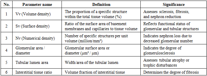
2.2. Morphometric Analysis
- Morphometric evaluation was performed using advanced digital image analysis systems, including ImageJ and QuPath, in conjunction with high-resolution slide scanning via the Hamamatsu NanoZoomer system. Histological sections were examined under light microscopy at magnifications ranging from ×200 to ×1000 to ensure both general architectural assessment and detailed cellular analysis.
|
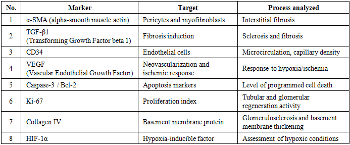
2.3. Immunohistochemical Analysis (IHC)
- Immunohistochemical (IHC) analysis was performed using monoclonal antibodies and standardized protocols based on the DAKO LSAB2 detection system. Paraffin-embedded sections (4 µm thick) were mounted on adhesive slides and incubated at 55–60°C to ensure proper adherence. Deparaffinization was carried out in xylene, followed by rehydration through graded ethanol solutions and rinsing in distilled water.Antigen retrieval was performed using heat-induced epitope retrieval in buffer solution at approximately 95–98°C for 30–40 minutes. Endogenous peroxidase activity was blocked using 3% hydrogen peroxide, and non-specific binding was minimized באמצעות protein blocking solution.Sections were then incubated with primary antibodies at room temperature for 60–120 minutes, followed by application of secondary antibodies conjugated with horseradish peroxidase. Visualization was achieved using chromogenic substrates such as DAB (diaminobenzidine), producing a brown-colored reaction product. Slides were counterstained with hematoxylin, dehydrated, and mounted for microscopic evaluation.
|
3. Results
3.1. Histochemical Findings
- Histochemical analysis demonstrated pronounced structural and biochemical alterations in renal tissue, which intensified with advancing age. The PAS (Periodic Acid–Schiff) reaction revealed progressive thickening of the glomerular basement membrane, along with increased deposition of PAS-positive substances within the mesangial matrix. These findings indicate enhanced accumulation of glycoproteins and mucopolysaccharides, reflecting early and progressive stages of glomerulosclerosis.Staining with Van Gieson highlighted a marked increase in collagen fiber deposition within the interstitial compartment, confirming the presence and progression of interstitial fibrosis. The fibrotic areas appeared more extensive and densely stained in older age groups, suggesting chronic and irreversible connective tissue remodeling.Sudan III/IV staining demonstrated lipid accumulation in tubular epithelial cells, indicative of degenerative processes such as lipidosis and tubular dysfunction. This lipid deposition was more prominent in advanced age groups, correlating with metabolic disturbances and impaired cellular homeostasis.In addition, Perls’ Prussian blue reaction identified focal iron deposition in certain cases, suggesting microhemorrhages and altered iron metabolism. Silver impregnation techniques further confirmed basement membrane irregularities and thickening.Overall, histochemical findings clearly showed age-dependent intensification of PAS-positive material, fibrosis, and degenerative changes, reflecting progressive renal structural damage in the context of CVD.
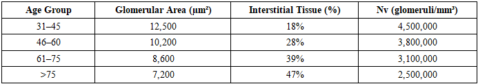
3.2. Morphometric Findings
- Quantitative morphometric analysis provided objective evidence of age-related structural deterioration in renal tissue.
|
3.3. Immunohistochemical Findings
- Immunohistochemical analysis provided important insights into the molecular mechanisms underlying the observed structural changes. A significant increase in α-SMA (alpha-smooth muscle actin) expression was observed in older age groups, indicating activation of myofibroblasts and progression of interstitial fibrosis. Similarly, elevated expression of TGF-β1, a key profibrotic cytokine, confirmed enhanced fibrogenic activity and extracellular matrix deposition.A progressive decrease in CD34 expression was noted with advancing age, reflecting a reduction in endothelial cell density and microvascular rarefaction. This finding suggests impaired renal perfusion and diminished capillary network integrity.In contrast, HIF-1α expression showed a marked increase, particularly in older groups, indicating a sustained hypoxic state within renal tissue. This hypoxia likely contributes to both fibrotic remodeling and cellular injury.Markers of apoptosis, such as Caspase-3, were significantly elevated, pointing to increased rates of programmed cell death. Concurrently, reduced expression of the proliferation marker Ki-67 suggests diminished regenerative capacity of renal cells. Furthermore, immunostaining for CD3 and CD20 revealed the presence of T- and B-lymphocyte infiltration within the interstitial areas, indicating an immunological component in the progression of renal damage.Overall, immunohistochemical findings confirm that age-related renal changes in CVD are mediated by a combination of fibrosis, hypoxia, vascular loss, apoptosis, and immune activation, providing a comprehensive understanding of the disease process at the molecular level.
4. Discussion
- The present study confirms that cerebrovascular diseases (CVD) exert significant systemic effects extending beyond the central nervous system, with notable structural and functional consequences for the kidneys. The findings support the concept of a brain–kidney axis, in which shared vascular risk factors and pathological mechanisms—such as endothelial dysfunction, arterial stiffness, and impaired autoregulation contribute to parallel damage in both organs. In this context, chronic hypoperfusion and sustained hypoxia appear to be central drivers of progressive renal injury.Histochemical analysis revealed substantial biochemical alterations, including accumulation of glycoproteins, lipids, and extracellular matrix components. These changes reflect metabolic imbalance and impaired cellular turnover, which are characteristic of chronic ischemic conditions. The observed increase in PAS-positive material and collagen deposition is indicative of glomerulosclerosis and interstitial fibrosis, both of which are key features of nephrosclerosis.Morphometric analysis provided objective, quantitative confirmation of these structural changes. The progressive decline in nephron density (Nv), reduction in glomerular size, and expansion of interstitial tissue collectively demonstrate significant architectural remodeling of the kidney with advancing age. These findings are consistent with the concept that nephron loss is a major determinant of age-related renal decline and is further accelerated in pathological conditions such as CVD.Immunohistochemical results offer important insights into the molecular mechanisms underlying these alterations. The increased expression of TGF-β1 and α-SMA reflects activation of fibrogenic pathways and myofibroblast proliferation, which are central to extracellular matrix accumulation and fibrosis. These findings are in agreement with previous studies that identify TGF-β1 as a key regulator of renal fibrogenesis [4,5].The observed decrease in CD34 expression suggests a reduction in microvascular density, supporting the presence of capillary rarefaction. This microvascular loss contributes to impaired oxygen delivery, thereby perpetuating hypoxic injury. Concurrently, increased expression of HIF-1α confirms the presence of a hypoxic microenvironment within renal tissue, which further stimulates fibrotic and apoptotic pathways.Elevated levels of apoptotic markers, such as Caspase-3, combined with reduced proliferative activity (Ki-67), indicate an imbalance between cell death and regeneration. This imbalance likely contributes to the progressive loss of functional renal units. Additionally, the presence of CD3- and CD20-positive lymphocytes suggests that immune-mediated mechanisms may also play a role in sustaining chronic inflammation and tissue damage.Importantly, the age-stratified analysis highlights that aging significantly amplifies these pathological processes. Structural degeneration, vascular compromise, and reduced regenerative capacity collectively enhance susceptibility to renal injury in older individuals. These findings are consistent with established literature demonstrating that aging is associated with nephron depletion, increased fibrosis, and diminished adaptive capacity [1].Overall, this study underscores the multifactorial nature of renal damage in CVD, involving hemodynamic, metabolic, hypoxic, and immunological mechanisms. The integration of histochemical, morphometric, and immunohistochemical methods provides a comprehensive framework for understanding the progression of renal pathology in this context.
5. Conclusions
- The combined application of histochemical, morphometric, and immunohistochemical analyses provides a comprehensive evaluation of age-related renal changes in cerebrovascular diseases. The findings of this study demonstrate that renal damage in CVD is progressive, multifactorial, and strongly age-dependent.Specifically, the study shows that:Ø Structural alterations such as nephrosclerosis, glomerular atrophy, and interstitial fibrosis increase significantly with advancing ageØ Chronic hypoxia and microvascular rarefaction play a central role in the progression of renal injuryØ Morphometric methods offer reliable and objective quantification of tissue remodeling, including nephron loss and fibrotic expansionØ Immunohistochemical analysis reveals key pathogenic mechanisms, including activation of fibrogenic pathways (TGF-β1, α-SMA), hypoxia-related responses (HIF-1α), vascular decline (CD34), apoptosis, and reduced cellular proliferationFrom a clinical perspective, understanding the mechanisms underlying age-related renal changes in CVD may support the development of targeted therapeutic strategies aimed at reducing fibrosis, improving microcirculation, and mitigating hypoxic injury. Ultimately, such approaches could improve patient outcomes by addressing not only neurological but also systemic consequences of cerebrovascular pathology.