-
Paper Information
- Next Paper
- Previous Paper
- Paper Submission
-
Journal Information
- About This Journal
- Editorial Board
- Current Issue
- Archive
- Author Guidelines
- Contact Us
American Journal of Medicine and Medical Sciences
p-ISSN: 2165-901X e-ISSN: 2165-9036
2026; 16(5): 2689-2692
doi:10.5923/j.ajmms.20261605.88
Received: Mar. 19, 2026; Accepted: Apr. 15, 2026; Published: May 27, 2026

Morphometric and Ki-67 Features of the Stomach in 6-Month-Old White Outbred Rats Under Combined Alimentary Deficiency of Magnesium, Iron, Zinc and Selenium
 Abstract
 Reference
 Full-Text PDF
 Full-text HTML
Abosov Azizbek Salim ugli1, Khasanova Dilnoza Akhrorovna2
1Independent Researcher, Bukhara State Medical Institute named after Abu Ali ibn Sino, Bukhara, Uzbekistan
2Doctor of Medical Sciences, Professor, Bukhara State Medical Institute named after Abu Ali ibn Sino, Bukhara, Uzbekistan
Correspondence to: Abosov Azizbek Salim ugli, Independent Researcher, Bukhara State Medical Institute named after Abu Ali ibn Sino, Bukhara, Uzbekistan.
| Email: |  |
Copyright © 2026 The Author(s). Published by Scientific & Academic Publishing.
This work is licensed under the Creative Commons Attribution International License (CC BY).
http://creativecommons.org/licenses/by/4.0/

The article presents an experimental morphological, morphometric and immunohistochemical study of the stomach in 6-month-old white outbred rats under combined alimentary deficiency of magnesium, iron, zinc and selenium. The study included two groups: a control group receiving a standard balanced diet and an experimental group receiving a combined Mg+Fe+Zn+Se-deficient diet. Morphometric analysis included the thickness of the gastric wall layers, depth of gastric pits, length and density of gastric glands, number of mucosal folds, parietal cells and chief cells. Ki-67 expression was evaluated immunohistochemically. In the control group, the gastric mucosa demonstrated morphofunctional maturity, with mucosal thickness of 800.6–880.4 µm and calculated mean value of 840.5 µm. Under combined microelement deficiency, mucosal thickness decreased to 648.3–702.1 µm, with calculated mean value of 675.2 µm. Gastric pit depth decreased from 122.1 µm to 88.7 µm, gland density from 25.0 to 16.5 per 100000 µm², parietal cells from 36.0 to 21.0 per 10000 µm², and chief cells from 54.0 to 34.0 per 10000 µm². Ki-67 expression increased from 6–10% in the control group to 22–32% in the combined-deficiency group. The obtained data indicate that combined deficiency of magnesium, iron, zinc and selenium in 6-month-old rats causes severe structural disorganization of the gastric mucosa, pronounced reduction of secretory cells and chaotic dysregulated epithelial proliferation.
Keywords: Stomach, Magnesium, Iron, Zinc, Selenium, Gastric mucosa, Morphometry
Cite this paper: Abosov Azizbek Salim ugli, Khasanova Dilnoza Akhrorovna, Morphometric and Ki-67 Features of the Stomach in 6-Month-Old White Outbred Rats Under Combined Alimentary Deficiency of Magnesium, Iron, Zinc and Selenium, American Journal of Medicine and Medical Sciences, Vol. 16 No. 5, 2026, pp. 2689-2692. doi: 10.5923/j.ajmms.20261605.88.
1. Introduction
- The gastric mucosa is a highly active tissue system characterized by continuous renewal, intensive secretion and dependence on adequate trophic, oxygen and antioxidant support. Magnesium, iron, zinc and selenium participate in different but interconnected biological mechanisms. Magnesium regulates cellular metabolism and smooth muscle activity. Iron supports oxygen transport and tissue respiration. Zinc is necessary for epithelial integrity and repair. Selenium is essential for antioxidant protection and prevention of oxidative tissue injury.Previous experimental studies have shown that deficiency of individual microelements can affect the stomach through different mechanisms. Magnesium deficiency may alter gastric mucosal structure [1]. Iron deficiency may lead to degenerative changes in gastric parietal cells [2]. Zinc deficiency impairs mucosal repair and delays ulcer healing [3,4]. Selenium deficiency may induce oxidative stress, inflammatory injury and cell death mechanisms in gastric mucosa [5].At the age of 6 months, the stomach of white outbred rats demonstrates more developed morphofunctional organization than in younger animals. The mucosa is thicker, gastric pits are deeper, and the glandular apparatus is denser. However, this higher functional load may make the gastric mucosa more vulnerable to combined nutritional deficiency.Ki-67 immunohistochemical analysis allows assessment of epithelial proliferation. In normal gastric mucosa, Ki-67-positive cells are located mainly in the neck zone of gastric glands. Under pathological conditions, Ki-67 expression may increase, become uneven or lose its normal localization, indicating dysregulation of epithelial renewal [6,7].The purpose of the study: to determine morphological, morphometric and Ki-67 immunohistochemical features of the stomach in 6-month-old white outbred rats under combined alimentary deficiency of magnesium, iron, zinc and selenium.
2. Materials and Methods
- The experimental study was carried out in 2025–2026 at the scientific experimental biomedical research center of Bukhara State Medical Institute named after Abu Ali ibn Sino and in the pathomorphological laboratory “Ipsum pathology”.The general experiment included 180 male white outbred rats distributed into 3- and 6-month age groups. For the present article, only the 6-month-old cohort was analyzed. The study included two groups:Group I — control group: 15 animals receiving a standard balanced diet.Group II — combined deficiency group: 15 animals receiving a diet deficient in magnesium, iron, zinc and selenium.The animals were kept under standard vivarium conditions with free access to water. Feeding was performed once daily according to the dose of 25 g of feed per 100 g of body weight per day.The diets were produced by Altromin Spezialfutter GmbH & Co. KG, Germany. The control group received Altromin C1000 containing Mg 684 mg/kg, Fe 178.58 mg/kg, Zn 29.30 mg/kg and Se 0.33 mg/kg. The combined-deficiency group received Altromin C1035 mod., containing Mg 97.7 mg/kg, Fe 3.82 mg/kg, Zn 2.02 mg/kg and Se 0.035 mg/kg. To prevent excessive exhaustion, no more than 10–15% of the total diet volume in the combined-deficiency group was supplemented with standard Altromin C1000.After completion of the experimental period, stomach tissue samples were collected and processed by routine histological methods. Sections were stained with hematoxylin and eosin. Morphometric analysis was performed on standardized microscopic fields. The number of folds and glands was recalculated per 100000 µm², while parietal and chief cells were calculated per 10000 µm².Immunohistochemical analysis was performed using the Ki-67 marker with DAB chromogen. Ki-67 expression was evaluated according to the proportion of positive cells.
3. Results and Discussion
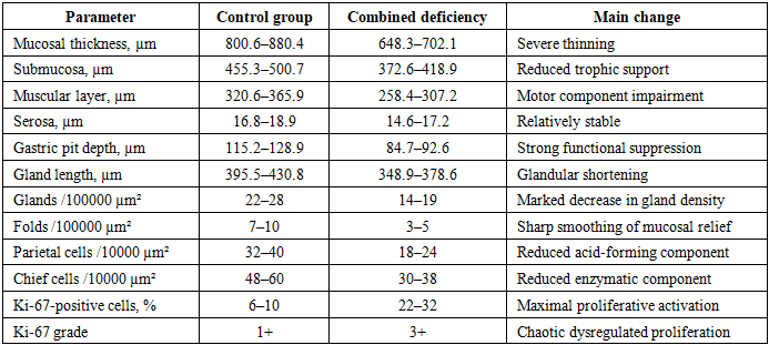
- In the control group of 6-month-old rats, the gastric mucosa showed preserved histological organization and signs of morphofunctional maturity. The surface epithelium was intact, gastric pits had regular contours, and the glandular apparatus was compact and structurally organized. Compared with younger animals, the mucosa was thicker, gastric pits were deeper, and the number of glands and secretory cells was higher.The mucosal thickness in the control group was 800.6–880.4 µm. Gastric pit depth was 115.2–128.9 µm. The number of mucosal folds was 7–10 per 100000 µm². Gastric gland length was 395.5–430.8 µm, and gland density was 22–28 per 100000 µm². The number of parietal cells was 32–40 per 10000 µm², while chief cells numbered 48–60 per 10000 µm². These parameters indicate preserved acid-producing and enzymatic functions of the stomach.Under combined Mg+Fe+Zn+Se deficiency, the stomach of 6-month-old rats showed sharply expressed morphological and morphometric changes. The mucosa was markedly thinned and uneven. The surface epithelium lost continuity over large areas. Extensive epithelial desquamation, cytoplasmic vacuolization and hydropic dystrophy were observed. In some microscopic fields, necrobiotic changes were present. The protective mucus layer was sharply reduced.Gastric pits were markedly shortened and deformed. Their contours were irregular and blurred. Morphometrically, gastric pit depth decreased to 84.7–92.6 µm, which was substantially lower than in the control group. The mucosal relief was sharply smoothed, and the number of folds decreased to 3–5 per 100000 µm². This indicates pronounced suppression of the functional surface of the gastric mucosa.The glandular apparatus was severely disorganized. Gastric glands were sparse, deformed and irregularly oriented. Many glands lost their parallel arrangement. Their length decreased to 348.9–378.6 µm, and their number decreased to 14–19 per 100000 µm². These changes indicate a significant reduction of the glandular component and structural reserve of the mucosa.The cellular composition of the mucosa underwent severe quantitative and qualitative changes. The number of parietal cells decreased to 18–24 per 10000 µm², while chief cells decreased to 30–38 per 10000 µm². Their distribution was sharply uneven. In some zones, dense focal accumulations of cells were observed, while in other zones the cells were rarefied. A significant part of the cells had signs of dystrophy and functional insufficiency.The mucosal thickness decreased to 648.3–702.1 µm, reflecting advanced atrophic and destructive changes. In the lamina propria, pronounced edema and lymphohistiocytic infiltration were found, indicating an active inflammatory process. The submucosa was thinned to 372.6–418.9 µm and showed vascular disturbances. The muscular layer decreased to 258.4–307.2 µm, with reduced density of muscle elements. The serosa remained relatively stable and measured 14.6–17.2 µm.
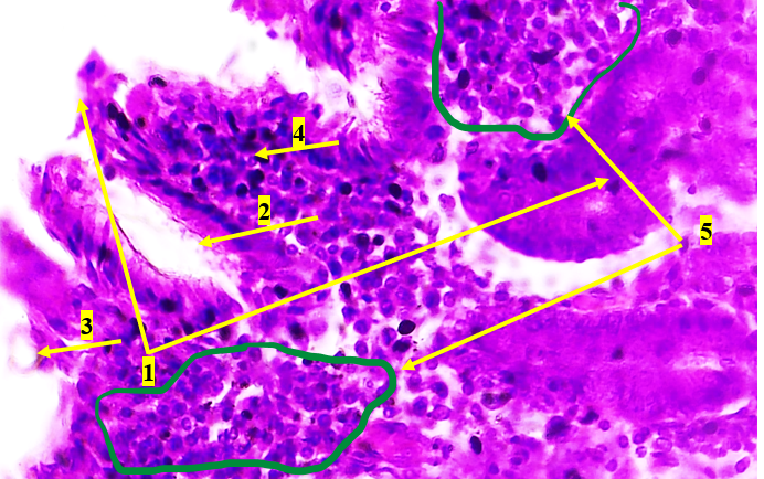 | Figure 1. Microscopic view of the stomach of a 6-month-old rat under combined Mg+Fe+Zn+Se deficiency. Histological appearance of the gastric mucosa in a 6-month-old rat under experimental combined deficiency of magnesium, iron, zinc and selenium. Hematoxylin and eosin staining. Eyepiece 10×, objective 40×. The following changes are observed: 1 — marked disorganization of the mucosa; 2 — deformation and dilatation of glandular lumina; 3 — epithelial cells with signs of vacuolization and hydropic degeneration; 4 — pronounced lymphocytic infiltration; 5 — formation of foci of lymphoid hyperplasia |
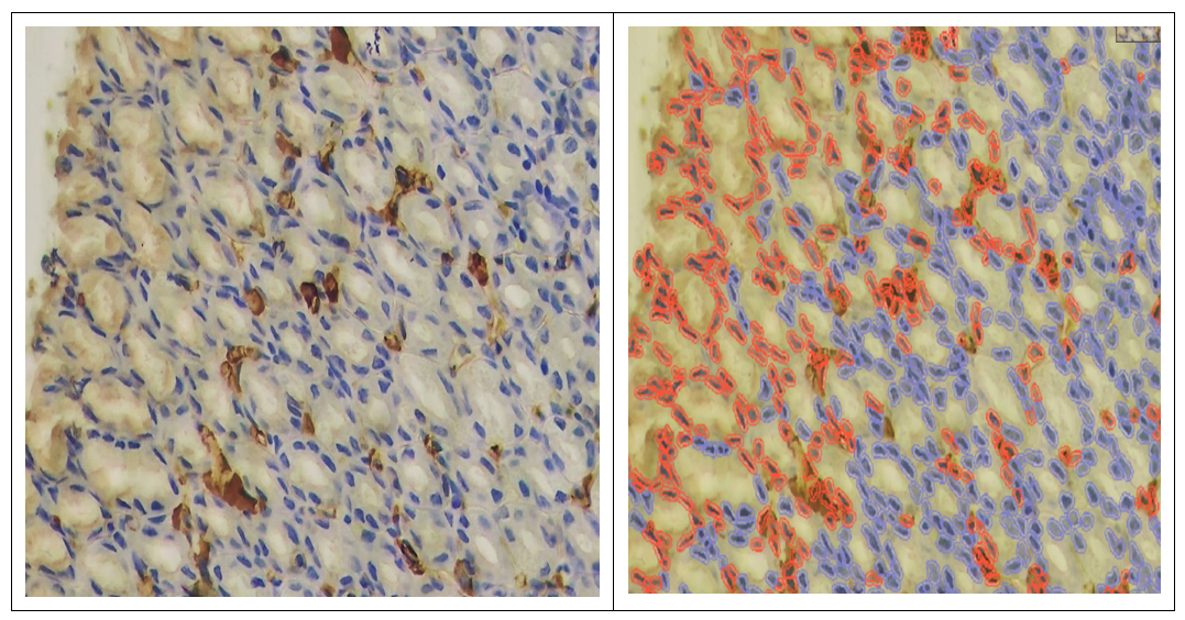 | Figure 2. Ki-67 expression in the gastric mucosa of a 6-month-old rat under combined microelement deficiency |
|
4. Conclusions
- In 6-month-old control rats, the stomach demonstrates morphofunctional maturity, with mucosal thickness of 800.6–880.4 µm, gastric pit depth of 115.2–128.9 µm, gland density of 22–28 per 100000 µm², parietal cell count of 32–40 per 10000 µm² and chief cell count of 48–60 per 10000 µm².Combined alimentary deficiency of magnesium, iron, zinc and selenium causes severe gastric mucosal injury characterized by epithelial desquamation, vacuolization, hydropic dystrophy, necrobiotic changes, edema, inflammatory infiltration and glandular disorganization.Under combined deficiency, mucosal thickness decreases to 648.3–702.1 µm, gastric pit depth to 84.7–92.6 µm, gland density to 14–19 per 100000 µm², parietal cells to 18–24 per 10000 µm² and chief cells to 30–38 per 10000 µm².The glandular apparatus undergoes severe structural disorganization: glands become sparse, deformed and irregularly oriented, while parietal and chief cells show uneven distribution and signs of functional insufficiency.Ki-67 expression increases to 22–32%, corresponding to 3+, and becomes chaotic and diffuse, indicating dysregulated epithelial proliferation rather than effective physiological regeneration.The most informative indicators of advanced gastric injury in 6-month-old rats under combined microelement deficiency are gastric pit depth, gland density, parietal cell number, chief cell number and the pattern of Ki-67 expression.The severity of changes in 6-month-old rats indicates a decrease in adaptive capacity of the gastric mucosa and progression of alimentary microelementosis from functional impairment to structural and inflammatory damage.