-
Paper Information
- Next Paper
- Previous Paper
- Paper Submission
-
Journal Information
- About This Journal
- Editorial Board
- Current Issue
- Archive
- Author Guidelines
- Contact Us
American Journal of Medicine and Medical Sciences
p-ISSN: 2165-901X e-ISSN: 2165-9036
2026; 16(5): 2685-2688
doi:10.5923/j.ajmms.20261605.87
Received: Apr. 23, 2026; Accepted: May 16, 2026; Published: May 27, 2026

Morphometric and Immunohistochemical Changes in the Myocardium Under Carbon Monoxide Exposure and Correction with Milk Thistle and Safflower Extracts
 Abstract
 Reference
 Full-Text PDF
 Full-text HTML
Mamedova Guletar Telman kizi1, Khasanova Dilnoza Akhrorovna2
1Assistant, Department of Anatomy and Clinical Anatomy, Bukhara State Medical Institute named after Abu Ali ibn Sino, Bukhara, Uzbekistan
2Doctor of Medical Sciences, Professor, Bukhara State Medical Institute named after Abu Ali ibn Sino, Bukhara, Uzbekistan
Correspondence to: Mamedova Guletar Telman kizi, Assistant, Department of Anatomy and Clinical Anatomy, Bukhara State Medical Institute named after Abu Ali ibn Sino, Bukhara, Uzbekistan.
| Email: |  |
Copyright © 2026 The Author(s). Published by Scientific & Academic Publishing.
This work is licensed under the Creative Commons Attribution International License (CC BY).
http://creativecommons.org/licenses/by/4.0/

Carbon monoxide poisoning causes systemic hypoxia and leads to structural myocardial damage [1,2]. The aim of this study was to assess morphometric, histochemical and immunohistochemical changes in the myocardium of white outbred rats after carbon monoxide exposure and correction with milk thistle and safflower extracts. The study was performed on 130 rats aged 6 and 18 months. Hematoxylin-eosin, Van Gieson staining and immunohistochemical detection of desmin were used. Carbon monoxide exposure caused cardiomyocyte hypertrophy, stromal remodeling, expansion of the pericapillary diffusion zone, increased collagen accumulation and decreased desmin expression. These changes were more pronounced in 18-month-old animals. Simultaneous correction with milk thistle and safflower extracts reduced fibrosis and betterpreserved desmin expression compared with post-exposure correction. The results indicate an age-dependent myocardial injury under carbon monoxide exposure and a protective effect of early plant-based correction.
Keywords: Morphology, Carbon monoxide, Myocardium, Cardiomyocytes, Morphometry, Desmin, Collagen fibers, Milk thistle, Safflower
Cite this paper: Mamedova Guletar Telman kizi, Khasanova Dilnoza Akhrorovna, Morphometric and Immunohistochemical Changes in the Myocardium Under Carbon Monoxide Exposure and Correction with Milk Thistle and Safflower Extracts, American Journal of Medicine and Medical Sciences, Vol. 16 No. 5, 2026, pp. 2685-2688. doi: 10.5923/j.ajmms.20261605.87.
1. Introduction
- Carbon monoxide poisoning remains an important medical and toxicological problem because carbon monoxide binds to hemoglobin with high affinity, forms carboxyhemoglobin and reduces oxygen delivery to tissues [1,2]. Myocardial injury in carbon monoxide poisoning develops through tissue hypoxia, microcirculatory disorders and direct cellular damage, while the heart is particularly vulnerable due to its high oxygen demand [1,3]. Clinical studies also indicate that myocardial damage during carbon monoxide poisoning may be associated with an unfavorable prognosis [2].The myocardium responds to hypoxic injury by a complex of structural changes, including cardiomyocyte hypertrophy, myofibrillar disorganization, interstitial edema, microcirculatory disorders and progressive stromal remodeling [3,4]. In chronic or repeated hypoxic conditions, these changes may be accompanied by increased collagen accumulation, impaired capillary-tissue exchange and disruption of intercellular contacts [4].Special attention in experimental morphology is given to cytoskeletal proteins of cardiomyocytes. Desmin, an intermediate filament protein, plays an important role in maintaining the spatial organization of myofibrils, mechanical stability of cardiomyocytes and their connection with the sarcolemma and intercellular junctions [5]. Therefore, changes in desmin expression may reflect the severity of myocardial cytoskeletal damage under hypoxic and toxic conditions [5].In recent years, plant-derived biologically active compounds have been studied as potential cardioprotective agents. Silymarin, the active complex of milk thistle, has demonstrated cardioprotective activity in experimental models of myocardial injury, including ischemia-reperfusion damage in rats [6]. Safflower extracts have also shown protective effects associated with antioxidant, anti-inflammatory and microcirculatory mechanisms in myocardial injury models [7].However, the morphological and immunohistochemical basis of myocardial changes after carbon monoxide exposure, especially with correction using milk thistle and safflower extracts, remains insufficiently studied. This determines the relevance of the present experimental study.The purpose of the study: was to determine morphometric, histochemical and immunohistochemical changes in the myocardium of 6- and 18-month-old white outbred rats under carbon monoxide exposure and to evaluate the effectiveness of correction with milk thistle and safflower extracts.
2. Materials and Methods
- The experimental study was carried out on 130 white outbred rats weighing 150–170 g, kept under standard vivarium conditions. The animals were divided into the following groups:Control group — rats without carbon monoxide exposure. Carbon monoxide group — animals exposed to carbon monoxide. Carbon monoxide + simultaneous correction group — animals receiving milk thistle and safflower extracts simultaneously with carbon monoxide exposure. Carbon monoxide + post-exposure correction group — animals receiving milk thistle and safflower extracts after carbon monoxide exposure. The myocardium of 6- and 18-month-old white outbred rats was studied. Histological examination was performed using hematoxylin-eosin staining. Histochemical evaluation of collagen fibers was performed using Van Gieson staining, which allowed clear visualization of collagen structures and quantitative assessment of their relative area in the myocardium. Digital morphometry was used to determine cardiomyocyte diameter and length, nuclear length, specific volume of cardiomyocytes and cytoplasm, stromal-parenchymal ratio, number of cardiomyocytes per 1 mg of myocardium, pericapillary diffusion zone, capillary diameter, and morphometric parameters of intercalated discs.Immunohistochemical examination was performed to assess desmin expression in cardiomyocytes.
3. Results and Discussion
- In the control group, the myocardium of 6-month-old white outbred rats had a preserved and well-organized structure. Cardiomyocytes were arranged mainly in parallel bundles, with clear transverse striation and centrally located oval nuclei. The stromal component was weakly expressed, and capillaries were evenly distributed between cardiomyocytes.Morphometric analysis showed that the diameter of cardiomyocytes in 6-month-old control rats ranged from 10.2 to 10.6 µm, and their length ranged from 107.3 to 114.8 µm. Nuclear length ranged from 5.3 to 5.9 µm. The specific volume of cardiomyocytes was 85.4–86.8 conventional units, while the specific volume of cytoplasm was 78.3–79.7 conventional units. The stromal-parenchymal ratio remained low and ranged from 6.2 to 7.8%, indicating predominance of the functionally active myocardial parenchyma. The number of cardiomyocytes per 1 mg of myocardium was 24.2–25.1 ×10³, and the pericapillary diffusion zone ranged from 81.4 to 89.6 µm.In 18-month-old control rats, signs of age-related myocardial remodeling were observed. Cardiomyocytes were enlarged, their arrangement became less regular, and the stromal component was more pronounced. Cardiomyocyte diameter increased to 11.3–11.7 µm, and cell length reached 112.6–123.7 µm. Nuclear length increased to 6.5–7.1 µm. At the same time, the specific volume of cardiomyocytes decreased to 75.6–78.9 conventional units, and the specific volume of cytoplasm decreased to 70.4–72.8 conventional units. The stromal-parenchymal ratio increased to 19.4–22.6%, which reflected the development of age-related interstitial remodeling. The number of cardiomyocytes per 1 mg of myocardium decreased to 16.3–20.8 ×10³, while the pericapillary diffusion zone increased to 101.8–118.7 µm.After carbon monoxide exposure, the myocardium of 6-month-old rats showed signs of hypoxic injury. Cardiomyocytes were moderately enlarged and partially disorganized. Interstitial spaces were expanded, and vascular dilation was observed. Cardiomyocyte diameter increased to 10.9–12.1 µm, and cell length increased to 110.5–121.3 µm. Nuclear length reached 6.2–6.6 µm, indicating functional stress of cardiomyocytes under hypoxic conditions (Figure 1).
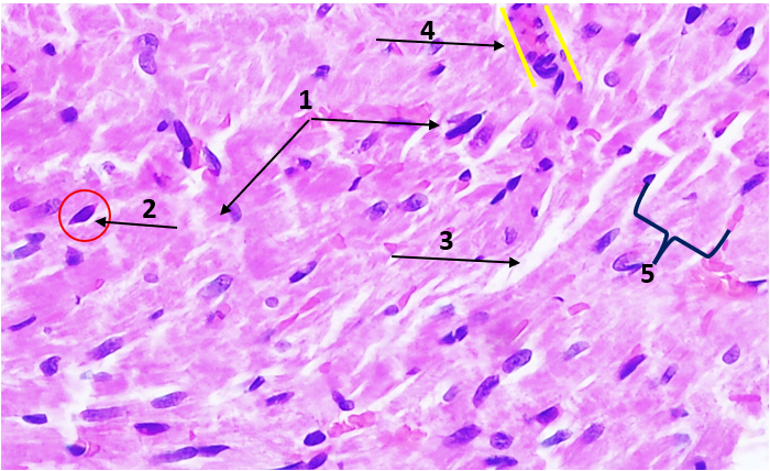 | Figure 1. Microscopic appearance of cardiomyocytes in a 6-month-old rat after carbon monoxide exposure (experimental group). Hematoxylin and eosin staining. Eyepiece ×10, objective ×40. 1 — cardiomyocytes with moderate disorganization and partially disturbed parallel orientation; 2 — predominantly centrally located nuclei, hyperchromic in some areas; 3 — expanded interstitial spaces between cardiomyocytes; 4 — dilated capillaries are visualized, reflecting vascular dilation and microcirculatory disturbance under hypoxic exposure; 5 — enlargement of the precapillary perfusion zone |
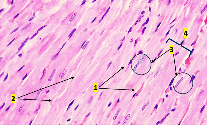 | Figure 2. Microscopic appearance of cardiomyocytes in an 18-month-old rat after carbon monoxide exposure (experimental group). Hematoxylin and eosin staining. Eyepiece ×10, objective ×40. 1 — signs of chronic hypoxic injury are observed; cardiomyocytes show a moderate increase in cytoplasmic eosinophilia; 2 — uneven thickness and focal disorganization of muscle fibers; 3 — in individual cardiomyocytes, hyperchromic elongated nuclei with signs of chromatin condensation are identified; 4 — enlargement of the precapillary perfusion zone |
4. Conclusions
- Carbon monoxide exposure causes pronounced age-dependent myocardial remodeling in white outbred rats. The main structural changes include cardiomyocyte hypertrophy, reduction of cellular density, expansion of the stromal component, capillary dilation and enlargement of the pericapillary diffusion zone.In 6-month-old rats, carbon monoxide exposure leads to moderate myocardial injury, while in 18-month-old rats the same exposure causes more severe and progressive damage, including pronounced stromal-parenchymal imbalance, fibrosis and disruption of intercellular contacts.Van Gieson staining demonstrated that carbon monoxide exposure significantly increases collagen fiber content in the myocardium. The increase was observed in both age groups, but was more pronounced in 18-month-old animals.Immunohistochemical analysis showed that carbon monoxide exposure sharply reduces desmin expression and causes fragmentation and disorganization of desmin-positive structures, indicating damage to the cytoskeleton of cardiomyocytes.Simultaneous correction with milk thistle and safflower extracts provides the most pronounced protective effect. It reduces collagen accumulation, preserves desmin expression and limits structural disorganization of the myocardium. Correction performed after carbon monoxide exposure has a partial restorative effect but is less effective than simultaneous correction, especially in 18-month-old animals.The obtained data indicate that early correction with milk thistle and safflower extracts may limit carbon monoxide-induced myocardial fibrosis and cytoskeletal damage, while age significantly reduces the adaptive and reparative capacity of the myocardium.