-
Paper Information
- Next Paper
- Previous Paper
- Paper Submission
-
Journal Information
- About This Journal
- Editorial Board
- Current Issue
- Archive
- Author Guidelines
- Contact Us
American Journal of Medicine and Medical Sciences
p-ISSN: 2165-901X e-ISSN: 2165-9036
2026; 16(5): 2613-2620
doi:10.5923/j.ajmms.20261605.73
Received: Apr. 23, 2026; Accepted: May 19, 2026; Published: May 25, 2026

Histomorphological Alterations of Lymph Nodes in Experimental Ulcerative Colitis
 Abstract
 Reference
 Full-Text PDF
 Full-text HTML
Mirshod Malikov1, Erali Sadiev2
1Independent Researcher, Department of Surgical Diseases in Family Medicine, Bukhara State Medical Institute named after Abu Ali ibn Sino, Bukhara, Uzbekistan
2PhD, Associate Professor, Department of Surgical Diseases in Family Medicine, Bukhara State Medical Institute named after Abu Ali ibn Sino, Bukhara, Uzbekistan
Copyright © 2026 The Author(s). Published by Scientific & Academic Publishing.
This work is licensed under the Creative Commons Attribution International License (CC BY).
http://creativecommons.org/licenses/by/4.0/

Background: Ulcerative colitis is a chronic inflammatory bowel disease characterized by destructive lesions of the intestinal mucosa and disturbances of immune regulation. Inflammatory processes developing in ulcerative colitis affect not only the intestinal wall but also peripheral immune organs, particularly lymph nodes. However, histomorphological and immunohistochemical alterations occurring in lymphatic tissues during experimental ulcerative colitis remain insufficiently investigated. Materials and Methods: The study was carried out on 120 white outbred rats aged 3, 6, and 12 months. Experimental ulcerative colitis was induced by intrarectal administration of 8% acetic acid. Histological, morphometric, and immunohistochemical methods were used to evaluate structural alterations in lymph nodes. Tissue sections were stained with hematoxylin-eosin and examined using CD-3 immunohistochemical markers. Quantitative analysis of immunopositive cells was performed using QuPath 0.5.1 software. Additionally, the biocorrective effect of milk thistle oil was evaluated in experimental animals. Results: Experimental ulcerative colitis caused significant structural remodeling of lymph nodes, including widening of medullary sinuses, enlargement of paracortical zones, stromal edema, and reduction of lymphocyte density. Immunohistochemical analysis demonstrated increased CD-3 expression and redistribution of T-lymphocytes in inflammatory conditions. Positive CD-3 expression increased from 9.50% in the control group to 33.39% in experimental animals. Biocorrection with milk thistle oil contributed to partial restoration of lymph node architecture and stabilization of T-cell activity, with positive expression reaching 40.22%. Conclusion: Experimental ulcerative colitis induces pronounced morphofunctional and immunohistochemical alterations in lymph nodes. Milk thistle oil demonstrated protective and immunomodulatory effects by improving lymphoid tissue organization and reducing inflammatory changes. The obtained findings expand current understanding of immunopathogenetic mechanisms involved in ulcerative colitis and may contribute to the development of new therapeutic approaches for inflammatory bowel diseases.
Keywords: Ulcerative colitis, Lymph nodes, Morphology, Morphometry, Immunohistochemistry, CD-3 marker, T-lymphocytes, Experimental study, Milk thistle oil, Biocorrection, Inflammatory bowel disease, White outbred rats
Cite this paper: Mirshod Malikov, Erali Sadiev, Histomorphological Alterations of Lymph Nodes in Experimental Ulcerative Colitis, American Journal of Medicine and Medical Sciences, Vol. 16 No. 5, 2026, pp. 2613-2620. doi: 10.5923/j.ajmms.20261605.73.
1. Introduction
- Ulcerative colitis (UC) is a chronic nonspecific inflammatory disease of the colon characterized by recurrent inflammation of the intestinal mucosa, ulcerative-destructive lesions, and dysregulation of the immune system [1,2]. In recent years, the incidence and prevalence of inflammatory bowel diseases, including ulcerative colitis, have been steadily increasing worldwide, making this pathology one of the most important problems in modern gastroenterology and experimental medicine [3]. The chronic recurrent course of the disease and the development of severe complications significantly reduce patients’ quality of life and increase the social and economic burden on healthcare systems [4]. Currently, the etiology and pathogenesis of ulcerative colitis remain incompletely understood. Modern studies indicate that the disease develops as a result of complex interactions between genetic predisposition, environmental factors, intestinal microbiota disturbances, and immune dysregulation [5,6]. Cytokine imbalance, excessive activation of T- and B-lymphocytes, and impaired immune tolerance play a central role in the progression of inflammatory processes in ulcerative colitis [7]. Persistent inflammation causes structural and functional alterations not only in the intestinal wall but also in regional lymphatic tissues. Lymph nodes are among the most important peripheral organs of the immune system and perform filtration, antigen presentation, lymphocyte proliferation, and immune regulatory functions [8]. During inflammatory diseases, lymph nodes undergo significant morphological and functional remodeling associated with activation of immune responses. Experimental and clinical studies have demonstrated that inflammatory bowel diseases are accompanied by lymphoid follicle hyperplasia, sinus histiocytosis, stromal edema, vascular disturbances, and alterations in immune cell populations within lymphatic tissues [9]. Although numerous investigations have focused on histopathological changes in the intestinal mucosa during ulcerative colitis, morphological alterations occurring in lymph nodes remain insufficiently studied [10,11]. Previous studies reported proliferative and degenerative changes in lymphoid structures, including disruption of normal lymph node architecture and alterations in T- and B-cell activity during experimental colitis [12]. Immunohistochemical markers such as CD-3 and CD-20 are considered informative indicators for evaluating changes in lymphocyte populations and immune activity in lymphatic tissues [13]. At present, studying the immunopathogenetic mechanisms of ulcerative colitis and identifying structural changes in lymphatic organs are among the priority directions of modern morphology and immunology [14]. In this regard, investigation of potential biocorrective agents with anti-inflammatory and antioxidant properties is of particular scientific interest. Milk thistle oil, known for its membrane-protective and antioxidant effects, may have a beneficial influence on morphological alterations developing in lymphatic tissues under inflammatory conditions [15]. Therefore, investigation of morphological, morphometric, and immunohistochemical changes in lymph nodes following experimental ulcerative colitis is important for understanding disease pathogenesis, improving diagnostic approaches, and increasing treatment effectiveness. The scientific novelty of the present study lies in the fact that, for the first time, comparative morphometric characteristics of lymph nodes in normal and experimental ulcerative colitis conditions were identified in white outbred rats, histomorphological and histochemical alterations in lymph nodes after experimental ulcerative colitis were comprehensively analyzed, and immunohistochemical changes using CD-3 and CD-20 markers were determined. In addition, the biocorrective effects of milk thistle oil on lymph node structures after experimental ulcerative colitis were experimentally substantiated, which expanded current understanding of the immunopathogenetic mechanisms of ulcerative colitis and the role of lymphatic tissue remodeling in inflammatory bowel diseases.
2. Materials and Methods
- This experimental investigation was performed at the scientific laboratory of the Bukhara State Medical Institute named after Abu Ali ibn Sino during 2024–2026. The study protocol was developed according to modern biomedical research standards, and all procedures involving laboratory animals were conducted in accordance with internationally accepted ethical principles.The study included 120 white outbred rats of both sexes aged 3, 6, and 12 months. Before inclusion in the experiment, all animals were kept under quarantine conditions and examined to exclude infectious and somatic diseases. Subsequently, the rats were housed in standard vivarium conditions with a controlled environment, standard nutrition, and free access to water.Experimental animals were divided into three groups. The first group consisted of intact rats and served as the control group. The second group included animals with experimentally induced ulcerative colitis. Experimental colitis was modeled by daily intrarectal administration of 1 ml of 8% acetic acid through a special probe for 20 days. The third group consisted of rats with induced ulcerative colitis that received milk thistle oil for biocorrection. During the experimental period, several animals died due to complications associated with severe inflammatory changes.Throughout the experiment, the animals’ body weight, physiological status, and behavioral activity were regularly monitored. At the end of the observation period, the rats were euthanized under ether anesthesia by decapitation according to international recommendations for biomedical studies using laboratory animals.Lymph node tissues collected from experimental and control animals served as the main material for morphological analysis. Tissue samples were fixed in 10% neutral buffered formalin, followed by washing and dehydration in ascending ethanol concentrations. After dehydration, the specimens were cleared in xylene and embedded in paraffin using standard histological techniques.Paraffin blocks were sectioned into 4–7 μm slices using a microtome. Histological preparations were stained with hematoxylin-eosin, Van Gieson, and Alcian blue methods for evaluation of tissue architecture and connective tissue alterations. Morphological observations and morphometric measurements were carried out using trinocular light microscopes equipped with digital imaging systems.Immunohistochemical analysis was performed to determine the expression of T- and B-lymphocyte markers within lymph node tissues. CD-3 and CD-20 antibodies were used for immunophenotypic evaluation. Tissue sections placed on adhesive-coated slides underwent deparaffinization, rehydration, antigen retrieval, and blocking of endogenous peroxidase activity before incubation with primary antibodies. Immunoreactivity was visualized using DAB chromogen staining.The immunohistochemical reaction was evaluated in several microscopic fields at ×200–×400 magnification. Quantitative analysis of positively stained cells was performed using QuPath 4.4.0 software. Depending on the proportion of immunopositive cells, expression intensity was categorized as low, moderate, or high.Statistical analysis of the obtained data was carried out using Microsoft Excel and STATGRAPH software packages. Arithmetic means, standard deviations, and standard errors were calculated for all quantitative variables. Student’s t-test was applied for normally distributed data, while the Mann–Whitney U-test was used for nonparametric analysis. Differences between groups were considered statistically significant at p<0.05.
3. Results
- The morphometric analysis of lymph nodes in the control group of white outbred rats demonstrated that their histological structure corresponded to normal physiological parameters. The capsule, cortical, and medullary regions of the lymph nodes were clearly differentiated from each other. The number, shape, and size of lymphoid follicles remained within normal limits, indicating preservation of the overall structural organization of the lymph nodes. Morphometric examination revealed stable values of the total lymph node area, the ratio between cortical and medullary zones, and the diameter of germinal centers. A balanced relationship between stromal and parenchymal components was maintained, while no inflammatory, degenerative, or reactive alterations were detected. These findings confirmed the normal immunomorphological condition of lymph nodes in the control group and served as a reliable basis for comparative evaluation with experimental groups.Histological examination of lymph nodes in 3-month-old white outbred rats from the control group also demonstrated age-related normal structural organization. Microscopic observations showed clear differentiation of the lymph node capsule, trabeculae, cortical and medullary regions, as well as lymphoid follicles. The capsule exhibited uniform thickness and consisted mainly of dense fibrous connective tissue. Lymphoid follicles located within the cortical zone were observed in normal numbers and sizes, while the proportion between primary and secondary follicles remained preserved. Germinal centers were well developed and distinctly expressed, indicating physiologically active lymphocyte proliferation. In the paracortical zone, the distribution of T-lymphocytes was arranged in a regular and organized manner. Structural integrity of the medullary sinuses and medullary cords was preserved, and cellular density remained within physiological limits. The obtained morphometric results confirmed that lymph nodes of 3-month-old white outbred rats possessed anatomically and histologically normal age-related features. No signs of inflammation, tissue destruction, or other pathological alterations were identified during the examination (Figure 1).
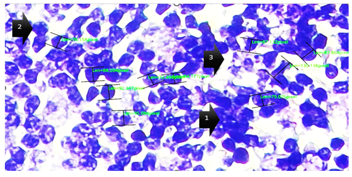 | Figure 1. Morphometric characteristics of lymph nodes in 3-month-old white outbred rats of the control group. Hematoxylin-eosin staining. Objective lens ×10, ocular lens ×100. (1) Germinal center of the lymph node; (2) lymphocyte; (3) sinusoidal space |
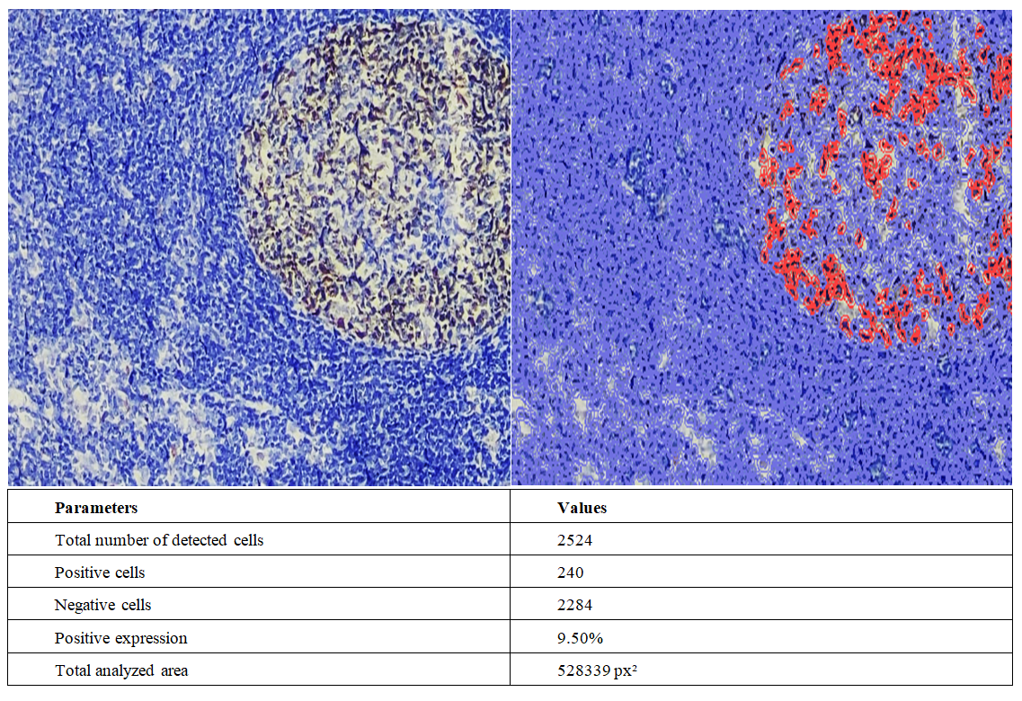 | Figure 2. Immunohistochemical evaluation of CD-3 marker expression in lymph nodes of 3-month-old white outbred rats from the control group. Stained using the DAB chromogen method. Magnification ×200. The image was scanned and analyzed using QuPath 0.5.1 software to determine the level of expression. Expressed cells are highlighted in red |
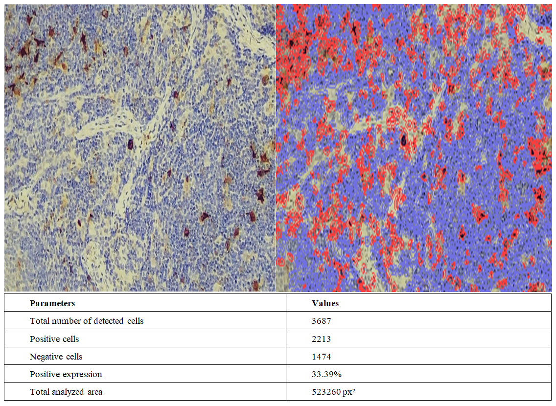 | Figure 3. Immunohistochemical evaluation of CD-3 marker expression in lymph nodes of 12-month-old white outbred rats under experimental conditions. Stained using the DAB chromogen method. Magnification ×200. The image was scanned and analyzed using QuPath 0.5.1 software to determine the level of expression. Expressed cells are highlighted in red |
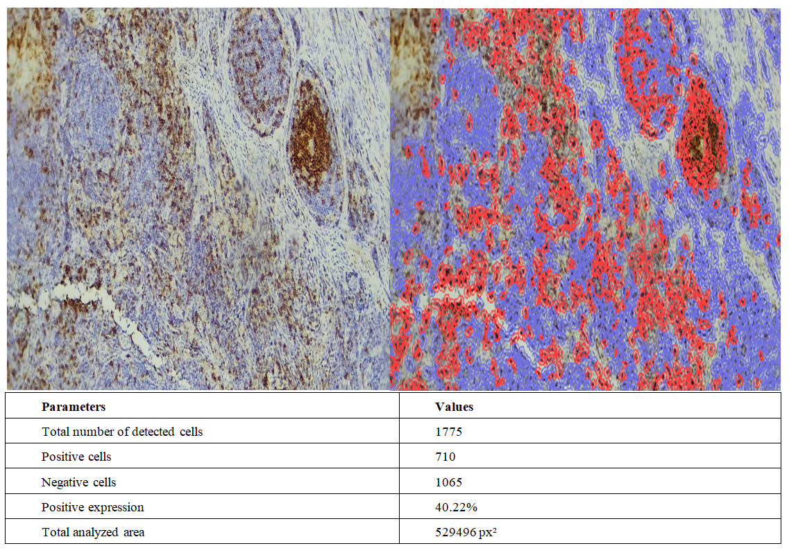 | Figure 4. Comparative evaluation of immunohistochemical alterations in lymph nodes of 3-month-old white outbred rats and biocorrected groups treated with milk thistle oil, with assessment of CD-3 marker expression. Stained using the DAB chromogen method. Magnification ×200. The image was scanned and analyzed using QuPath 0.5.1 software to determine the level of expression. Expressed cells are highlighted in red |
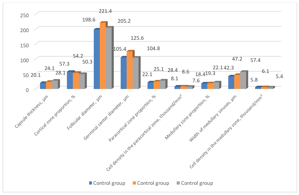 | Figure 5. Morphometric parameters of lymph nodes in white outbred rats of the control group |
4. Discussion
- The results of the present study demonstrated that experimental ulcerative colitis leads to significant morphofunctional and immunohistochemical alterations in lymph nodes of white outbred rats. Comparative analysis between control and experimental groups revealed that inflammatory processes induced structural remodeling of lymphoid tissue, disturbances in zonal organization, and changes in immune cell distribution. These findings confirm that ulcerative colitis affects not only the intestinal wall but also peripheral immune organs involved in inflammatory regulation [1,2].Morphometric investigations showed that lymph nodes of control animals preserved normal histological architecture characterized by clear differentiation of cortical, paracortical, and medullary regions, stable follicular structure, and physiologically balanced cellular density. In contrast, experimental groups demonstrated enlargement of paracortical and medullary zones, widening of medullary sinuses, and reduction of lymphocyte density. Such changes indicate activation of immune-inflammatory reactions accompanied by stromal edema and progressive disorganization of lymph node architecture.Immunohistochemical analysis using CD-3 markers demonstrated alterations in T-lymphocyte distribution within lymph node tissues. In control animals, CD-3 positive cells were mainly localized within paracortical regions and exhibited physiologically normal expression levels. Under experimental conditions, increased expression and redistribution of CD-3 positive lymphocytes were observed, reflecting activation of cellular immune responses associated with ulcerative colitis [3]. The decrease in cellular density in older experimental animals may indicate gradual weakening of proliferative and adaptive immune activity.An important finding of the study was the positive effect of milk thistle oil on lymph node structures. Animals receiving biocorrection demonstrated partial restoration of lymph node architecture, reduction of inflammatory infiltration, and more organized distribution of immunopositive cells. Normalization of CD-3 expression patterns suggests stabilization of T-lymphocyte activity and improvement of immune regulation under inflammatory conditions. These findings may be associated with the antioxidant and anti-inflammatory properties of milk thistle components [4].Overall, the obtained results indicate that experimental ulcerative colitis causes pronounced structural and immunological alterations in lymph nodes, whereas biocorrection with milk thistle oil contributes to partial restoration of lymphoid tissue organization and cellular immune balance. The findings expand current understanding of immunopathogenetic mechanisms involved in ulcerative colitis and confirm the important role of lymphatic tissue remodeling in chronic inflammatory bowel diseases [5].
5. Conclusions
- 1. In experimental ulcerative colitis, significant morphometric alterations were identified in lymph nodes of white outbred rats. In 12-month-old animals, capsule thickness increased from 20.1±0.54 μm to 28.1±1.7 μm, while the cortical zone proportion decreased from 57.3±0.64% to 50.3±2.4%. At the same time, the medullary zone proportion increased to 22.1±1.9%, indicating age-related structural remodeling of lymphoid tissue.2. Experimental ulcerative colitis caused pronounced histomorphological disturbances in lymph nodes characterized by widening of medullary sinuses, stromal edema, disorganization of lymphoid follicles, and reduction of cellular density. The width of medullary sinuses increased from 42.3±3.5 μm in 3-month-old control animals to 57.4±4.2 μm in 12-month-old experimental rats, reflecting progressive inflammatory and degenerative alterations in lymphoid structures.3. Immunohistochemical analysis using CD-3 markers demonstrated activation of T-lymphocytic immune responses in experimental ulcerative colitis. Positive CD-3 expression increased from 9.50% in the control group to 33.39% in experimental animals, while redistribution of immunopositive cells within paracortical and interfollicular zones confirmed disturbances in cellular immune regulation.4. Biocorrection with milk thistle oil exerted a protective effect on lymph node tissues by improving structural organization and stabilizing T-lymphocyte activity. In the biocorrected group, CD-3 positive expression reached 40.22%, accompanied by reduction of inflammatory infiltration and partial restoration of physiological lymphoid architecture, indicating immunomodulatory and anti-inflammatory properties of milk thistle oil.