Gafurov Adham Anvarovich1, Nazirov Nodirbek2, Nematjonov Farrukh Zokirjon Ugli3
1Doctor of Medical Science, Professor of the Department of Pediatric Surgery, Andijan State Medical Institute, Andijan, Uzbekistan
2Doctor of the Andijan Multidisciplinary Children's Medical Center, Andijan, Uzbekistan
3Assistant of the Department of Pediatric Surgery1, Andijan State Medical Institute, Andijan, Uzbekistan
Correspondence to: Gafurov Adham Anvarovich, Doctor of Medical Science, Professor of the Department of Pediatric Surgery, Andijan State Medical Institute, Andijan, Uzbekistan.
| Email: |  |
Copyright © 2026 The Author(s). Published by Scientific & Academic Publishing.
This work is licensed under the Creative Commons Attribution International License (CC BY).
http://creativecommons.org/licenses/by/4.0/

Abstract
This article presents the results of evaluating modern radiation imaging methods in the diagnosis of inguinal and abdominal forms of cryptorchidism in children. From 2020 to 2025, 253 patients aged from 8 months to 17 years with various forms of cryptorchidism were examined and treated at the multidisciplinary children's clinic of the Andijan State Medical Institute. The diagnostic complex included ultrasound scanning, Dopplerography, magnetic resonance imaging (MRI), and digital radiography. The study assessed the localization, structural condition, blood circulation, and viability of undescended testes. Ultrasound scanning was identified as the primary noninvasive diagnostic method, allowing accurate determination of gonadal size and echostructural changes. Dopplerography provided important information about testicular blood flow and hemodynamic disorders, while MRI demonstrated high effectiveness in detecting non-palpable gonads and evaluating morphological changes in testicular tissue. The sensitivity, specificity, and diagnostic accuracy of each imaging method were comparatively analyzed. The obtained results showed that the combined step-by-step use of radiation imaging methods significantly increases diagnostic accuracy and improves preoperative assessment in children with cryptorchidism. The study confirms the necessity of an integrated diagnostic approach for timely surgical management and prevention of complications associated with undescended testes.
Keywords:
Cryptorchidism, Undescended testis, Inguinal cryptorchidism, Abdominal cryptorchidism, Ultrasound scanning, Dopplerography, Magnetic resonance imaging, Pediatric surgery, Radiation imaging, Testicular blood flow, Gonadal hypoplasia, Non-palpable testis
Cite this paper: Gafurov Adham Anvarovich, Nazirov Nodirbek, Nematjonov Farrukh Zokirjon Ugli, Radiation Imaging of Inguinal and Abdominal Forms of Cryptorchidism in Children, American Journal of Medicine and Medical Sciences, Vol. 16 No. 5, 2026, pp. 2609-2612. doi: 10.5923/j.ajmms.20261605.72.
1. Introduction
Cryptorchidism in children remains one of the urgent problems that pediatric surgeons and urologists very often face, since most of the parents' complaints are related to the main complaints about the absence of a testicle in its proper place in the scrotum, while almost all of these children are physically not lagging behind in their development. Despite a sufficient number of scientific papers, many of which contradict each other, it is necessary to understand one thing, that in most cases the revealed abdominal ectopia is one of the variants of the non-palpable testicle. According to many researchers, among children with cryptorchidism, testicular ectopia is detected in only 5% of cases, which are difficult to detect and even more difficult to palpate on the normal path of their migration from the abdominal cavity. In turn, a perineally ectopic testicle can be visualized by careful examination of the patient, but in some situations it cannot be palpated, while the diagnosis is established using imaging techniques or intraoperatively. [3,7,9,10].Modern highly informative radiation research methods have high resolution capabilities, which makes it possible to identify the absence or presence of a gonad, as well as accurately determine its localization, viability, echostructure and blood flow. These diagnostic methods also make it possible to determine the parameters of the undescended gonad not only on the side of the missing testicle, but also to visualize the condition of the contralateral gonad with an assessment of hemodynamics, which allows an indirect assessment of the condition of the testicular parenchyma. Despite their advantages, many radiation research methods have limited sensitivity and specificity in imaging non-palpable gonads, especially in determining the testicular volume of the gonad in the preoperative period. Each of the existing imaging methods has its advantages and disadvantages, but their consistent application allows accurate and correct diagnosis in most cases. This helps to increase the effectiveness of complex treatment of cryptorchidism in children, reduce the risk of complications and improve the results of surgical interventions for lowering the testicle into the scrotum. [1,2,4,5,6,8,11].Many key questions about the diagnosis of cryptorchidism are presented in various scientific publications, but they do not always reflect the true characteristics of radiation examinations of undescended gonads in children, and therefore the search for answers to these questions determined the purpose of this study.The purpose of the study. Evaluation of the effectiveness of modern imaging methods for cryptorchidism in children.
2. Research Materials and Methods
From 2020 to 2025, 253 patients with various forms of cryptorchidism aged from 8 months to 17 years were examined and treated at the multidisciplinary children's clinic of the Andijan State Medical Institute. Depending on the form of cryptorchidism and the age of the patients, we performed various radiation research methods, the list of which included: X-ray imaging, ultrasound scanning, color Doppler mapping and magnetic resonance imaging.
3. The Results of the Study
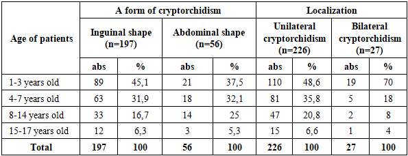
Our observations revealed a significant difference in the incidence of cryptorchidism on the affected side, while localization of unilateral cryptorchidism was detected in 226 (89.3%) cases, bilateral in 27 (10.7%) patients. In unilateral forms of cryptorchidism, retention of the right testicle was more often observed in 144 (56.9%) patients, and left-sided gonadal non-prolapse in unilateral forms of cryptorchidism was detected in 82 (32.4%) patients. The distribution of patients by age, form, and localization of cryptorchidism is shown in Table 1.Table 1. Distribution of patients by age, shape, and location of the undescended testicle
 |
| |
|
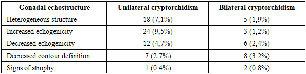
The data presented in Table 1 indicate the predominant number of hospitalized patients under the age of 7 years, which indicates the caution of parents when determining the absence of a testicle in the scrotum. The main clinical signs of cryptorchidism in children were the absence of a testicle in the corresponding half of the scrotum, while 98% of patients showed signs of scrotal underdevelopment, and 63 (24.9%) patients were diagnosed with concomitant inguinal hernia, which was most often detected with a right-sided lesion.Ultrasound scanning was the primary and main noninvasive method of diagnosing cryptorchidism in children in our studies. It made it possible to determine the size of an undescended testicle by using the geometric formula of the volume of an ellipsoid in order to conduct a comparative parameter with the contralateral gonad. The volume was calculated in the following parameters V=0.523×L×W×H (L-length, W-width, H-thickness). Based on this formula, the ultrasound scan data showed that in 4 (1.6%) patients under the age of 5, the gonadal volume was reduced by 12% and averaged up to 0.6 ml, while pronounced testicular hypoplasia was detected in 9 (3.5%) patients over the age of 5 years. The characteristic echostructural changes of the undescended testicle of inguinal localization are presented in Table 2.Table 2. Significant echostructural changes revealed in inguinal cryptorchidism in children
 |
| |
|
The determination of the linear dimensions of the undescended gonad in inguinal cryptorchidism also showed a significant difference in relation to age norms. The parameters we obtained allowed us to establish that in 28 (11.1%) children, the gonadal length was 4-5 mm lower than normal, and a decrease in the longitudinal diameter of the testicle was detected in 17 (6.7%) patients, while a decrease in testicular thickness was up to 0.6 cm, which was 1.5-2 times lower than normal. During ultrasound scanning, in 197 (77.8%) patients with inguinal cryptorchidism in 127 cases, the testicle was localized in the lower third of the inguinal canal, in 31 patients in the middle third and in 37 patients in the upper third of the inguinal canal.Ultrasound scanning with Dopplerography was of indisputable importance in the diagnosis of cryptorchidism in children, especially in the abdominal form of the disease. This research method is based on the principle of measuring the speed and direction of blood flow in the vascular pedicle of the spermatic cord feeding the testicle and its parenchyma, and also allows the identification of areas of circulatory disorders and ischemic areas in the undescended gonad. The Doppler study allowed not only to determine the presence of intratesticular blood flow, but also to calculate the resistance index (RI) by comparing the symmetry of blood supply in the contralateral gonad. Ultrasound scanning with Dopplerography was important for assessing the functionality of an undescended testicle, this examination method is not a painful procedure for children and does not require special training (Fig. 1.).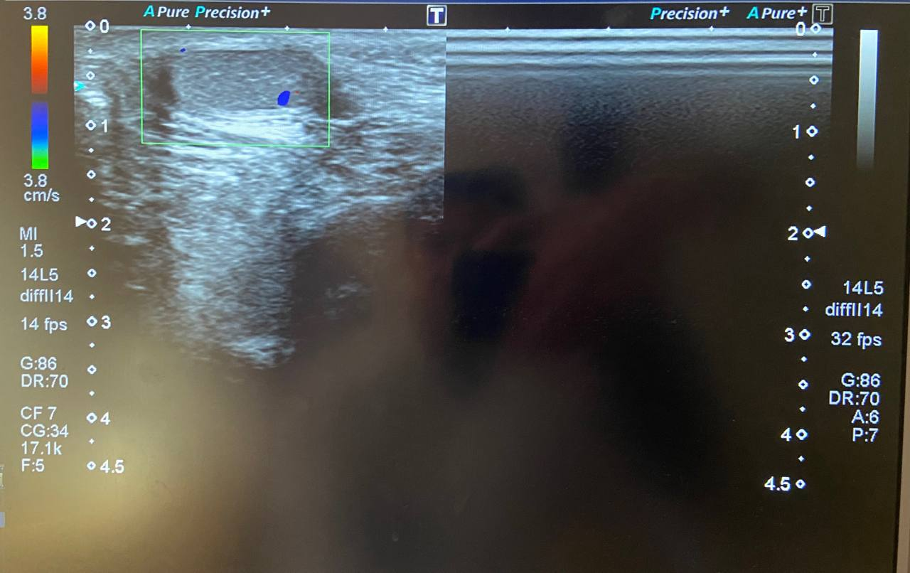 | Figure 1. Ultrasound scanning with Dopplerography (areas of circulatory disorders in the undescended gonad) |
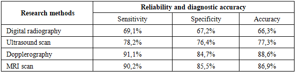
The Doppler resistance index (RI) was calculated using the calculation formula: RI=(Vmax-Vmin)/Vmax, where Vmax is the peak systolic rate, and Vmin is the final diastolic rate. 0.5-0.7 (cm/s) was considered the norm of the resistance index (RI) during Dopplerography, and an increased value in the range from 0.7 to 0.9 (cm/s) was considered a deviation from the norm, which indicated a violation of perfusion in children with cryptorchidism. According to calculations based on the index of resistance (RI) in Dopplerography, it should be assumed that signs of degeneration in children with cryptorchidism are established in the presence of a decrease in testicular volume by more than 25%, a decrease in its blood flow and structural heterogeneity, as well as an increase in the index of resistance from 0.7 to 0.9 (cm/s). As noted above, the ultrasound scan data we obtained showed that in the inguinal form of cryptorchidism in most cases - 178 (70.4%) observations, signs of a preserved testicle were revealed. However, 13 (5.1%) patients with inguinal cryptorchidism showed signs of hypoplasia, decreased blood supply and testicular vascularization; in these patients, the resistance index (RI) ranged from 0.7 to 0.9 (cm/s). In 6 (2.4%) patients with torsion of the inguinal shape of the undescended testicle, a significant slowdown in blood flow was noted. According to Dopplerography data, 14 (5.5%) children with abdominal cryptorchidism showed a decrease and depletion of the vascular pattern; in these patients, the resistance index (RI) ranged from 0.5 to 0.7 (cm/s). In 8 (3.2%) cases of abdominal cryptorchidism, a decrease in linear blood flow velocity was found, which served as a direct indication for diagnostic laparoscopy in all these patients.The use of ultrasound scanning with Dopplerography in the diagnosis of inguinal and abdominal cryptorchidism allowed us to accurately determine not only the location and volume of the undescended testicle, but also to identify signs of circulatory disorders and ischemic areas in the gonad itself. A decrease and depletion of the vascular pattern, a decrease in the linear velocity of blood flow, and a slowdown and absence of blood flow, revealed during ultrasound scanning with Dopplerography, were direct indications for diagnostic laparoscopy in this cohort of patients. The diagnostic information value of Dopplerography in determining the reliability and accuracy of the study showed that the sensitivity of the method was 91.1%, the specificity was 84.7%, and the accuracy of the method was 88.6%.Magnetic resonance imaging was performed in 22 (8.6%) patients with bilateral absence of gonads in the scrotum, it was performed in a strict and specific scanning sequence in standard T1 and T2 modes. The standard T1 and T2 modes used on MRI displayed axial and coronal planes, which made it possible to determine the structure of the testicular body, the presence of anatomical landmarks, signal intensity compared to surrounding tissues, and visualization of soft tissues and blood vessels. The interpretation of the images included an assessment of tissue uniformity and signs of limited diffusion to determine viable tissue. The MRI parameters we obtained showed that in 10 (3.9%) cases of bilateral abdominal cryptorchidism and in 7 (2.8%) cases of inguinal cryptorchidism, in the T1 and T2 modes, a uniform signal was detected, which corresponded to normal and preserved oval-shaped gonads, which were clearly visualized in relation to surrounding structures, while The measurements of their sizes testified to their safety, without signs of destruction. In 5 (1.9%) cases of non-palpated gonads in T1 mode, MRI showed a hypointensive low signal, which indicated testicular hypoplasia, and a decrease in the cell density marker was also noted in these patients. Of the 15 (5.8%) patients with abdominal cryptorchidism who underwent magnetic resonance imaging, in 10 cases the localization of undescended gonads was determined at the entrance to the inner ring of the inguinal canal, which was an indication for laparoscopic reduction. The advantage of this research method was that MRI allowed not only to assess the condition of the inguinal canal and abdominal cavity in which the testicle was located, but also helped to identify morphological changes in the undescended gonad. The effectiveness of MRI was not only in determining the topographic and anatomical location of the testicle and its volume, but also contributed to a reliable assessment of structural and morphological changes in the parenchyma of the undescended gonad.Modern and highly informative methods of radiation imaging have shown that the reliability of the information obtained depends on many factors, such as the form of cryptorchidism, location of the undescended testicle, the age of the patient and the presence of complications. At the same time, each of these methods has its own advantage, and if necessary, they complement each other in obtaining reliable research results, which is very important when choosing tactics, method and method of surgical intervention. In addition to their informative value, they are radiologically safe methods for diagnosing cryptorchidism in children, which allows their use for dynamic monitoring in the postoperative period. We compared their significance in assessing the diagnostic information content of our studies (Table 3.).Table 3. Diagnostic information value of radiation research methods in the diagnosis of acute pleural empyema in children
 |
| |
|
4. Conclusions
Thus, the conducted radiation examination methods in the preoperative period in 253 children with various forms of cryptorchidism allowed the identification of diagnostic features of the disease with the determination of structural and clinical signs of an undescended testicle. The step-by-step complex of radiation imaging methods used has shown high informative value in detecting an undescended testicle in children. However, the revealed existing difference in the sensitivity and specificity of these studies obliges them to be conducted in combination. Since they have certain advantages that facilitate their use both independently and in addition to each other in order to obtain complete information and take priority measures for surgical intervention to reduce an undescended testicle.
References
| [1] | S. N. Blokhin and A. A. Ivanov, “Early orchidopexy in cryptorchidism: Indications, technique and outcomes,” Pediatric Surgery, vol. 25, no. 6, pp. 12–18, 2021. |
| [2] | P. A. Goncharov, S. N. Kirillov, and I. I. Baranov, “Optimization of laparoscopic orchidopexy in non-palpable cryptorchidism,” Pediatric Surgery (Khhirurgiya Detskogo Vozrasta), no. 4, pp. 33–40, 2022. |
| [3] | M. N. Ekimov, N. A. Tsap, and S. Y. Komarova, “Non-palpable testis syndrome: History and current state of the issue (literature review),” Pediatric Surgery, vol. 29, no. 1, pp. 22–32, 2025. |
| [4] | A. A. Ivanov, “Modern methods of diagnosis and treatment of cryptorchidism in children,” Bulletin of Pediatric Surgery, vol. 27, no. 1, pp. 14–22, 2021. |
| [5] | S. N. Kirillov, “Surgical management of cryptorchidism in children: Analysis of long-term outcomes,” Pediatric Surgery, no. 5, pp. 27–33, 2020. |
| [6] | A. G. Makarov, “Surgical treatment of palpable forms of cryptorchidism in children,” Ph.D. dissertation abstract, Rostov-on-Don, 2023. |
| [7] | S. M. Alaqeel, A. H. Hakeem, and J. O. Almaary, “Testicular ectopia in a child’s anterior abdominal wall: A case report and literature review,” American Journal of Case Reports, vol. 21, p. e927495, 2020. |
| [8] | S. W. Leslie, H. Sajjad, and C. A. Villanueva, “Cryptorchidism,” in StatPearls [Internet]. Treasure Island (FL): StatPearls Publishing, 2025. |
| [9] | A. Priam, N. Chabani, C. Klein, and E. Haraux, “Scrotal orchidopexy for perineal ectopic testis,” Archives de Pédiatrie, vol. 29, no. 5, pp. 404–406, 2022. |
| [10] | V. V. Punwani, J. S. Y. Wong, C. Y. H. Lai, et al., “Testicular ectopia: Why does it happen and what do we do?” Journal of Pediatric Surgery, vol. 52, no. 11, pp. 1842–1847, 2017. |
| [11] | J. Vikraman, S. Donath, and J. M. Hutson, “Undescended testes: Diagnosis and timely treatment in Australia (1995–2014),” Australian Family Physician, vol. 46, no. 3, pp. 152–158, 2017. |

 Abstract
 Reference
 Full-Text PDF
 Full-text HTML