
-
Paper Information
- Previous Paper
- Paper Submission
-
Journal Information
- About This Journal
- Editorial Board
- Current Issue
- Archive
- Author Guidelines
- Contact Us
American Journal of Medicine and Medical Sciences
p-ISSN: 2165-901X e-ISSN: 2165-9036
2026; 16(5): 3209-3213
doi:10.5923/j.ajmms.20261605.181
Received: Apr. 16, 2026; Accepted: May 10, 2026; Published: May 30, 2026

Pathomorphological Changes in the Kidneys During the Post–Acute Myocardial Infarction Period
 Abstract
 Reference
 Full-Text PDF
 Full-text HTML
Shakirov Sardor Abdusaminovich
PhD in Medical Sciences, Associate Professor, Fergana Medical Institute of Public Health, Fergana, Uzbekistan
Correspondence to: Shakirov Sardor Abdusaminovich, PhD in Medical Sciences, Associate Professor, Fergana Medical Institute of Public Health, Fergana, Uzbekistan.
Copyright © 2026 The Author(s). Published by Scientific & Academic Publishing.
This work is licensed under the Creative Commons Attribution International License (CC BY).
http://creativecommons.org/licenses/by/4.0/

The main substrate of pathomorphological changes developing in the kidneys during the post–acute myocardial infarction period is represented by ischemic lesions of the renal parenchyma associated with hemodynamic disturbances. These lesions are characterized by glomerular congestion, hyaline droplet degeneration in the proximal and distal tubules, plasmolysis, desquamation of epithelial cells, collapse of the tubular lumen, and the absence of primary urine traces. In the renal stroma, plasma imbibition and uneven vascular congestion in the paratubular vessels are observed. Microscopic examination revealed that the renal changes associated with myocardial infarction were manifested by vascular congestion, ischemic injury, and multifocal necrotic nephrosis.
Keywords: Acute myocardial infarction, Ischemia, Glomerular apparatus, Pathomorphology
Cite this paper: Shakirov Sardor Abdusaminovich, Pathomorphological Changes in the Kidneys During the Post–Acute Myocardial Infarction Period, American Journal of Medicine and Medical Sciences, Vol. 16 No. 5, 2026, pp. 3209-3213. doi: 10.5923/j.ajmms.20261605.181.
Article Outline
1. Introduction
- An exponential increase in the number of patients with end-stage renal failure is being observed worldwide. In many cases, renal dysfunction is considered secondary, developing against the background of cardiovascular pathology. At the same time, approximately 5–8% of men aged 20 to 44 years and 18–24.5% of men aged 45 to 69 years suffer from ischemic heart disease (IHD). Among women, the prevalence of ischemic heart disease is slightly lower and, in the older age group, usually does not exceed 13–15% [1]. The aim of this study is to investigate the frequency of development of renal dysfunction in various forms of ischemic heart disease and to assess its dynamics during prospective follow-up.Pathogenesis of Renal Damage in Arterial Hypertension: In the formation of chronic renal failure, changes in myocardial and nephron function should be considered through the prism of the evolution of arterial hypertension (AH) [2]. The concept of the “continuum” refers to a continuous chain of disease progression from risk factors to the patient’s death. Cardiovascular and renal continuums develop in parallel and are closely interconnected. The initial signs of cardiac involvement are associated with the early manifestations of renal involvement. The emergence and progression of changes in the heart and kidneys are largely determined by similar hemodynamic, neurohumoral, and genetic mechanisms [3].A significant similarity of morphological changes in the heart and kidneys is noted during the development of cardiovascular diseases and the renal continuum [4]. In patients with essential arterial hypertension, a decrease in glomerular filtration is associated with a significant increase in the frequency of complications such as ischemic heart disease, atrial fibrillation, as well as diabetic and urate nephropathies [5], and ischemic renal disease [6]. Hypertensive nephropathy results from impairment of renal hemodynamic mechanisms that protect the glomeruli from the damaging effects of high blood pressure (BP) [7]. As a result, two fundamentally different pathological processes develop in the kidneys: ischemic and hypertrophic glomerular injury [8]. These processes lead to the development of focal segmental glomerulosclerosis and progressive loss of renal function [9]. The cause of the first process is an excessive autoregulatory response complicated by obstructive hyalinosis of afferent arterioles, ischemic glomerular injury, and loss of a portion of functioning nephrons [10].Aim of the Study: To study the morphological changes occurring in the kidneys during the period after acute myocardial infarction.
2. Materials and Methods of the Study
- The material for the dissertation consisted of 191 autopsy cases examined at the Republican Pathological Anatomy Center during 2020–2025, in which the diagnosis of ischemic heart disease was confirmed on the basis of clinical and morphological data. The following methods were used to conduct the study: analysis of clinical and laboratory data of patients who died from ischemic heart disease. The patients’ medical histories and autopsy protocols were analyzed. The frequency of the main renal changes in IHD was assessed, and the cases were divided into groups according to age and sex. The following distribution by age was identified.
3. Results and Discussion
- During the period after acute myocardial infarction, the essence of the main microscopic changes occurring in the kidneys is related to ischemia of the cortical region and recovery following hemodynamic disturbances. It was found that renal glomerular congestion developed, leading to severe dystrophic changes in mesangial cells within the glomerular capillary network.According to the WHO classification, the period after acute myocardial infarction is divided as follows:Acute period: from day 1 to day 10, sometimes up to 14 days. During this period, renal changes reach their peak.Subacute period: from day 10–14 to 4–8 weeks, up to 2 months. During this period, renal function may recover or progress to chronic changes.Post-infarction period: beginning from 2 months and continuing for several years. At this stage, chronic cardiorenal changes, such as fibrosis and sclerosis, are more commonly formed in the kidneys.In our study, the acute period was specifically investigated, taking into account the first 1–10 days after myocardial infarction. During this period, the main microscopic characteristics of renal changes in relation to age and sex after myocardial ischemia included renal congestion and, due to the absence of filtration processes in the glomeruli for 1–3 days, focal proliferation of mesangial cells in the glomerular capillary network. It was determined that hyaline droplet dystrophy occurring in the proximal tubules and foci of clasmolysis of varying degrees, together with desquamation of epithelial cells into the tubular lumen, led to thickening of the tubular basement membrane.
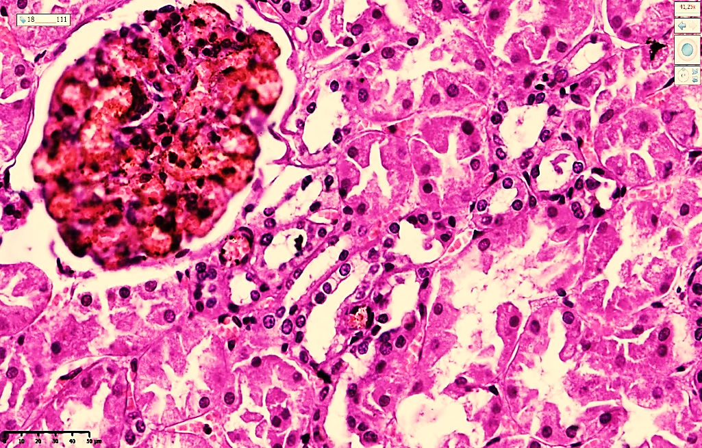 | Figure 1. Microscopic image of the kidneys during days 1–3 of acute myocardial infarction. Marked massive congestion is observed in the glomerular capillary network, and focal proliferation of mesangial cells is detected in some dark-stained areas. Foci of desquamation and necrobiosis are identified in the proximal and distal tubules. Stain: H&E. Magnification: 20×10 |
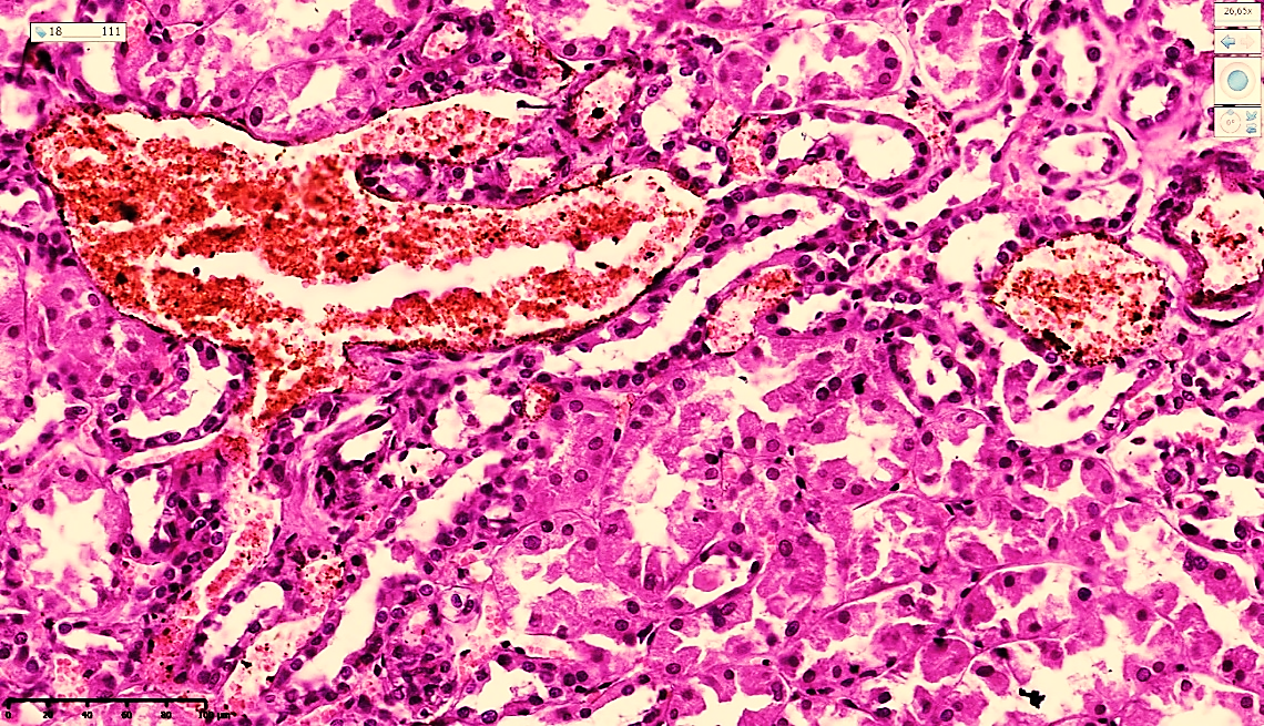 | Figure 2. Microscopic image of the kidneys on day 4 of acute myocardial infarction in a 44-year-old male. Venous congestion in the blood vessels around the proximal tubules, plasma swelling of the stroma, and diapedetic hemorrhages are observed. Stain: H&E. Magnification: 20×10 |
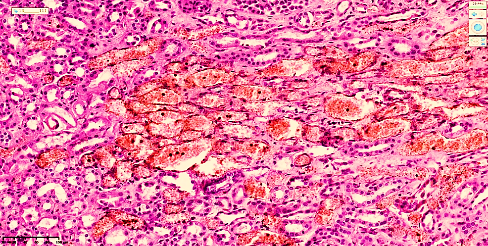 | Figure 3. Microscopic image of the kidneys on day 4 of acute myocardial infarction. Venous congestion in the blood vessels around the distal tubules, plasma swelling of the stroma, and diapedetic hemorrhages are observed. Stain: H&E. Magnification: 20×10 |
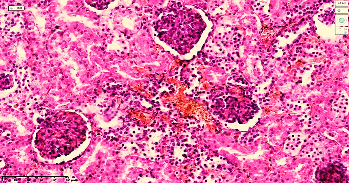 | Figure 4. Microscopic image of the kidneys on days 4–7 after acute myocardial infarction. Congestion is preserved in the glomeruli. Epithelial cells undergoing necrobiosis and desquamation are observed in the proximal and distal tubules. Homogeneous protein substrates are detected in the tubular lumens. Stain: H&E. Magnification: 20×10 |
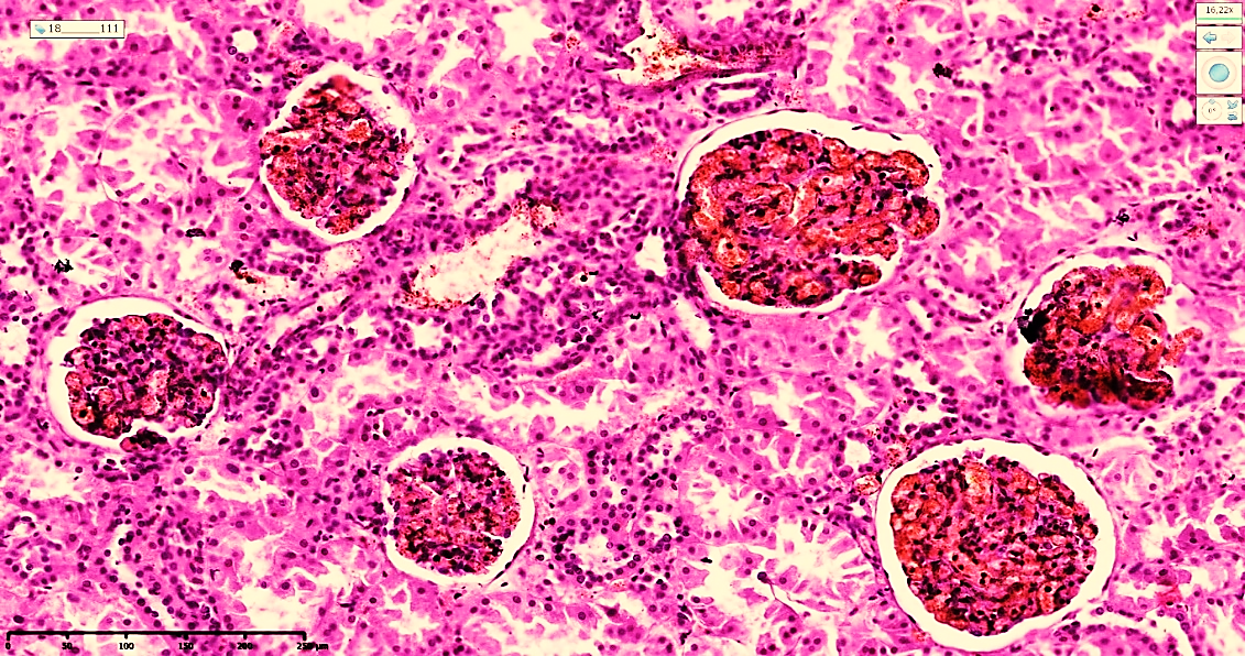 | Figure 5. Kidneys on day 8 after acute myocardial infarction. Congestion is preserved in the glomeruli; the glomeruli vary in size, and focal proliferation of mesangial cells is observed. Foci of necrobiosis and desquamation are detected in the proximal and distal tubules. Stain: H&E. Magnification: 20×10 |
4. Conclusions
- Primary effect of hemodynamic disturbances and ischemia. In the period following acute myocardial infarction, the main substrate of pathomorphological changes in the kidneys is represented by ischemic foci caused by hemodynamic disturbances. This process is characterized by congestion/hyperemia in the renal glomeruli and severe dystrophic changes in mesangial cells.Destructive changes in the tubular system. During the acute phase of myocardial infarction, especially within the first 1–10 days, significant damage is observed in the proximal and distal tubules of the kidneys. These changes include hyaline-droplet dystrophy, plasmolysis, desquamation of epithelial cells, and foci of necrobiosis.Functional inactivity and “collapse” state of the kidneys. During the first 1–3 days after ischemia, cessation of the filtration process is observed in the kidneys. This condition is manifested by collapse of the tubular lumen and the absence of traces of primary and secondary urine, indicating a temporary functionally inactive state of the kidneys.Morphological risks of diuretic use. During the first 72 hours after infarction, when filtration is at its lowest and most critical point, the use of diuretics may further increase glomerular congestion. This process may lead to precipitation of large protein fractions in the tubules and serves as a major factor contributing to the development of multifocal necrotic nephrosis in the proximal tubules.