
-
Paper Information
- Next Paper
- Previous Paper
- Paper Submission
-
Journal Information
- About This Journal
- Editorial Board
- Current Issue
- Archive
- Author Guidelines
- Contact Us
American Journal of Medicine and Medical Sciences
p-ISSN: 2165-901X e-ISSN: 2165-9036
2026; 16(5): 3205-3208
doi:10.5923/j.ajmms.20261605.180
Received: Apr. 15, 2026; Accepted: May 12, 2026; Published: May 30, 2026

Age-Related Pathomorphological Features of the Kidneys in Ischemic Heart Disease
 Abstract
 Reference
 Full-Text PDF
 Full-text HTML
Shakirov Sardor Abdusaminovich
PhD in Medical Sciences, Associate Professor, Fergana Medical Institute of Public Health, Fergana, Uzbekistan
Correspondence to: Shakirov Sardor Abdusaminovich, PhD in Medical Sciences, Associate Professor, Fergana Medical Institute of Public Health, Fergana, Uzbekistan.
Copyright © 2026 The Author(s). Published by Scientific & Academic Publishing.
This work is licensed under the Creative Commons Attribution International License (CC BY).
http://creativecommons.org/licenses/by/4.0/

In ischemic heart disease, the main morphological substrate of age-related changes in the kidneys is considered to be alterations in the renal glomeruli. According to the WHO classification, ischemic heart disease is most commonly observed among individuals aged 45–59 years. It is precisely at this age that a reduction in the surface area of renal glomeruli occurs, along with the development of atrophic and sclerotic changes in the functional units of the kidneys. In particular, hypertension, atherosclerosis, diabetes mellitus, and metabolic syndrome, which serve as major background conditions in ischemic heart disease, are manifested by various significant pathological changes. In hypertension, persistent narrowing of the arterial vessels leads to impaired filtration in the renal glomeruli, resulting in the development of glomerulosclerosis and making the process irreversible.
Keywords: Ischemic heart disease, Glomerulosclerosis, Hypertension, Blood pressure, Pathomorphology
Cite this paper: Shakirov Sardor Abdusaminovich, Age-Related Pathomorphological Features of the Kidneys in Ischemic Heart Disease, American Journal of Medicine and Medical Sciences, Vol. 16 No. 5, 2026, pp. 3205-3208. doi: 10.5923/j.ajmms.20261605.180.
1. Introduction
- Worldwide, there is an exponential increase in the number of patients suffering from end-stage renal disease. In many cases, renal dysfunction is secondary and develops against the background of cardiovascular pathology. At the same time, approximately 5–8% of men aged 20–44 years and 18–24.5% of men aged 45–69 years suffer from ischemic heart disease (IHD). Among women, the prevalence of ischemic heart disease is slightly lower and, in the older age group, usually does not exceed 13–15% [1].The aim of this study is to investigate the frequency of renal dysfunction development in various forms of ischemic heart disease and to evaluate its dynamics during prospective follow-up. In the formation of chronic renal failure, changes in myocardial and nephron functions should be considered through the prism of the evolution of arterial hypertension (AH) [2]. The concept of the “continuum” refers to a continuous chain of disease progression, beginning with risk factors and ending with the patient’s death [10]. Cardiovascular and renal continuums develop in parallel and are closely interconnected. The earliest signs of cardiac involvement are associated with the initial manifestations of renal involvement. The onset and progression of changes in the heart and kidneys largely depend on similar hemodynamic, neurohumoral, and genetic mechanisms [3]. During the development of the cardiovascular and renal continuum, a significant similarity is observed in the morphological changes occurring in the heart and kidneys [4]. In patients with essential arterial hypertension, a decrease in glomerular filtration is associated with a significant increase in the frequency of complications such as ischemic heart disease, atrial fibrillation, diabetic and urate nephropathies [5], as well as ischemic kidney disease [6]. Hypertensive nephropathy is the result of impaired renal hemodynamic mechanisms that normally protect the glomeruli from the damaging effects of high blood pressure (BP) [7]. As a result, two fundamentally different pathological processes begin in the kidneys: ischemic and hypertrophic damage to the glomeruli. These processes lead to the development of focal segmental glomerulosclerosis and progressive loss of renal function [8]. The cause of the first process is an excessive autoregulatory response complicated by obstructive hyalinosis of the afferent arterioles, ischemic injury to the glomeruli, and the loss of a portion of functioning nephrons [9].Aim of the Study: The aim of the study was to investigate age-related morphological changes and characteristic features of the kidneys in chronic ischemic heart disease.
2. Materials and Methods
- The material of the dissertation included 191 autopsy cases examined at the Republican Center of Pathological Anatomy during 2020–2025. In all cases, the diagnosis of ischemic heart disease was confirmed on the basis of clinical and morphological data.The following methods were used to conduct the study: clinical and laboratory data of patients who died from ischemic heart disease were analyzed. The patients’ medical histories and autopsy protocols were also examined. The frequency of the main renal changes in ischemic heart disease was analyzed according to age and sex. According to age, the cases were divided into the following groups:Group 1 — 18–44 years: 15 cases;Group 2 — 45–59 years: 72 cases;Group 3 — 60–74 years: 26 cases;Group 4 — 75–90 years: 38 cases.
3. Results and Discussion
- In chronic ischemic heart disease, particularly against the background of hypertension and atherosclerosis, which are among the initial predictor pathologies, the main pathological processes in the kidneys were primarily identified in the microscopic changes of the glomeruli, which are important functional structures of the nephron. In the first group of our study, consisting of individuals aged 18–44 years, the earliest critical changes in the glomeruli were observed mainly among patients aged 41–44 years. These changes were characterized by the initial stages of fibrosis and sclerosis in the glomeruli. Thickening of the walls of the glomerular capillary network was detected, along with transformation of mesangial cells into fibroblasts, resulting in the development of glomerulofibrosis. Microscopic examination at ×200 magnification revealed that some glomeruli were reduced in size and showed thickening of the basement membrane. These findings indicate that, against the background of hypertension and atherosclerosis, hemodynamic disturbances in chronic ischemic heart disease led to a decrease in glomerular functional activity. This was mainly manifested by congestion in the capillary network of the Shumlyansky–Bowman capsule. Persistent ischemic conditions contributed to thickening of the basement membrane. In addition, metaplastic changes were observed in the proximal and distal tubules, with transformation from a cylindrical epithelial appearance toward a cuboidal form. As a result of the slowing of the filtration process in the glomeruli, the walls of small-caliber arterioles located around the proximal and distal tubules were found to be thickened, with signs of functional impairment. Note: In the age-group distribution, the numbers given are 15 + 72 + 26 + 38 = 151 cases, while the total number of cases is stated as 191. This should be checked and corrected before publication.
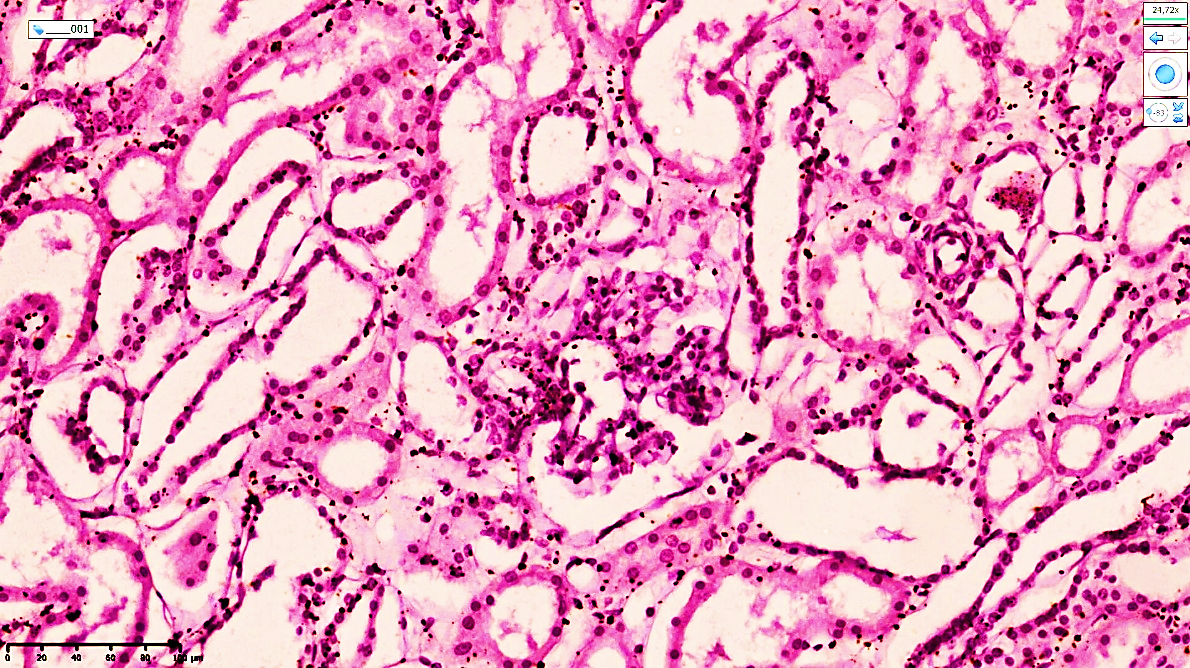 | Figure 1. A 43-year-old man. Kidney tissue from a patient who died of myocardial infarction in acute ischemic heart disease. Uneven acute impairment of filtration is observed in the glomeruli. Developing foci of glomerulofibrosis are identified. Stain: H&E. Magnification: 20×10 |
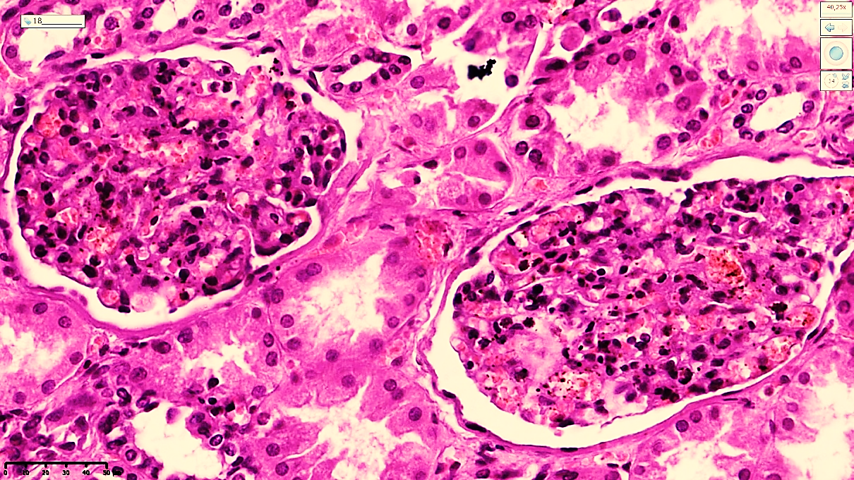 | Figure 2. A 44-year-old man. Foci of glomerulofibrosis are identified in the central capillary tuft of the glomerulus as irregular fibrous structures located between the mesangial cells. The epithelium of the proximal and distal tubules shows relatively flattened metaplastic changes, which are regarded as a consequence of chronic ischemia. Stain: H&E. Magnification: 20×10 |
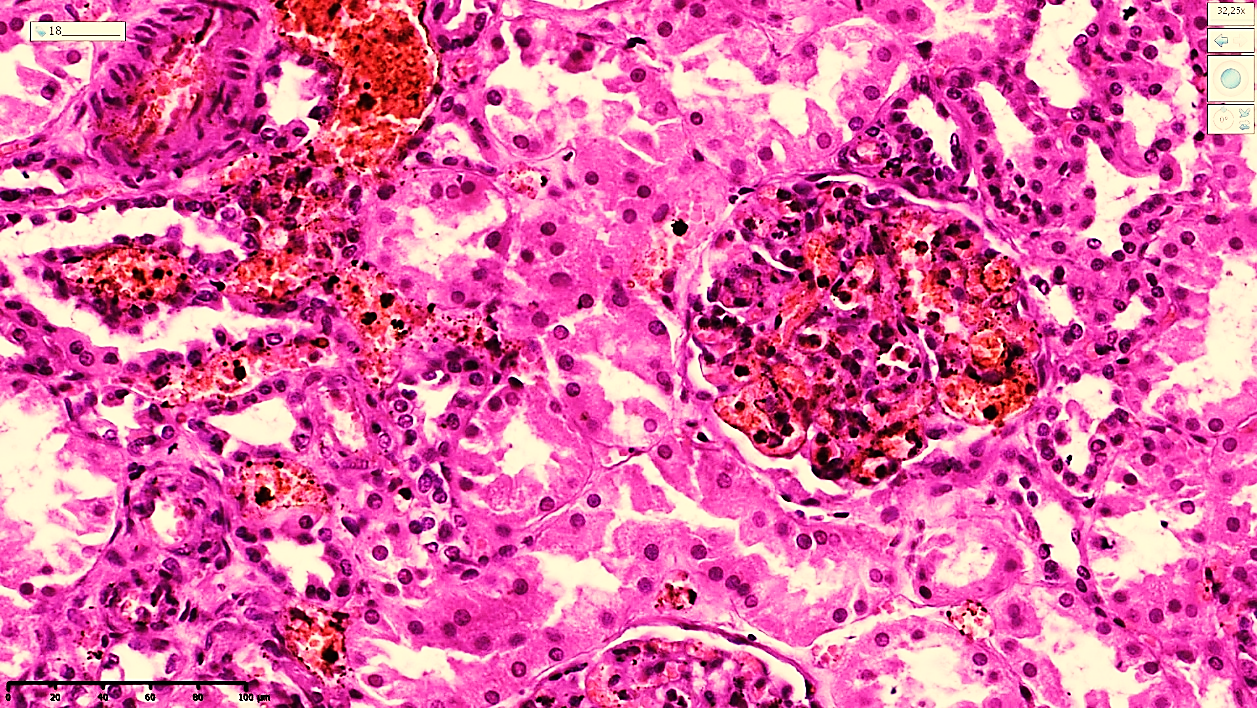 | Figure 3. Patient aged 55 years. Morphological changes in the kidneys in acute myocardial infarction: congestion of the glomeruli and uneven congestion around the proximal tubules. Acute dystrophic and necrobiotic changes are identified in the epithelium of the proximal tubules. Stain: H&E. Magnification: 20×10 |
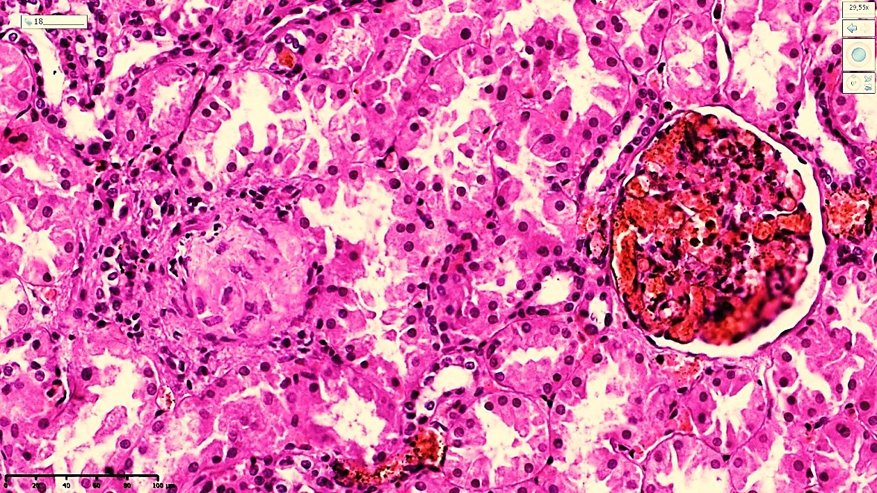 | Figure 4. Patient aged 60 years. Renal cortical region in recurrent myocardial infarction. Glomerulosclerosis and congestion of the glomeruli are observed. A homogeneous thickened basement layer is identified in the mesangial area. Foci of necrobiosis and desquamation are detected in the proximal tubules. Stain: H&E. Magnification: 20×10 |
4. Conclusions
- Main substrate of renal changes. In ischemic heart disease (IHD), the main morphological substrate of renal pathological changes is the renal glomeruli. Under the influence of the disease, a decrease in glomerular size, as well as atrophic and sclerotic changes, is observed. Age-related features. The study results showed that renal pathologies associated with IHD occur most frequently in patients aged 45–59 years. In this age group, glomerulosclerosis progresses more rapidly and becomes irreversible. Relationship between cardiac and renal pathologies. Pathological changes in the cardiovascular system and kidneys develop through common pathogenetic mechanisms, such as arterial hypertension, atherosclerosis, and hemodynamic disorders. Prolonged ischemia and narrowing of the renal arteries in IHD impair the filtration process and create conditions for the development of chronic renal failure.