
-
Paper Information
- Next Paper
- Previous Paper
- Paper Submission
-
Journal Information
- About This Journal
- Editorial Board
- Current Issue
- Archive
- Author Guidelines
- Contact Us
American Journal of Medicine and Medical Sciences
p-ISSN: 2165-901X e-ISSN: 2165-9036
2026; 16(5): 3199-3204
doi:10.5923/j.ajmms.20261605.179
Received: Apr. 22, 2026; Accepted: May 17, 2026; Published: May 30, 2026

Comparative Features of CD-20 Marker Expression in the Thymus During Experimental Intestinal Fibrosis
 Abstract
 Reference
 Full-Text PDF
 Full-text HTML
Oblokulov Abdusattar Abdurashidovich1, Xasanova Dilnoza Axrorovna2
1Bukhara Regional Infectious Diseases Hospital, Uzbekistan
2Bukhara State Medical Institute named after Abu Ali ibn Sina, Uzbekistan
Correspondence to: Oblokulov Abdusattar Abdurashidovich, Bukhara Regional Infectious Diseases Hospital, Uzbekistan.
| Email: |  |
Copyright © 2026 The Author(s). Published by Scientific & Academic Publishing.
This work is licensed under the Creative Commons Attribution International License (CC BY).
http://creativecommons.org/licenses/by/4.0/

The intestinal cicatricial process is associated with complex immune and morphofunctional alterations that may affect the cellular composition of the thymus. Particular interest is given to the B-cell component of the thymic tissue, which can be characterized by immunohistochemical detection of the CD-20 marker. The aim of this study was to assess the features of CD-20 immunohistochemical expression in the thymus of white outbred rats during experimental intestinal cicatrization. Materials and methods. The study included white outbred rats of different age groups. An experimental intestinal cicatricial process was modeled, followed by morphological and immunohistochemical examination of thymic tissue. Histological sections were prepared and immunohistochemical staining for the CD-20 marker was performed. The number and distribution of CD-20-positive cells in the thymic tissue were evaluated, and the obtained quantitative data were subjected to statistical analysis. Results. The investigation demonstrated age-related differences in the representation and distribution of CD-20-positive cells in the thymus. Under conditions of experimental intestinal cicatrization, changes in CD-20 expression were observed, indicating a restructuring of the B-cell component of the thymic microenvironment in response to the pathological process. The character and degree of these changes differed depending on the age of the animals. Conclusion. Experimental intestinal cicatrization is accompanied by changes in CD-20 immunohistochemical expression in the thymus, reflecting age-dependent remodeling of its immune cellular composition. Assessment of CD-20 expression may serve as an informative additional approach for evaluating immunomorphological changes in the thymus during chronic intestinal pathology.
Keywords: Thymus, CD-20, Immunohistochemistry, B-lymphocytes, Immunomorphology, Rats, Age-related changes, Experimental study
Cite this paper: Oblokulov Abdusattar Abdurashidovich, Xasanova Dilnoza Axrorovna, Comparative Features of CD-20 Marker Expression in the Thymus During Experimental Intestinal Fibrosis, American Journal of Medicine and Medical Sciences, Vol. 16 No. 5, 2026, pp. 3199-3204. doi: 10.5923/j.ajmms.20261605.179.
Article Outline
1. Introduction
- Chronic inflammatory and fibrotic-scarring processes in the intestine can affect not only the body's local tissue structures but also the central organs of the immune system. Intestinal fibrosis is typically associated with long-term inflammatory and reparative processes, and its development involves a complex interplay between immune cells, cytokines, fibroblasts, and tissue remodeling [1,2]. Modern research has placed a special focus on the immunological link between the intestine and the thymus. In particular, it has been noted that chronic immune dysregulation in the intestine can affect the structure and function of the thymus [3].As a central lymphoid organ, the thymus plays a key role in the development and selection of T-lymphocytes and the establishment of immunological tolerance [4]. However, the thymus is not a structure composed solely of T-lymphocytes. Scientific research has shown that a small number of B-lymphocytes are also present in the thymic medulla. They may participate in antigen presentation and the establishment of central immunological tolerance within the thymic microenvironment [5,6].Thymic B-cells differ morphologically and functionally to some extent from peripheral B-lymphocytes [9]. It has been established that the majority are located in the medullary and perivascular regions of the thymus. Some studies have indicated that the number of B-lymphocytes in a normal thymus is relatively low, whereas in pathological conditions, their number and distribution can change significantly [7,8].The development of immunohistochemical methods has expanded the ability to identify B-cells in the thymus and assess their distribution patterns. Specifically, CD20 is one of the most widely used markers for identifying B-lymphocytes. Immunohistochemical studies have shown that the localization of CD20-positive cells in the thymic medulla, as well as their quantity and distribution, can vary depending on pathological conditions [9,10].The significance of thymic B-lymphocytes in the body is not limited to antibody production. As antigen-presenting cells, they may participate in the central selection of T-lymphocytes and contribute to establishing immunological tolerance to self-antigens. Therefore, assessing the quantity and distribution of CD20-positive cells in the thymus serves as an additional important indicator for studying the morphofunctional state of the immune system [11].Age-related factors also influence the B-cell population in the thymus. According to scientific data, the involutional processes of the thymus intensify with age, and its cellular composition is remodeled. The accumulation of B-cells in perivascular areas may also have age-related characteristics. Therefore, a comparative study of CD20 expression in animals of different ages allows for the assessment of age-related immunomorphological restructuring of the thymus [12,13].Changes that occur in the immune system against the backdrop of chronic inflammatory and scarring processes of the intestine are one of the key manifestations of the "gut-immune system" axis. The contemporary view that intestinal immune dysregulation can affect the immunological function of the thymus and alter mechanisms of central immune tolerance underscores the importance of research in this area [14].At the same time, age-related changes in the expression of CD20-positive cells in the thymus in the context of experimental intestinal scarring have not been sufficiently studied. In particular, an immunohistochemical assessment of the pathological scarring process's impact on the B-lymphocytic component of the thymus is crucial for determining the systemic immunomorphological consequences of this process.
2. Purpose of the Research
- To study the characteristics of CD20-immunohistochemical expression in the thymus of white purebred rats during experimental intestinal scarring, as well as to evaluate the age dynamics of the identified changes and their morphological characteristics.
3. Materials and Methods
- The study was conducted on 180 outbred white rats aged 4, 7, and 10 months, kept under standard vivarium conditions. The animals were divided into control and experimental groups based on age and the type of intervention.The first group (control) consisted of 30 intact animals, with 10 rats in each of the 4-, 7-, and 10-month age groups.The second group (experimental) consisted of 30 animals with an induced intestinal cicatricial process, with 10 rats from each age group.The third group (experimental) consisted of 60 animals with experimental intestinal scarring that were treated with silymarin extract, with 20 rats in each of the 4-, 7-, and 10-month age groups.Morphometric, cytomorphological, and immunohistochemical methods were used to assess the state of the thymus and bone marrow. In the thymus, key morphometric parameters were determined, and the expression of immunohistochemical markers CD-20 was evaluated.
4. Results
- The findings of the study on the immunohistochemical expression levels in the thymus of 4-, 7-, and 10-month-old rats with an experimental intestinal cicatricial process demonstrate that chronic cicatricial and sclerotic changes in the intestinal tissues affect not only the local morphology of the damaged organ but also the systemic immune homeostasis, particularly the morphofunctional state of the thymus as a central immune organ.The development of intestinal cicatricial changes is accompanied by a persistent inflammatory response, disturbances in immune regulation, alterations in reparative processes, and functional adaptation of the immune system. These systemic changes are reflected in the altered immunohistochemical expression patterns within the thymic tissue. The obtained results contribute to a better understanding of the morphofunctional relationship between chronic intestinal cicatricial changes and the thymus, the central organ of immunogenesis. The findings provide a theoretical basis for further investigation of the immunopathogenetic mechanisms underlying chronic intestinal disorders, assessment of systemic immune disturbances, and development of promising approaches for their prevention and correction.Based on the results of immunohistochemical analysis, the characteristics of CD-20 marker expression were studied to assess the state of B-lymphocyte cells in the thymus of 4-month-old white outbred rats under conditions of cicatricial intestinal processes. During this age period, the body's immune system is formed, and response reactions to a pathological factor are primarily carried out through compensatory and adaptive mechanisms (Fig. 1).
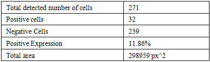
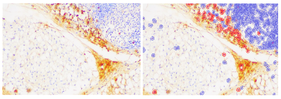 | Figure 1. Representative immunohistochemical images of CD20 expression in the thymus of 4-month-old outbred rats during experimental intestinal cicatricial processes: original micrograph (left) and computer-assisted identification of CD20-positive and CD20-negative cells (right). Magnification ×200 |
|
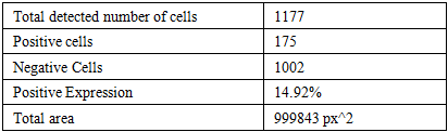
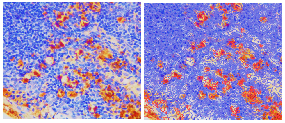 | Figure 2. Representative immunohistochemical images of CD20 expression in the thymus of 7-month-old outbred rats during experimental intestinal cicatricial processes: original micrograph (left) and computer-assisted identification of CD20-positive and CD20-negative cells (right). Magnification ×200 |
|
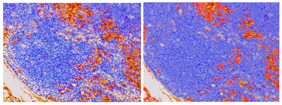 | Figure 3. Representative immunohistochemical images of CD20 expression in the thymus of 10-month-old outbred rats during experimental intestinal cicatricial processes: original micrograph (left) and computer-assisted identification of CD20-positive and CD20-negative cells (right). Magnification ×200 |
|
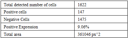
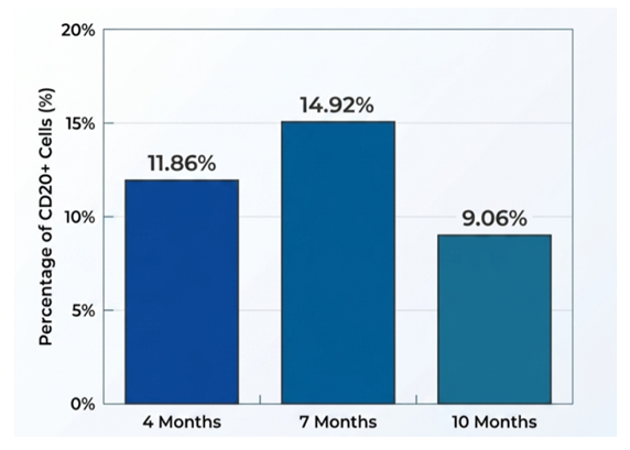 | Figure 4. Age-related dynamics of CD20 expression in the thymus Level of CD20-positive cells in the rat thymus with experimental intestinal scarring |
5. Discussion
- The present study demonstrated age-related differences in the distribution and immunohistochemical expression of CD20-positive cells in the thymus of white outbred rats during experimental intestinal cicatrization. The findings indicate that chronic cicatricial and sclerotic changes in the intestine are associated not only with local structural alterations but also with remodeling of the immune microenvironment of the thymus.The relatively higher representation of CD20-positive cells in 7-month-old rats may reflect a period of comparatively stable functional activity of the immune system, during which adaptive mechanisms are actively maintained in response to chronic pathological stimulation. The observed distribution of CD20-positive cells suggests that the B-cell component of the thymic microenvironment is not constant and may undergo compensatory and adaptive changes depending on both the duration of pathological exposure and the age-related functional state of the organism.In 4-month-old animals, the immune response was characterized by greater cellular plasticity. The observed CD20-positive cell distribution may be associated with the developmental and functional maturation of the immune system. In contrast, the changes observed in 7-month-old rats appeared to reflect a more coordinated adaptive response to chronic intestinal injury. This may explain the relatively higher proportion of CD20-positive cells in this age group.The decrease in CD20 expression observed in 10-month-old animals may indicate a reduction in the adaptive potential of the thymic immune microenvironment. Age-related changes in lymphoid tissues are known to be accompanied by gradual remodeling of cellular composition and reduced functional plasticity. In the context of chronic intestinal cicatrization, these age-dependent changes may limit the ability of the immune system to generate an adequate cellular response and may result in greater reliance on compensatory mechanisms involving previously established immune cell populations.The morphological distribution of CD20-positive cells also demonstrated that their localization within the thymic tissue was heterogeneous. Areas of accumulation and relative dispersion of immunopositive cells may represent different functional states of the lymphoid microenvironment. Such redistribution can be interpreted as an adaptive response to persistent inflammatory and fibrotic stimulation originating from the intestine.The findings obtained support the concept that chronic intestinal cicatrization has systemic immunomorphological consequences. Although the primary pathological process is localized in the intestinal tissue, prolonged inflammatory and sclerotic changes may influence central immune organs, including the thymus. The age-dependent differences identified in CD20 expression indicate that the magnitude and character of this response depend on the functional maturity of the immune system.The results also provide a morphological basis for considering the thymus as an important component of the systemic response to chronic intestinal pathology. Assessment of CD20 expression complements conventional morphological examination and makes it possible to identify changes in the B-cell component of the thymic microenvironment that may not be evident from routine histological evaluation alone.Overall, the results demonstrate that experimental intestinal cicatrization is accompanied by dynamic remodeling of CD20-positive cells in the thymus, with distinct age-related patterns. The observed changes can be regarded as manifestations of adaptive and compensatory restructuring of the immune system in response to chronic pathological stimulation.
6. Conclusions
- Experimental intestinal cicatrization is accompanied by significant age-dependent changes in the immunohistochemical expression and distribution of CD20-positive cells in the thymus of white outbred rats.The identified redistribution of CD20-positive cells indicates remodeling of the B-cell component of the thymus under conditions of chronic intestinal cicatrization. These changes appear to represent adaptive and compensatory reactions of the central immune system to persistent pathological stimulation.Thus, immunohistochemical assessment of CD20 expression can be considered an informative additional method for characterizing age-related immunomorphological changes in the thymus during experimental chronic intestinal pathology. The obtained findings emphasize the systemic nature of the immune response to intestinal cicatricial processes and demonstrate the importance of considering the age of the organism when evaluating thymic immunomorphological changes.