
-
Paper Information
- Next Paper
- Paper Submission
-
Journal Information
- About This Journal
- Editorial Board
- Current Issue
- Archive
- Author Guidelines
- Contact Us
American Journal of Medicine and Medical Sciences
p-ISSN: 2165-901X e-ISSN: 2165-9036
2026; 16(5): 3195-3198
doi:10.5923/j.ajmms.20261605.178
Received: Mar. 22, 2026; Accepted: Apr. 12, 2026; Published: May 30, 2026

Morphological and Morphometric Characteristics of Rat Myocardium in Experimental Combined Deficiency of Magnesium, Iron, Zinc, and Selenium
 Abstract
 Reference
 Full-Text PDF
 Full-text HTML
Irina L. Kim, A. B. Radjabov
Bukhara State Medical Institute named after Abu Ali ibn Sino, Bukhara, Uzbekistan
Correspondence to: Irina L. Kim, Bukhara State Medical Institute named after Abu Ali ibn Sino, Bukhara, Uzbekistan.
| Email: |  |
Copyright © 2026 The Author(s). Published by Scientific & Academic Publishing.
This work is licensed under the Creative Commons Attribution International License (CC BY).
http://creativecommons.org/licenses/by/4.0/

This study presents the morphological, morphometric, and immunohistochemical characteristics of the myocardium in 6-month-old male outbred rats subjected to experimental combined dietary deficiency of magnesium, iron, zinc, and selenium. Combined micronutrient deficiency resulted in reduced heart mass and linear dimensions, decreased cardiomyocyte diameter and cross-sectional area, a marked reduction in the proportion of binucleated cells, thickening of intermuscular connective tissue septa, and rarefaction of the capillary network. Histological examination revealed pronounced cardiomyocyte dystrophy, vascular congestion, interstitial edema, and disruption of myocardial architecture. α-SMA expression decreased to a score of 1 and was retained only in isolated vascular and interstitial structures. These findings demonstrate profound cellular, vascular, and stromal remodeling of the myocardium under combined micronutrient deficiency.
Keywords: Myocardium, Combined micronutrient deficiency, Magnesium, Iron, Zinc, Selenium, Cardiomyocytes, α-SMA, Remodeling
Cite this paper: Irina L. Kim, A. B. Radjabov, Morphological and Morphometric Characteristics of Rat Myocardium in Experimental Combined Deficiency of Magnesium, Iron, Zinc, and Selenium, American Journal of Medicine and Medical Sciences, Vol. 16 No. 5, 2026, pp. 3195-3198. doi: 10.5923/j.ajmms.20261605.178.
1. Introduction
- Magnesium, iron, zinc, and selenium are involved in energy metabolism, mitochondrial respiration, antioxidant defense, regulation of vascular tone, and maintenance of cardiomyocyte structural integrity. Simultaneous insufficiency of several essential elements may affect multiple interconnected metabolic pathways and aggravate myocardial injury [1,2,3].Magnesium deficiency disrupts ionic homeostasis and myocardial contractility; iron deficiency impairs cellular respiration and promotes tissue hypoxia; zinc deficiency weakens antioxidant defense; and selenium deficiency compromises selenoprotein function [4,5,6,7]. Under combined deficiency, these mechanisms act concurrently, creating conditions for progressive dystrophic, vascular, and interstitial changes in the heart.Myocardial remodeling during chronic injury involves alterations in cardiomyocytes, the microcirculatory bed, and the extracellular matrix. Expansion of the interstitial compartment and changes in α-smooth muscle actin (α-SMA) expression reflect disturbed vascular-stromal organization of the myocardium [8]. Therefore, an integrated morphological, morphometric, and immunohistochemical assessment of the myocardium under simultaneous Mg, Fe, Zn, and Se deficiency is of considerable interest.Although the cardiac effects of individual micronutrient deficiencies have been described, the structural consequences of their combined deficiency remain less well characterized. Quantitative assessment of heart organometry, cardiomyocyte size and nuclear characteristics, intermuscular connective tissue septa, capillary density, and α-SMA expression within a single experimental model is particularly important.ObjectiveTo characterize the morphological, morphometric, and immunohistochemical features of the myocardium in 6-month-old male outbred rats with experimental combined dietary deficiency of magnesium, iron, zinc, and selenium.
2. Materials and Methods
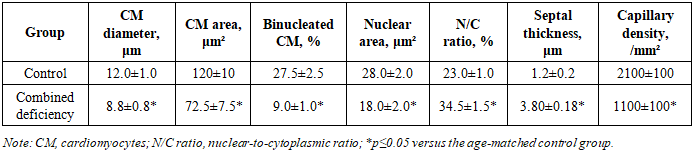
- Hearts from 6-month-old male outbred rats were examined. The age-matched control group and the combined Mg, Fe, Zn, and Se deficiency group each comprised 15 animals. Six months of age corresponded to the period of morphofunctional maturity of the rat myocardium.Combined deficiency was induced using the modified Altromin C 1035 diet containing 97.7 mg/kg magnesium, 3.82 mg/kg iron, 2.02 mg/kg zinc, and 0.035 mg/kg selenium. Control animals received Altromin C 1000 containing 684, 178.58, 29.30, and 0.33 mg/kg of the respective micronutrients. Animals were maintained under standard vivarium conditions with free access to food and water.At the end of the experiment, body mass, heart mass, heart length and width, and the heart-to-body mass index (SSI) were recorded. Tissue samples were fixed in 10% neutral formalin, processed through ascending concentrations of ethanol, embedded in paraffin, and sectioned at 3–5 μm. Sections were stained with hematoxylin and eosin.Digital micrographs were analyzed using ImageJ. Cardiomyocyte diameter and cross-sectional area, the proportion of binucleated cardiomyocytes, nuclear area, nuclear-to-cytoplasmic ratio, thickness of intermuscular connective tissue septa, and capillary density were measured. α-SMA immunoreactivity was evaluated semiquantitatively as follows: 0, no staining; 1, weak; 2, moderate; and 3, strong expression. The individual animal was used as the statistical unit. Data are presented as mean ± standard deviation (M±SD); differences were considered statistically significant at p≤0.05.
3. Results and Discussion
- Combined micronutrient deficiency caused a marked reduction in organometric parameters. Body mass decreased from 206.4±4.5 to 166.0±11.1 g, heart mass from 1.22±0.05 to 0.78±0.04 g, heart length from 10.0±0.3 to 8.0±0.3 mm, heart width from 6.5±0.2 to 5.1±0.2 mm, and SSI from 0.60±0.02 to 0.47±0.02 (Table 1). The concurrent decrease in heart mass and linear dimensions indicates marked suppression of cardiac growth and adaptive processes.
|

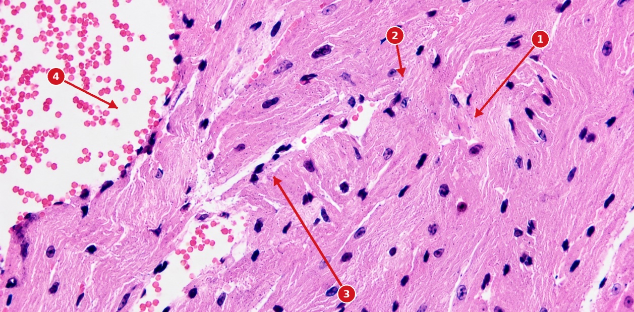 | Figure 1. Histological structure of the myocardium in a 6-month-old rat with combined Mg, Fe, Zn, and Se deficiency: 1, cardiomyocytes with pronounced dystrophic changes; 2, hyperchromatic nuclei; 3, widened intermuscular spaces (interstitial edema); 4, a dilated blood vessel containing formed blood elements (vascular congestion). Visible changes include cardiomyocyte dystrophy and separation, nuclear hyperchromasia, interstitial edema, vascular congestion, and disruption of myocardial architecture. Hematoxylin and eosin staining. Original magnification ×200 |
|
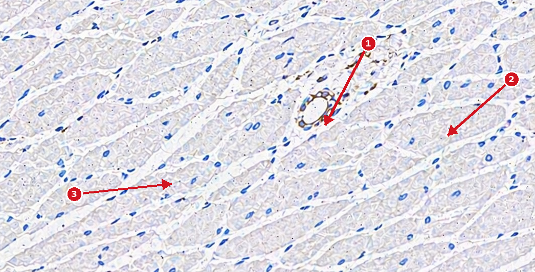 | Figure 2. α-SMA expression in the myocardium of a 6-month-old rat with combined Mg, Fe, Zn, and Se deficiency: 1, vascular smooth muscle cells with weak positive α-SMA expression; 2, myocardial interstitial tissue with markedly reduced α-SMA expression; 3, myocardial area with minimal immunopositive reaction. Visible changes include weak, focal staining of vascular structures, a pronounced reduction in interstitial immunoreactivity, and predominance of α-SMA-negative myocardial areas. α-SMA immunohistochemistry, DAB chromogen. Original magnification ×200 |
|

4. Conclusions
- Experimental combined deficiency of magnesium, iron, zinc, and selenium causes profound structural remodeling of the myocardium in 6-month-old male outbred rats. It is characterized by reduced heart mass and linear dimensions; decreased cardiomyocyte diameter, cross-sectional area, proportion of binucleated cells, and nuclear area; an increased nuclear-to-cytoplasmic ratio; thickened intermuscular connective tissue septa; and capillary rarefaction. Histological changes include cardiomyocyte dystrophy and disorganization, interstitial edema, and vascular congestion. Reduction of α-SMA expression to a score of 1 reflects profound disruption of the vascular-interstitial organization of the myocardium.