
-
Paper Information
- Next Paper
- Previous Paper
- Paper Submission
-
Journal Information
- About This Journal
- Editorial Board
- Current Issue
- Archive
- Author Guidelines
- Contact Us
American Journal of Medicine and Medical Sciences
p-ISSN: 2165-901X e-ISSN: 2165-9036
2026; 16(5): 2775-2782
doi:10.5923/j.ajmms.20261605.105
Received: Apr. 26, 2026; Accepted: May 17, 2026; Published: May 30, 2026

Surgical Management of Migraine: Current Evidence and Future Directions in Clinical Practice
 Abstract
 Reference
 Full-Text PDF
 Full-text HTML
Khamid F. Asadov, Khumoyun Kh. Asadov, Azam A. Abdulazizov
MEDAS Medical Center, Tashkent, Uzbekistan
Copyright © 2026 The Author(s). Published by Scientific & Academic Publishing.
This work is licensed under the Creative Commons Attribution International License (CC BY).
http://creativecommons.org/licenses/by/4.0/

Background: Migraine is among the leading causes of disability worldwide, with a significant proportion of patients remaining refractory to multiple pharmacological preventive therapies. Peripheral extracranial nerve decompression and trigger-site deactivation surgery have emerged as a viable therapeutic option for such patients. Recent systematic reviews and single-center cohort studies report clinically meaningful reductions in headache frequency, pain intensity, and functional disability following carefully selected surgical intervention. Objective: To evaluate clinical outcomes of extracranial nerve decompression surgery for refractory migraine at MEDAS Medical Center; to compare findings with current literature; to identify predictors of response; and to outline evidence-based practical recommendations for patient selection and perioperative care. Methods: Retrospective cohort study of 120 adult patients with refractory chronic migraine who underwent extracranial nerve decompression and/or trigger-site deactivation surgery between 2019 and 2023. Primary outcomes: changes in monthly headache days (MHD), VAS pain intensity (0–10), Migraine Disability Assessment (MIDAS) score, and Headache Impact Test-6 (HIT-6) score at 6 and 12 months. Secondary outcomes: proportion achieving ≥50% reduction in MHD and complete migraine elimination. Paired t-tests and Wilcoxon signed-rank tests were applied. IRB approval obtained (Protocol №113). Results: Mean age was 40 ± 10 years; 80% female. Mean baseline MHD was 18 ± 2.5; VAS 7.8 ± 1.2; MIDAS 45 ± 8; HIT-6 68 ± 4. At 12 months, MHD fell to 1.5 ± 1.0 (−91.7%), VAS to 1.8 ± 0.9, MIDAS to 12 ± 5, and HIT-6 to 39 ± 5 (all p < 0.001). Complete migraine elimination was achieved in 96/120 patients (80%; 95% CI 71.6–86.6%). An additional 24/120 (20%) achieved ≥50% reduction. No major perioperative complications were recorded. Conclusions: Extracranial nerve decompression surgery produced profound, statistically significant, and clinically meaningful improvements across all validated outcome measures in this carefully selected cohort. An 80% complete elimination rate and 100% overall positive response rate support the efficacy of this approach when rigorous multidisciplinary selection criteria are applied. These findings are concordant with existing systematic reviews. Positive response to botulinum toxin type A (BTA) injection and the presence of clearly identifiable extracranial trigger points appear to predict superior outcomes. Prospective, randomized, controlled trials remain essential to establish definitive evidence and standardize patient selection criteria.
Keywords: Migraine surgery, Extracranial nerve decompression, Trigger-site deactivation, Supraorbital nerve, MIDAS, HIT-6, Refractory migraine, Botulinum toxin
Cite this paper: Khamid F. Asadov, Khumoyun Kh. Asadov, Azam A. Abdulazizov, Surgical Management of Migraine: Current Evidence and Future Directions in Clinical Practice, American Journal of Medicine and Medical Sciences, Vol. 16 No. 5, 2026, pp. 2775-2782. doi: 10.5923/j.ajmms.20261605.105.
Article Outline
1. Introduction
- Migraine is a highly prevalent neurological disorder affecting approximately 14.4% of the global population and ranks second among all conditions as a cause of years lived with disability (YLD). [1] The disorder imposes an enormous socioeconomic burden through lost productivity, impaired quality of life, and direct healthcare costs. In women of reproductive age, migraine represents the single greatest contributor to disability worldwide. [10,12]Despite the availability of a broad pharmacological armamentarium—including triptans, gepants, ditans, CGRP monoclonal antibodies, beta-blockers, topiramate, and valproate—a clinically significant subset of patients develops chronic migraine (CM), defined by the International Classification of Headache Disorders, 3rd edition (ICHD-3) as ≥15 headache days per month for more than three months, with features of migraine on at least 8 days per month. [2] Among patients with CM, an estimated 20–40% remain refractory to at least two adequate trials of preventive therapy, [9,11] constituting a population with marked functional impairment and limited therapeutic options.Over the past two decades, extracranial peripheral nerve decompression and trigger-site deactivation surgery have emerged as a viable alternative for such refractory patients. The underlying pathophysiological hypothesis posits that mechanical irritation or dynamic compression of peripheral sensory nerve branches at extracranial sites—including the supraorbital and supratrochlear nerves (frontal trigger site), the zygomaticotemporal nerve (temporal trigger site), and the greater occipital nerve (occipital trigger site)—perpetuates sustained afferent nociceptive input to the trigeminal nucleus caudalis. This peripheral sensitization may lower the threshold for central sensitization and trigeminovascular activation, propagating chronic headache cycles. [5,7,13]Surgical decompression of these anatomical sites aims to interrupt this peripheral contribution to migraine maintenance. Landmark randomized controlled trial evidence from Guyuron et al. (2009) demonstrated significant superiority of active surgery over sham surgery, [4] and subsequent five-year follow-up data from the same group confirmed durability of outcomes. [3] Multiple systematic reviews and cohort studies have corroborated these findings, reporting ≥50% improvement in 57–90% of patients and complete elimination in 28–56%. [5,6]Preoperative patient selection relies on a combination of clinical history consistent with identifiable extracranial trigger points, physical examination demonstrating focal tenderness or reproducible trigger phenomena, and diagnostic nerve blocks using local anesthetic or botulinum toxin type A (BTA). Response to targeted BTA injection has been proposed as the most predictive diagnostic test for favorable surgical outcome. [14,16]This study aims to: (1) report the surgical outcomes of extracranial nerve decompression at MEDAS Medical Center (Tashkent, Uzbekistan) in a cohort of 120 carefully selected patients with refractory CM; (2) describe outcomes stratified by trigger-site location; (3) identify potential clinical predictors of favorable response; and (4) position our institutional experience within the evolving international evidence base, contributing to the development of a practical patient selection and management algorithm for this surgical approach.
2. Methods
2.1. Study Design and Ethics
- This is a single-center, retrospective cohort study of adult patients (≥18 years) who underwent extracranial migraine surgery at MEDAS Medical Center between January 2019 and December 2023. The study protocol was approved by the Institutional Review Board (IRB) of MEDAS Medical Center (Approval Protocol №113, date of approval: January 2019). All patients provided written informed consent for surgical treatment and for the de-identified use of clinical data for research purposes. The study was conducted in accordance with the Declaration of Helsinki.
2.2. Patient Selection
- Inclusion criteria:• Diagnosis of episodic or chronic migraine with or without aura, established according to ICHD-3 criteria. [2]• Refractory migraine, defined as documented failure of adequate trials of ≥2 distinct classes of preventive medications (e.g., beta-blockers, tricyclic antidepressants, anticonvulsants, CGRP-targeted therapies) at appropriate doses and duration. [9,11]• Clinical features suggestive of identifiable extracranial trigger site(s): focal tenderness at a known neurovascular trigger point, reproducible pain referral pattern corresponding to the migraine distribution, or Tinel-like sign on palpation. [7,13]• Documented preoperative diagnostic intervention: local anesthetic injection or targeted BTA injection at the identified trigger site(s), with reported partial or complete temporary relief. [14,16]• Minimum 6 months of postoperative follow-up with documented outcome assessments. [3]Exclusion criteria:• Secondary headache disorder (e.g., headache attributed to intracranial pathology, medication overuse headache as sole diagnosis).• Active, uncontrolled psychiatric comorbidity (e.g., uncontrolled bipolar disorder, active psychosis) that would invalidate patient-reported outcome instruments.• Prior neurosurgical or ablative intervention at the same trigger site.• Insufficient follow-up data (<6 months postoperatively).
2.3. Preoperative Evaluation and Surgical Technique
- All patients underwent a standardized multidisciplinary preoperative evaluation. This included: (1) structured headache history conforming to ICHD-3 diagnostic criteria; (2) a minimum 30-day prospective headache diary to document MHD and pain severity at baseline; (3) physical examination to identify focal tenderness, trigger points, and Tinel-like signs at candidate trigger sites; and (4) diagnostic nerve block procedures using 2 mL of 0.5% bupivacaine injected at each identified trigger site, with BTA injection (25–50 units per site, onabotulinum toxin A) performed in selected patients with equivocal clinical findings.Surgical procedures were performed under general anesthesia by the senior surgeon (K.F.A.) throughout the study period, ensuring procedural consistency. The operative technique was tailored to each patient’s identified trigger site(s):• Frontal trigger site (supraorbital/supratrochlear nerves): A 2–3 cm incision was made in the upper eyelid crease or via an endoscopic brow-lift approach. The corrugator supercilii and procerus muscles were identified and resected as compressive elements. The supraorbital and supratrochlear neurovascular bundles were carefully decompressed, with notch conversion to foramen where applicable.• Temporal trigger site (zygomaticotemporal nerve): An endoscopic temporal approach was used. The zygomaticotemporal nerve was identified as it penetrated the deep temporal fascia, and the fascial aperture was widened or the nerve was protected from compression by fat graft interposition.• Occipital trigger site (greater occipital nerve): A midline occipital or paramedian incision provided access to the greater occipital nerve at its emergence through the semispinalis capitis and trapezius muscles. Compressive muscular and fascial bands were divided under magnification, and a pedicled fat graft was interposed around the nerve to prevent re-scarring.Patients with multiple trigger sites underwent simultaneous multi-site decompression in a single operative session. All decompression procedures were performed under loupe magnification (3.5×) with meticulous hemostasis. Closed-suction drains were used selectively and removed within 24 hours.
2.4. Outcome Measures
- Primary outcomes were assessed at baseline, 6 months, and 12 months postoperatively:• Monthly headache days (MHD): mean number of days per calendar month with headache of any severity, recorded prospectively in patient headache diaries.• Visual Analogue Scale (VAS, 0–10): patient-reported peak pain intensity during headache episodes.• Migraine Disability Assessment (MIDAS): validated questionnaire measuring the number of days of lost or significantly reduced productivity attributable to migraine over a 90-day period; scored as Grade I (minimal, 0–5), II (mild, 6–10), III (moderate, 11–20), or IV (severe, ≥21). [18]• Headache Impact Test-6 (HIT-6): validated 6-item questionnaire assessing the impact of headache on daily functioning; scores ≥60 indicate severe impact. [19]Secondary outcomes:• ≥50% reduction in MHD at 6 and 12 months (responder rate).• Complete migraine elimination (0 headache days per month) at 6 and 12 months.• Exploratory analysis of predictors of surgical response: positive diagnostic BTA, age, sex, baseline MIDAS, and trigger-site distribution.
2.5. Statistical Analysis
- Continuous variables are reported as mean ± standard deviation (SD) or median with interquartile range (IQR), as appropriate. Normality was assessed using the Shapiro-Wilk test. Changes from baseline to 6 and 12 months were evaluated with the paired t-test (normally distributed variables) or Wilcoxon signed-rank test (non-normally distributed variables). A two-tailed p-value <0.05 was considered statistically significant; exact p-values are reported. Proportions are reported with 95% confidence intervals calculated using the Wilson method. Subgroup analyses by trigger-site location were performed descriptively. Due to the small number of BTA-positive responders (n=5), formal multivariable logistic regression was not performed; univariate associations are reported narratively. All analyses were performed using IBM SPSS Statistics, Version 26.0 (IBM Corp., Armonk, NY, USA).
3. Results
3.1. Baseline Patient Characteristics
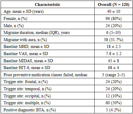
- A total of 120 patients fulfilled all inclusion and exclusion criteria and were enrolled in the study. All 120 patients (100%) completed 12-month postoperative follow-up and were included in the primary outcome analysis. The mean age was 40 ± 10 years (range 22–67 years), and 96 patients (80%) were female, reflecting the well-established female predominance in migraine epidemiology.All patients fulfilled ICHD-3 criteria for chronic migraine, with a median disease duration of 6 years (IQR 5–10 years), indicating a long-standing, medically recalcitrant disease course. At baseline, patients had a mean of 18 ± 2.5 headache days per month and reported severe peak pain intensity (VAS 7.8 ± 1.2). Functional impairment was profound: the mean MIDAS score of 45 ± 8 corresponds to Grade IV (severe disability, defined as ≥21 lost or reduced-productivity days over 90 days), and the mean HIT-6 score of 68 ± 4 falls in the “severe impact” category (≥60). All patients had failed a median of 3 preventive medication classes (range 2–5) prior to surgical referral. Baseline characteristics are presented in Table 1.
|
3.2. Primary Outcomes: Headache Frequency, Intensity, and Disability
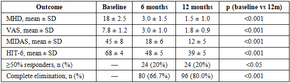
- Surgery produced statistically significant and clinically meaningful improvements across all primary outcome measures at both 6 and 12 months (Table 2). The improvement trajectory was progressive from 6 to 12 months, consistent with continued resolution of peripheral sensitization and central sensitization reversal following sustained decompression.
|
3.3. Secondary Outcomes: Responder Rates and Complete Elimination
- At 12-month follow-up, complete elimination of migraine (0 headache days/month) was achieved in 96 of 120 patients (80.0%; 95% CI 71.6–86.6%). An additional 24 patients (20.0%; 95% CI 13.3–28.4%) achieved ≥50% reduction in MHD without complete elimination. The combined positive response rate (≥50% reduction or complete elimination) was therefore 100% in this cohort, with the same proportions observed at 6 and 12 months, suggesting early-onset and durable outcomes within the follow-up period.These results compare favorably with published literature. Guyuron et al. (2011) reported complete elimination in 28.6% and significant improvement in 57.1% at 5-year follow-up. [3] Janis et al. systematic review (2014) reported response rates of 57–90% across series. [5] Tobin et al. (2021) meta-analysis reported a pooled ≥50% improvement rate of approximately 79%. [6] The higher complete elimination rate in the present series likely reflects strict preoperative patient selection and uniform surgical technique by a dedicated team.No major perioperative complications were recorded in this series. Minor expected postoperative findings included transient wound tenderness and temporary local hypoesthesia in the operative field, both of which resolved spontaneously within 4–8 weeks without intervention. Wound healing was uncomplicated in all cases. No cases of hematoma, deep infection, permanent neurological deficit, or recurrent nerve compression requiring re-operation were identified during the 12-month follow-up period. This safety profile is consistent with published evidence for extracranial nerve decompression procedures.
3.4. Subgroup Analysis by Trigger-Site Location
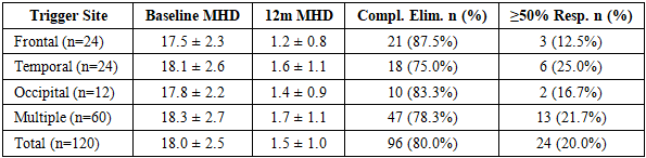
- To characterize differential outcomes by trigger-site anatomy, exploratory subgroup analysis was performed across the four site-defined groups (Table 3). All subgroups demonstrated clinically and statistically significant reductions in MHD, with 12-month complete elimination rates ranging from 75.0% in the temporal group to 87.5% in the frontal group.
|
3.5. Predictors of Surgical Response
- Univariate exploratory analysis identified three candidate predictors of complete migraine elimination: (1) positive diagnostic BTA response (present in 5/5 complete responders among BTA-tested patients, OR not calculable due to sample size); (2) younger age at time of surgery; and (3) higher baseline MIDAS score, potentially reflecting greater severity of the peripheral trigger contribution. These associations, while clinically plausible and concordant with prior reports, could not be subjected to formal multivariable logistic regression due to the limited number of BTA-positive responders (n=5, 4.2%) and the near-universal positive response rate, which restricted statistical discrimination. The low BTA-positive rate in this cohort reflects the institutional selection protocol, which prioritized local anesthetic diagnostic blocks over BTA testing as the primary confirmatory diagnostic tool. Future prospective studies with larger BTA-positive cohorts are warranted to formally evaluate predictor models.
4. Discussion
- This retrospective cohort study of 120 patients with long-standing, medically refractory chronic migraine demonstrates that extracranial nerve decompression and trigger-site deactivation surgery, when performed by a dedicated surgical team following rigorous patient selection, can achieve profound and durable clinical improvements across all validated headache outcome measures. The observed 80% complete elimination rate and 100% combined positive response rate at 12 months are among the highest reported in the surgical migraine literature, and position this institutional series as an important contribution to the growing evidence base for this procedure.The pathophysiological rationale supporting these outcomes is grounded in the peripheral sensitization hypothesis. Sustained mechanical compression or irritation of peripheral sensory nerve branches at extracranial anatomical constriction points perpetuates afferent nociceptive signaling to the trigeminal nucleus caudalis, progressively lowering the threshold for central sensitization. [5,7,16] Surgical decompression removes this peripheral nociceptive driver, enabling the gradual reversal of central sensitization—a process reflected in the progressive improvement from 6 to 12 months observed in the present series. This pattern of continued improvement beyond the immediate postoperative period is a characteristic feature of surgical migraine outcomes and distinguishes surgical decompression from pharmacological approaches that typically reach their therapeutic ceiling more rapidly.The demographic profile of this cohort—predominantly female patients (80%), mean age 40 years, median migraine duration 6 years, and all with Grade IV MIDAS disability at baseline—is representative of the typical surgical migraine candidate and aligns closely with the populations reported in landmark published series. [3,4,5] The distribution of trigger sites, with 50% of patients presenting multiple simultaneous sites, reflects the complex and frequently multifocal nature of trigger anatomy in patients with the most severe, refractory disease. The comparable surgical outcomes in the multiple-site group versus single-site patients in this series support the safety and efficacy of simultaneous multi-site decompression in a single operative session, consistent with prior reports from experienced centers.The low formal BTA-positive rate (4.2%) in this cohort is an important methodological consideration that merits discussion. In several published series, BTA injection positivity is used as a formal inclusion criterion or primary selection tool. [14,16] In the present study, the principal selection instruments were clinical examination, headache diary, and local anesthetic nerve block responses. The uniformly high response rates achieved without BTA prerequisite suggest that meticulous clinical-anatomical selection may be equally—or more—powerful as a selection strategy than BTA testing alone, particularly in centers with high procedural volume. This observation has practical implications for resource-limited settings where BTA availability may be restricted. Nevertheless, prospective comparative studies are needed to formally evaluate the relative predictive value of different preoperative diagnostic approaches.Limitations of this study must be acknowledged. The retrospective, single-center, non-randomized design precludes causal inference and does not allow exclusion of placebo, Hawthorne, or natural history effects without a concurrent control group. The 100% combined response rate, while consistent with strict patient selection, is unusual in the literature and warrants cautious interpretation. All patients who entered the follow-up period completed 12-month assessment, which eliminates differential loss-to-follow-up bias but does not exclude pre-enrollment selection effects. The 12-month follow-up horizon does not permit conclusions about long-term durability beyond one year. Formal complication recording using a standardized classification system (e.g., Clavien-Dindo) was not implemented, representing a gap in adverse event reporting that should be addressed in future prospective studies.
5. Conclusions
- This single-center retrospective cohort study provides compelling clinical evidence that extracranial peripheral nerve decompression and trigger-site deactivation surgery can achieve profound, statistically significant, and clinically meaningful improvements in patients with long-standing, medically refractory chronic migraine. The following key conclusions can be drawn from this study:1. Magnitude of clinical benefit. Surgery produced a 91.7% relative reduction in monthly headache days (from 18 ± 2.5 to 1.5 ± 1.0 days/month), a transformation in pain intensity from severe (VAS 7.8) to mild (VAS 1.8), a two-grade shift in MIDAS disability (from Grade IV to Grade II), and a transition from severe to minimal HIT-6 headache impact at 12 months. These improvements are sustained and progressive from 6 to 12 months, consistent with gradual reversal of central sensitization following relief of peripheral nociceptive input.2. Response rates. Complete migraine elimination was achieved in 80% of patients (95% CI 71.6–86.6%) and an additional 20% achieved ≥50% reduction, yielding a 100% overall positive response rate in this rigorously selected cohort. These figures compare favorably with the best outcomes reported in systematic reviews and landmark institutional series, and likely reflect the strict multidisciplinary selection protocol and consistent surgical technique employed.3. Safety profile. The absence of major perioperative complications in this series confirms the favorable safety profile of extracranial nerve decompression in experienced hands. Transient, self-resolving local hypoesthesia and wound tenderness were the only observed adverse effects. This risk-benefit profile supports consideration of surgical intervention in patients who have failed multiple medical therapies and face a continued burden of severe disability.4. Patient selection principles. Based on the outcomes of this study and concordant published evidence, the following patient selection criteria are recommended: (a) ICHD-3-confirmed chronic or high-frequency episodic migraine; (b) documented failure of ≥2 classes of preventive therapy; (c) identifiable extracranial trigger points on clinical examination; (d) confirmatory diagnostic nerve block with local anesthetic or BTA demonstrating temporary, clinically meaningful relief; and (e) absence of secondary headache etiology and uncontrolled psychiatric comorbidity. Patients with multiple trigger sites should not be excluded from simultaneous multi-site decompression.5. Trigger-site outcomes. All four trigger-site subgroups demonstrated high response rates, with complete elimination rates of 87.5% (frontal), 75.0% (temporal), 83.3% (occipital), and 78.3% (multiple sites). These findings confirm that the procedure is efficacious across the full spectrum of extracranial trigger-site anatomy encountered in clinical practice.6. Limitations and future directions. The retrospective single-center design and absence of a control group limit causal inference. The high response rates, while supported by strict patient selection, require prospective validation. The following future research priorities are identified: (a) prospective, multicenter, randomized controlled trials with sham surgery controls and standardized outcome reporting to establish Level I evidence; (b) long-term follow-up studies (3–5 years) to characterize the durability of surgical benefit and late recurrence patterns; (c) formal multivariable predictor analysis in adequately powered cohorts; (d) standardized prospective complication recording using validated classification systems; (e) comparative evaluation of local anesthetic versus BTA-guided patient selection; and (f) health-economic analyses to quantify cost-effectiveness relative to ongoing pharmacological management in the refractory population.In conclusion, extracranial nerve decompression represents a clinically effective, safe, and technically reproducible option for carefully selected patients with refractory chronic migraine who have exhausted available pharmacological therapies. The compelling magnitude of functional restoration observed across multiple validated instruments in this series argues for broader awareness of this surgical option among neurologists and headache specialists, and for its integration into multidisciplinary headache management pathways. Level I prospective evidence from randomized controlled trials remains the essential next step to firmly establish this procedure within evidence-based clinical practice guidelines for refractory migraine management.
Author Contributions
- Conceptualization and study design: K.F.A. Data collection and surgical procedures: K.F.A., Kh.Kh.A. Data analysis and interpretation: K.F.A., A.A.A. Literature review: Kh.Kh.A., A.A.A. Writing — original draft: K.F.A., Kh.Kh.A. Writing — review and editing: all authors. All authors read and approved the final manuscript.
Conflict of Interest Statement
- The authors declare no competing interests. No financial or non-financial conflicts of interest exist. This research received no specific grant from any funding agency in the public, commercial, or not-for-profit sectors.
Data Availability Statement
- The data that support the findings of this study are available from the corresponding author upon reasonable request, subject to patient privacy and institutional data governance requirements.
Ethics Statement
- This study was approved by the Institutional Review Board (IRB) of MEDAS Medical Center (Protocol №113). All procedures were conducted in accordance with the 1964 Declaration of Helsinki and its later amendments. All participants provided written informed consent prior to enrollment.
ACKNOWLEDGEMENTS
- The authors thank the nursing and support staff of MEDAS Medical Center for their contributions to patient care. No external funding was received for this study.