
-
Paper Information
- Next Paper
- Paper Submission
-
Journal Information
- About This Journal
- Editorial Board
- Current Issue
- Archive
- Author Guidelines
- Contact Us
American Journal of Medicine and Medical Sciences
p-ISSN: 2165-901X e-ISSN: 2165-9036
2026; 16(5): 2769-2774
doi:10.5923/j.ajmms.20261605.104
Received: Feb. 15, 2026; Accepted: Mar. 10, 2026; Published: May 30, 2026

Improving Diagnostic Accuracy in Reactive Arthritis: The Clinical and Immunogenetic Role of IL-17A and TLR-2
 Abstract
 Reference
 Full-Text PDF
 Full-text HTML
Pulatov Ulugbek Sunatovich1, Shopulotov Shokhrukh Asliddinovich2, Nematullayev Javlon Erkinovich3
1PhD, Associate Professor, Samarkand State Medical University, Samarkand, Uzbekistan
2Free Applicant, Samarkand State Medical University, Head of the Department of Quality Control of Emergency Medical Care and Monitoring of the Samarkand Branch of the Republican Center for Emergency Medical Care, Samarkand, Uzbekistan
3Free Applicant, Samarkand State Medical University, Samarkand, Uzbekistan
Copyright © 2026 The Author(s). Published by Scientific & Academic Publishing.
This work is licensed under the Creative Commons Attribution International License (CC BY).
http://creativecommons.org/licenses/by/4.0/

Reactive arthritis is an immune-mediated inflammatory joint disease that develops after extra-articular infection and remains a significant diagnostic challenge due to heterogeneous clinical presentation and the absence of specific biomarkers. Urogenital infections, particularly those caused by Chlamydia trachomatis, represent one of the leading etiological factors and are associated with a higher risk of persistent and chronic disease. The present prospective comparative clinical–immunogenetic study aimed to improve diagnostic accuracy in reactive arthritis by assessing the clinical and immunological relevance of interleukin-17A (IL-17A) and Toll-like receptor 2 (TLR-2). The study was conducted in 2025 at Samarkand State Medical University and included 90 participants: 30 patients with urogenital reactive arthritis, 30 patients with enterogenic reactive arthritis, and 30 controls with non-inflammatory joint pain. All patients underwent standardized clinical evaluation, laboratory testing, microbiological and molecular diagnostics, imaging studies, and immunological analysis. Serum IL-17A levels were measured using enzyme-linked immunosorbent assay, while TLR-2 gene expression was assessed by reverse transcription polymerase chain reaction. All patients with urogenital reactive arthritis had confirmed Chlamydia trachomatis infection. Both reactive arthritis groups demonstrated significantly elevated inflammatory markers compared with controls. IL-17A levels were significantly higher in urogenital reactive arthritis than in enterogenic forms and showed moderate correlations with disease activity indices, inflammatory markers, and disease duration. Increased TLR-2 expression was observed in both reactive arthritis groups and correlated with systemic inflammation. These findings indicate that IL-17A and TLR-2 are closely associated with inflammatory activity and disease persistence in reactive arthritis and may serve as valuable immunogenetic markers to enhance diagnostic precision and early risk stratification for chronic disease progression.
Keywords: Reactive arthritis, Urogenital infection, Chlamydia trachomatis, IL-17A, TLR-2, Immunogenetics, Diagnosis, Chronicity risk
Cite this paper: Pulatov Ulugbek Sunatovich, Shopulotov Shokhrukh Asliddinovich, Nematullayev Javlon Erkinovich, Improving Diagnostic Accuracy in Reactive Arthritis: The Clinical and Immunogenetic Role of IL-17A and TLR-2, American Journal of Medicine and Medical Sciences, Vol. 16 No. 5, 2026, pp. 2769-2774. doi: 10.5923/j.ajmms.20261605.104.
1. Introduction
- Reactive arthritis (ReA) is an immune-mediated inflammatory joint disease belonging to the spectrum of seronegative spondyloarthritides and developing as a sterile inflammatory response following an extra-articular infection [1,2]. Over the past decade, ReA has remained a significant clinical challenge due to its heterogeneous presentation, lack of specific diagnostic biomarkers, and high risk of progression to chronic inflammatory arthritis [3,4].Epidemiological data indicate that the global incidence of reactive arthritis ranges from 3 to 40 cases per 100,000 population annually, with considerable regional variability depending on the prevalence of triggering infections and genetic susceptibility [5,6]. Urogenital infections represent one of the leading etiological pathways, with Chlamydia trachomatis consistently identified as the most common causative agent [2,7]. Despite advances in antimicrobial therapy, the incidence of urogenital reactive arthritis has not declined substantially, largely due to asymptomatic or latent infection and delayed diagnosis [8].Current pathogenetic concepts emphasize the role of persistent bacterial antigens rather than viable microorganisms in the maintenance of joint inflammation [9]. Chlamydia trachomatis has been shown to persist intracellularly in a metabolically inactive form, evading immune clearance and continuously stimulating innate and adaptive immune responses [7,10]. This mechanism explains the frequent discordance between clinical disease activity and negative conventional microbiological cultures.From a diagnostic perspective, reactive arthritis remains a diagnosis of exclusion. No universally accepted diagnostic criteria exist, and diagnosis relies on a combination of clinical features, laboratory markers of inflammation, evidence of preceding infection, and exclusion of other inflammatory arthritides [1,11]. Standard laboratory parameters such as erythrocyte sedimentation rate and C-reactive protein reflect inflammatory activity but lack etiological specificity [12].In recent years, molecular diagnostic techniques, particularly nucleic acid amplification tests (NAATs), have become the cornerstone for detecting urogenital pathogens associated with reactive arthritis [2,13]. Polymerase chain reaction (PCR) testing for Chlamydia trachomatis is now considered the diagnostic gold standard, especially for identifying latent or persistent infections [14]. Nevertheless, microbiological confirmation alone does not fully explain disease severity, chronicity, or interindividual variability.Consequently, growing attention has been directed toward immunogenetic and immunological biomarkers that may refine diagnostic accuracy and risk stratification. The association between reactive arthritis and HLA-B27 has been well established, with carriage rates reported in 40–70% of patients and a strong correlation with severe and chronic disease courses [6,15]. However, HLA-B27 lacks diagnostic specificity and cannot serve as an independent diagnostic marker.Recent experimental and clinical studies highlight the pivotal role of the IL-17/Th17 axis in the pathogenesis of reactive arthritis and related spondyloarthritides [16,17]. Elevated IL-17A expression has been linked to increased inflammatory activity, joint damage, and persistence of disease, suggesting its potential utility as a biomarker of disease activity and chronicity risk [18]. In parallel, innate immune receptors such as Toll-like receptor 2 (TLR-2) have been implicated in the recognition of chlamydial components and the amplification of inflammatory signaling cascades [19].Despite increasing knowledge regarding immune mechanisms underlying reactive arthritis, translation of these findings into clinical diagnostic algorithms remains limited. Most current diagnostic strategies do not integrate immunogenetic markers, resulting in delayed diagnosis and suboptimal risk stratification [20]. Therefore, further investigation of IL-17A and TLR-2 as clinically relevant biomarkers may contribute to improving diagnostic precision, early identification of patients at risk for chronic disease, and optimization of clinical management.The aim of this study was to improve the diagnostic accuracy of reactive arthritis by evaluating the clinical, laboratory, and immunogenetic significance of interleukin-17A (IL-17A) and Toll-like receptor-2 (TLR-2) in patients with urogenital reactive arthritis, and to assess their potential role in early disease detection and risk stratification for chronic disease progression.
2. Materials and Methods
- This prospective comparative clinical–immunogenetic study was conducted in 2025 at the Department of Internal Diseases No. 3 of Samarkand State Medical University, based in a university-affiliated city hospital in Samarkand, Uzbekistan. A total of 90 participants aged 18 years and older were enrolled and divided into three groups: 30 patients with reactive arthritis of urogenital origin, 30 patients with reactive arthritis of enterogenic origin, and 30 patients with non-inflammatory joint pain serving as the control group. The diagnosis of reactive arthritis was established using a combined approach that included characteristic clinical manifestations of inflammatory arthritis, documented evidence of a preceding infectious trigger, and exclusion of other inflammatory and infectious rheumatic diseases. Patients with reactive arthritis were further stratified according to etiological factors into urogenital and enterogenic subgroups based on microbiological and molecular findings.Inclusion criteria comprised age ≥18 years, presence of asymmetric oligoarthritis or polyarthritis, disease duration not exceeding six months, and evidence of recent or current infection; for patients with urogenital reactive arthritis, polymerase chain reaction (PCR) confirmation of Chlamydia trachomatis infection was mandatory. Exclusion criteria included rheumatoid arthritis, psoriatic arthritis, septic arthritis, crystal-induced arthritis, inflammatory bowel disease–associated arthritis, current or prior biologic therapy, pregnancy, and active malignancy.All participants underwent standardized clinical assessment, which included evaluation of pain intensity using the Visual Analog Scale, measurement of morning stiffness duration in minutes, tender and swollen joint counts, assessment of disease activity using the Bath Ankylosing Spondylitis Disease Activity Index and the Disease Activity Score 28, and physician global assessment of disease activity. Routine laboratory investigations were performed in all patients and included complete blood count, erythrocyte sedimentation rate measured by the Westergren method, high-sensitivity C-reactive protein, liver function tests, and renal function tests to assess systemic inflammation and exclude alternative diagnoses.Microbiological and molecular diagnostics were focused on identifying infectious triggers of reactive arthritis. PCR-based nucleic acid amplification tests and multiplex PCR panels were used for pathogen detection. Biological specimens included urethral swabs in men, cervical swabs in women, and first-void urine samples. Detection of Chlamydia trachomatis by PCR was obligatory for the diagnosis of urogenital reactive arthritis, whereas enterogenic pathogens were identified using multiplex PCR assays in accordance with standard laboratory protocols.Immunogenetic and molecular analyses were performed under a contractual agreement at the Institute of Immunology and Human Genomics of the Academy of Sciences of the Republic of Uzbekistan. Serum levels of interleukin-17A were determined using enzyme-linked immunosorbent assay, while expression of the Toll-like receptor 2 gene was assessed in peripheral blood samples by reverse transcription polymerase chain reaction. Total RNA was isolated, reverse transcribed into complementary DNA, and amplified using specific primers, with gene expression normalized to housekeeping reference genes.Imaging studies included conventional radiography of affected joints in all patients. Ultrasound examination with power Doppler was performed to assess synovitis and inflammatory activity when clinically indicated. Magnetic resonance imaging was conducted in 15 patients from each study group to detect early inflammatory changes not visible on standard radiographs. The study was conducted in accordance with the Declaration of Helsinki, and written informed consent was obtained from all participants prior to enrollment. Statistical analysis was carried out using Statistica software version 8. Quantitative variables were expressed as mean ± standard deviation, intergroup comparisons were performed using the Student’s t-test, and differences were considered statistically significant at a p-value of less than 0.05.The study was conducted in accordance with the principles of the Declaration of Helsinki. Written informed consent was obtained from all participants prior to inclusion in the study.
3. Results
- A total of 90 participants were included in the analysis: 30 patients with urogenital reactive arthritis, 30 patients with enterogenic reactive arthritis, and 30 control subjects with non-inflammatory joint pain. The mean age of patients with urogenital reactive arthritis was 32.5 ± 2.4 years, those with enterogenic reactive arthritis 36.4 ± 3.2 years, and controls 33.2 ± 2.4 years. Male patients predominated in all study groups. No statistically significant differences in age or sex distribution were observed between groups (p > 0.05), confirming their comparability.
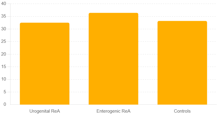 | Figure 1. Mean age distribution of the study groups |
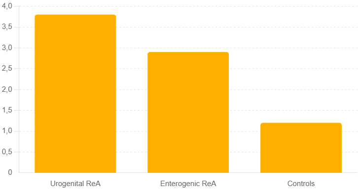 | Figure 2. Serum IL-17A levels in patients with reactive arthritis and controls |
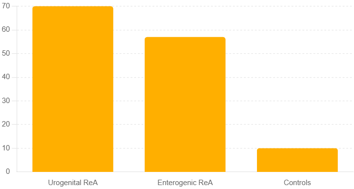 | Figure 3. Prevalence of ultrasound-detected synovitis among study groups |
4. Discussion
- The present prospective comparative study provides additional evidence supporting the concept that reactive arthritis represents a heterogeneous immune-mediated condition in which the nature of the triggering infection significantly influences clinical presentation, inflammatory activity, and immunogenetic profiles. Our findings confirm that urogenital reactive arthritis, predominantly associated with Chlamydia trachomatis, is characterized by a more pronounced systemic and local inflammatory response compared with enterogenic forms, despite comparable demographic characteristics among study groups.Consistent with previously published epidemiological data, male predominance and the young working-age profile of patients were observed in both reactive arthritis subgroups [1,4,6]. The absence of significant age and sex differences between the urogenital, enterogenic, and control groups supports the internal validity of the comparative analysis and reduces the likelihood of confounding demographic effects on immunological outcomes.In agreement with international guidelines and cohort studies, Chlamydia trachomatis was identified as the triggering pathogen in all patients with urogenital reactive arthritis, underscoring its central etiological role [2,7,13]. The detection of enterogenic pathogens such as Yersinia enterocolitica, Campylobacter jejuni, and Salmonella enteritidis in the enterogenic subgroup is consistent with earlier observations that gastrointestinal infections remain an important but heterogeneous cause of reactive arthritis [5,8]. Importantly, the frequent absence of active infectious symptoms at the time of arthritis onset highlights the diagnostic challenge emphasized in recent reviews and supports the need for molecular diagnostic approaches [9,14].Clinically, joint involvement predominated in all patients, with preferential involvement of the knee, ankle, and foot joints, as well as a substantial prevalence of sacroiliitis. These findings mirror those reported in contemporary clinical reviews, which describe reactive arthritis as a predominantly peripheral arthritis with frequent axial involvement in a subset of patients [1,3]. The relatively high frequency of mucocutaneous and ocular manifestations in urogenital reactive arthritis observed in our cohort further supports the concept of broader systemic immune activation associated with Chlamydia-triggered disease [11,12].Inflammatory laboratory markers, including ESR and hs-CRP, were significantly elevated in both reactive arthritis subgroups compared with controls, confirming their utility as indicators of disease activity but not etiology. This observation aligns with previous reports emphasizing the limited specificity of conventional inflammatory markers in reactive arthritis [4,12]. The lack of significant differences in these markers between urogenital and enterogenic forms suggests that additional biomarkers are required to refine etiological differentiation and prognostic assessment.A key finding of this study is the significantly elevated serum IL-17A levels in patients with reactive arthritis, particularly in those with urogenital etiology. This result is consistent with growing evidence implicating the IL-17/Th17 axis as a central driver of inflammation in seronegative spondyloarthritides [16–18]. The observed moderate correlations between IL-17A levels and DAS28, hs-CRP, and disease duration support the hypothesis that IL-17A reflects both inflammatory burden and disease persistence. These data extend previous observations by suggesting that IL-17A may serve not only as a marker of disease activity but also as a potential indicator of chronicity risk in reactive arthritis.The upregulation of TLR-2 gene expression in both reactive arthritis subgroups further underscores the importance of innate immune activation in disease pathogenesis. Toll-like receptors are known to play a critical role in recognizing bacterial components and initiating inflammatory signaling cascades [19]. Our findings of a positive correlation between TLR-2 expression and systemic inflammatory markers corroborate experimental data demonstrating sustained innate immune stimulation in reactive arthritis, even in the absence of active infection [7,9]. Notably, the lack of significant differences in TLR-2 expression between urogenital and enterogenic forms suggests that innate immune activation represents a shared pathogenic mechanism across etiologies.Imaging results reinforce current views on the limited sensitivity of conventional radiography in early reactive arthritis and highlight the value of ultrasound and MRI for detecting subclinical inflammation [1,14]. The higher frequency of power Doppler activity in urogenital reactive arthritis corresponds with the more pronounced immunological activation observed in this subgroup and supports the integration of advanced imaging modalities into diagnostic algorithms.Taken together, our findings support a multidimensional diagnostic approach to reactive arthritis that extends beyond traditional clinical and laboratory parameters. The integration of immunological markers such as IL-17A and molecular indicators of innate immune activation such as TLR-2 may improve diagnostic accuracy, facilitate etiological differentiation, and enable early identification of patients at increased risk of chronic disease. These results align with recent calls in the literature for biomarker-driven diagnostic strategies and underscore the need for further longitudinal studies to validate the prognostic value of immunogenetic markers in reactive arthritis [6,16,20].
5. Conclusions
- This prospective comparative study demonstrates that reactive arthritis is characterized by distinct immunological profiles that are closely related to the etiology of the triggering infection. Urogenital reactive arthritis, predominantly associated with Chlamydia trachomatis, is accompanied by a more pronounced inflammatory response compared with enterogenic forms, despite similar demographic and clinical characteristics.The findings highlight the diagnostic limitations of conventional clinical assessment and routine inflammatory markers in reactive arthritis and emphasize the added value of immunological and molecular biomarkers. Elevated serum IL-17A levels were strongly associated with disease activity, inflammatory burden, and disease duration, suggesting a pivotal role of Th17-mediated immune mechanisms in the pathogenesis and persistence of reactive arthritis. Increased expression of TLR-2 further supports the contribution of innate immune activation to sustained inflammation across different etiological forms.Importantly, the combined assessment of IL-17A and TLR-2 provides additional diagnostic insight beyond standard laboratory parameters and may facilitate improved etiological differentiation and early identification of patients at risk for chronic disease progression. These results support the integration of immunogenetic markers into diagnostic algorithms for reactive arthritis and underscore the potential of biomarker-guided approaches to optimize early diagnosis and clinical management.Further longitudinal studies with larger cohorts are warranted to validate the prognostic significance of IL-17A and TLR-2 and to explore their potential role in personalized diagnostic and therapeutic strategies for reactive arthritis.