-
Paper Information
- Next Paper
- Previous Paper
- Paper Submission
-
Journal Information
- About This Journal
- Editorial Board
- Current Issue
- Archive
- Author Guidelines
- Contact Us
American Journal of Medicine and Medical Sciences
p-ISSN: 2165-901X e-ISSN: 2165-9036
2026; 16(4): 1863-1865
doi:10.5923/j.ajmms.20261604.64
Received: Mar. 13, 2026; Accepted: Apr. 6, 2026; Published: Apr. 15, 2026

Hemodynamics in the Central Retinal Vein in Intracranial Hypertension
 Abstract
 Reference
 Full-Text PDF
 Full-text HTML
Muxamadiev Raxmon Omonovich1, Alimova Zebiniso Farxod qizi2
1Termez Branch of the Tashkent Medical Academy, Uzbekistan
2Medical Faculty of the Termez University of Economics and Service, Uzbekistan
Correspondence to: Muxamadiev Raxmon Omonovich, Termez Branch of the Tashkent Medical Academy, Uzbekistan.
| Email: |  |
Copyright © 2026 The Author(s). Published by Scientific & Academic Publishing.
This work is licensed under the Creative Commons Attribution International License (CC BY).
http://creativecommons.org/licenses/by/4.0/

Objective. To determine the blood flow velocity in the central retinal vein (CRV) in hypertensive syndrome. Material and Methods: The study involved 36 patients at various stages of optic disc edema (papilledema) associated with hypertensive syndrome. Results: It was noted that in the initial stage of hypertensive syndrome development, the blood flow velocity in the central retinal vein decreased by 1.0 cm/s. In cases of pronounced optic disc edema, the blood flow velocity in the central vein decreased by 2.0 cm/s. Conclusion: The blood flow velocity in the central retinal vein is a determining factor in the diagnosis of intracranial hypertension.
Keywords: Optic disc, Blood flow velocity in the central retinal vein
Cite this paper: Muxamadiev Raxmon Omonovich, Alimova Zebiniso Farxod qizi, Hemodynamics in the Central Retinal Vein in Intracranial Hypertension, American Journal of Medicine and Medical Sciences, Vol. 16 No. 4, 2026, pp. 1863-1865. doi: 10.5923/j.ajmms.20261604.64.
1. Introduction
- An increase in intracranial pressure, which affects the circulation of cerebrospinal fluid, is not only accompanied by dysmetabolic changes in brain neurons and impairment of hemodynamic parameters but also, in the vast majority of cases, leads to the development of optic disc edema (papilledema) [1].According to the literature, the incidence of optic disc edema (papilledema) in various brain pathologies reaches up to 93% [5,6]. Numerous methods exist for studying ocular microcirculation, allowing for the assessment of circulatory disorders within the eyeball. Among these, Dopplerographic studies, which measure blood flow velocity in the venous system, are considered the most informative [2,5,6].Optic disc edema (papilledema) serves as a direct indicator of intracranial hypertension. The digital equivalents of papilledema progression during intracranial hypertension are objectively evaluated by measuring the blood flow velocity in the central retinal vein [3,4].By measuring the blood flow velocity in the central retinal vein (CRV), it is possible to obtain objective information regarding the state of intracranial hypertension. Purpose and Objectives of the Study. The study aims to determine the blood flow velocity in the central retinal vein (CRV) using ultrasound Dopplerography in patients with intracranial hypertension across various stages of optic disc edema (papilledema).
2. Material and Methods
- The study material consisted of the examination results of 36 patients presenting with various degrees of optic disc edema (papilledema) resulting from intracranial hypertension. The age of the patients ranged from 35 to 61 years; the cohort included 21 males and 15 females.According to the clinical findings, 11 patients were diagnosed with cerebral arachnoiditis, 10 with brain tumors, and 15 with post-traumatic brain pathology. For a comparative analysis of blood flow velocity in the central retinal vein (CRV), a control group of 12 healthy individuals (6 males and 6 females) aged 35 to 60 years was included.Initial-stage optic disc edema was detected in 12 patients, while pronounced (advanced) edema was observed in 24 patients. We utilized highly informative diagnostic methods, including Optical Coherence Tomography (OCT) of the optic nerve head and combined ultrasound examination with Doppler imaging mode. In this study, blood flow registration is based on the shift in ultrasound signal frequency reflected from moving blood particles, primarily erythrocytes [4,5].
3. Results and Discussion
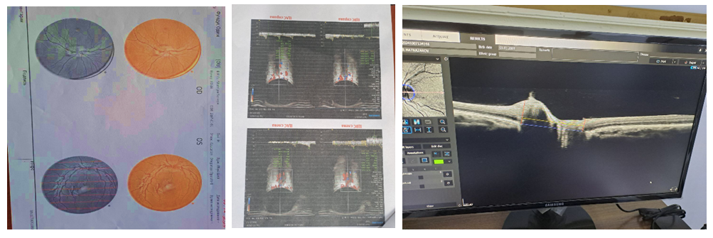
- During the study, all patients were found to present with optic disc edema (papilledema) of varying severity resulting from increased intracranial pressure. In 11 patients, the initial stage of papilledema was characterized by blurred optic disc margins, slight prominence (elevation) of the disc, and dilation of the retinal veins. In 25 patients, the optic disc edema was identified in the advanced stage.According to our findings, visual acuity and visual fields remained unaffected in 24 patients with early and developed stages of optic disc edema. In cases of severely pronounced edema, the mean visual acuity decreased to 0.7. Notably, the peripheral visual fields remained unchanged in all examined cases.Hemodynamic analysis of the central retinal vein (CRV) demonstrated a slowing of venous outflow of varying degrees in all patients with papilledema associated with intracranial hypertension (Fig. 1).
|
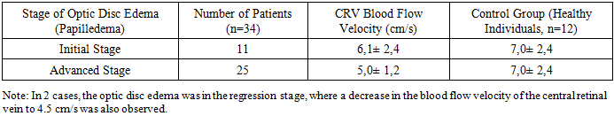
 | Figure 1. Initial stage of papilledema development. Blood flow velocity in the central retinal vein (CRV) is 6 cm/s. Disappearance of the optic disc excavation |
 | Figure 2. Advanced stage of papilledema. Blood flow velocity in the central retinal vein (CRV) is 2 cm/s |
4. Conclusions
- One of the primary indicators of the initial stage of intracranial hypertension syndrome is a decrease in blood flow velocity by 1.0 cm/s. As the hypertension syndrome progresses to the advanced stage of papilledema, accompanied by prominent optic disc edema, the blood flow velocity in the central retinal vein decreases by 2.0 cm/s. This reduction serves as a critical clinical signal, necessitating an immediate and comprehensive neurological examination. In such cases, the treatment of the underlying pathological process in the brain is of paramount and urgent importance.