-
Paper Information
- Next Paper
- Previous Paper
- Paper Submission
-
Journal Information
- About This Journal
- Editorial Board
- Current Issue
- Archive
- Author Guidelines
- Contact Us
American Journal of Medicine and Medical Sciences
p-ISSN: 2165-901X e-ISSN: 2165-9036
2026; 16(4): 1728-1733
doi:10.5923/j.ajmms.20261604.38
Received: Mar. 11, 2026; Accepted: Mar. 28, 2026; Published: Apr. 9, 2026

Pathogenetic Mechanisms of Venous Wall Remodeling in Varicose Disease: A Histochemical Study
 Abstract
 Reference
 Full-Text PDF
 Full-text HTML
Kosimov Sherzodbek Khursanali ogli, Azimova Gulnoza Ravshanovna, Deepak Sharma
Fergana Medical Institute of Public Health, Fergana, Republic of Uzbekistan
Correspondence to: Kosimov Sherzodbek Khursanali ogli, Fergana Medical Institute of Public Health, Fergana, Republic of Uzbekistan.
| Email: |  |
Copyright © 2026 The Author(s). Published by Scientific & Academic Publishing.
This work is licensed under the Creative Commons Attribution International License (CC BY).
http://creativecommons.org/licenses/by/4.0/

Background: Varicose disease of the lower extremities is associated with structural remodeling of the venous wall and valvular apparatus, leading to venous reflux and chronic venous insufficiency. Objective: To investigate and refine the histochemical characteristics of venous vessels in lower extremity varicose disease. Methods: A qualitative histochemical study was conducted using venous wall specimens obtained during surgical treatment. Periodic acid–Schiff (PAS) reaction, Schiff reaction, and Van Gieson staining were applied. Microscopic examination was performed at magnifications of ×100–×400. Results: Uneven accumulation of PAS- and Schiff-positive structures, thickening and irregularity of the basement membrane, plasma imbibition, and mucoid swelling were identified. Disorganization and fragmentation of fibrous components were observed. Van Gieson staining revealed focal proliferation of fuchsinophilic fibers in all layers of the venous wall and valvular structures. Endothelial damage and thrombotic deposits were detected. Conclusions: The identified changes reflect metabolic injury of the venous wall with development of phlebofibrosis and collagen remodeling, contributing to valvular insufficiency and thrombogenesis.
Keywords: Varicose disease, Chronic venous insufficiency, Venous wall, Histochemistry, PAS reaction, Van Gieson staining
Cite this paper: Kosimov Sherzodbek Khursanali ogli, Azimova Gulnoza Ravshanovna, Deepak Sharma, Pathogenetic Mechanisms of Venous Wall Remodeling in Varicose Disease: A Histochemical Study, American Journal of Medicine and Medical Sciences, Vol. 16 No. 4, 2026, pp. 1728-1733. doi: 10.5923/j.ajmms.20261604.38.
Article Outline
1. Introduction
- Varicose disease of the lower extremities is one of the most prevalent vascular pathologies worldwide, significantly contributing to morbidity and reduced quality of life. According to epidemiological data, the prevalence of chronic venous disorders reaches 30–40% in the adult population and continues to increase globally [1–3].Despite advances in diagnostics and treatment, varicose disease remains a clinically significant condition due to its progressive course, high recurrence rates, and associated complications. The disease imposes a considerable socio-economic burden, particularly in regions with limited access to specialized vascular care [4,5].Current concepts consider varicose disease as a systemic disorder involving structural and functional changes of the venous wall. Remodeling of the extracellular matrix, including alterations in collagen and elastin components, is regarded as a key pathogenetic mechanism underlying venous dilation and valvular insufficiency [6–8].The aim of this study was to investigate and refine the histochemical characteristics of venous vessels in lower extremity varicose disease.
2. Materials and Methods
- Study design and participantsThe study was conducted on venous specimens obtained from 175 patients undergoing surgical treatment for lower extremity varicose disease between 2022 and 2024. Inclusion criteria comprised clinically and instrumentally confirmed varicose disease. Exclusion criteria included acute inflammatory conditions and systemic connective tissue disorders.Data collection instrumentsHistological and histochemical analysis was performed using light microscopy. Standard staining techniques included periodic acid–Schiff (PAS) reaction, Schiff reaction, and Van Gieson staining. Microscopic examination was carried out at magnifications ranging from ×100 to ×400.ProceduresVenous wall fragments and biopsy samples from the muscle–venous pump region were collected intraoperatively. Tissue samples were fixed in formalin, processed according to standard histological protocols, embedded in paraffin, and sectioned for staining and microscopic evaluation.Ethical considerationsThe study was conducted in accordance with the Declaration of Helsinki. Ethical approval was obtained from the Local Ethics Committee of the Fergana Medical Institute of Public Health. Written informed consent was obtained from all participants.Data analysisThe study employed qualitative histochemical analysis. Structural alterations were assessed descriptively based on staining characteristics and tissue morphology.
3. Results and Discussion
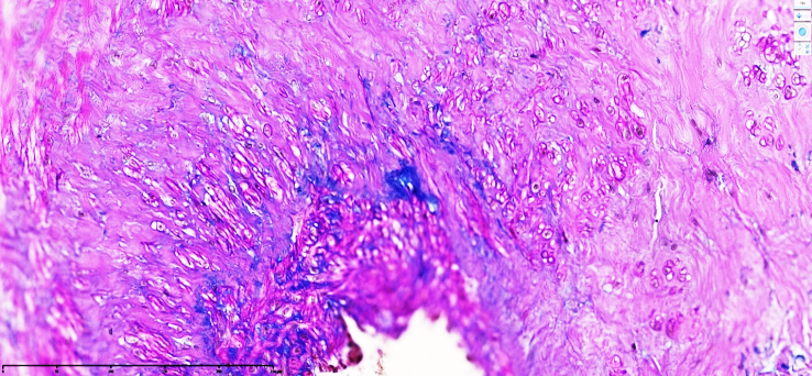
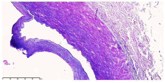
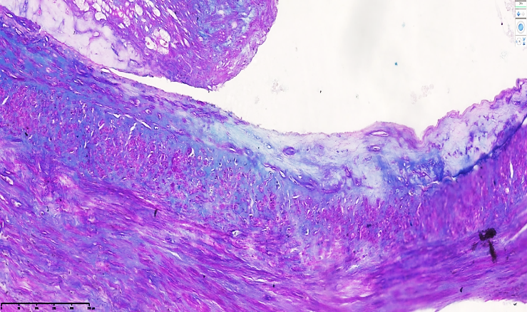
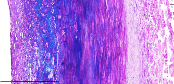
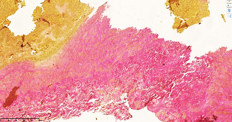
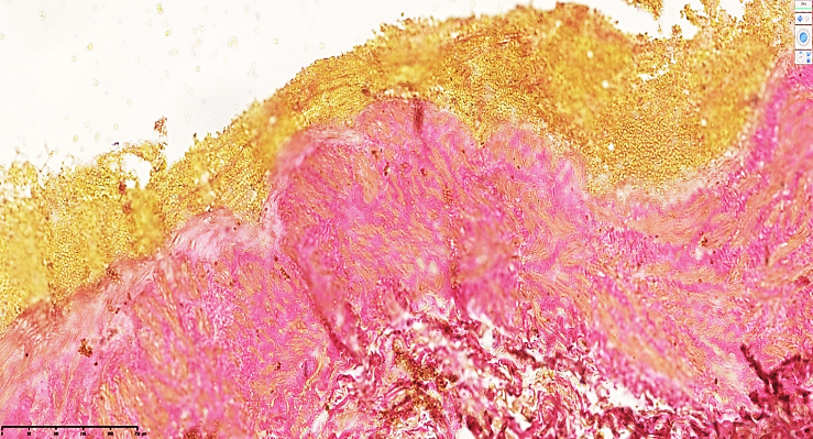
- Histochemical examination revealed pronounced structural alterations in the venous wall of varicose veins.Accumulation of PAS- and Schiff-positive structures was predominantly localized in the subendothelial and medial layers, demonstrating an uneven distribution pattern. Thickening and irregularity of the basement membrane were consistently observed.Pronounced plasma imbibition and mucoid swelling were identified within the vascular wall, indicating increased hydrophilicity of the extracellular matrix.Fragmentation and disorganization of collagen and elastic fibers were detected across all layers of the venous wall. These alterations were accompanied by dilation of the vascular lumen.Van Gieson staining demonstrated focal proliferation of fuchsinophilic fibers, particularly within the medial and valvular regions. Structural deformation of venous valves and microfissure-type damage were identified.Endothelial injury and focal thrombotic deposits were observed on the luminal surface.The presence of dark homogeneous pink inclusions surrounding Schiff-positive structures reflects pronounced accumulation of neutral mucopolysaccharides, manifested as PAS-positive formations (see Fig. 1).
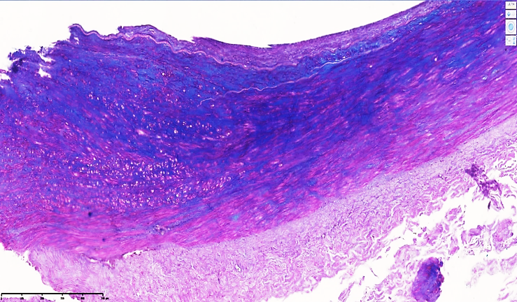
 | Figure 1. Venous vessel. The intimal surface is of uneven thickness (1); interlaminar spaces of the subendothelial layer are variably expanded (2); developing edematous changes are observed around Schiff-positive structures (3); the basement membrane is thickened with an irregular contour. Schiff staining. Magnification ×40×10 |
 | Figure 2. Venous vessel. The intimal surface is of uneven thickness (1); interlaminar spaces of the subendothelial layer are variably expanded (2); developing edematous changes are observed around Schiff-positive structures (3); the basement membrane is thickened with an irregular contour. Schiff staining. Magnification ×10×10 |
 | Figure 3. Superficial vein of the middle third of the lower leg. Massive simultaneous accumulation of PAS- and Schiff-positive structures is observed in the muscular layer of the vascular wall. The vessel wall demonstrates plasma imbibition and fragmentation of fibrous structures of different origin. Along the endothelium, the basement membrane boundaries are altered with irregular thickening. PAS + Schiff staining. Magnification ×20×10 |
 | Figure 4. The medial layer demonstrates focal destruction and fragmentation of fibrous structures (1), as well as PAS- and Schiff-positive formations of varying intensity with irregular contours. Intervening areas reveal a disorganized distribution of sparse, weakly stained fibers (2). Schiff staining. Magnification ×20×10 |
 | Figure 5. Medial layer of the lower leg vein. Foci of destruction and fragmentation of fibrous structures are identified (1), together with PAS- and Schiff-positive formations of varying intensity with irregular contours. The intervening areas demonstrate a disorganized arrangement of sparse, weakly stained fibers (2). Schiff staining. Magnification ×20×10 |
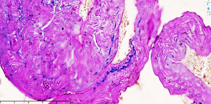 | Figure 6. Venous valve of the lower leg. Linear distribution of Schiff-positive structures is observed within the valve tissue. In most areas, a substantial number of PAS-positive formations are detected, indicating pronounced plasma imbibition of the valve (intense dark-pink staining). PAS + Schiff staining. Magnification ×40×10 |
 | Figure 7. Venous vessel of the middle third of the lower leg. The vascular wall demonstrates irregular, focal distribution of fuchsinophilic fibers. The luminal surface exhibits traces of thrombotic deposits and areas of structural damage. Van Gieson staining. Magnification ×40×10 |
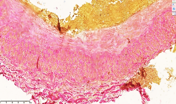
 | Figure 8. Venous vessel of the upper third of the lower leg. The vascular wall demonstrates massive accumulation of fuchsinophilic fibers. A fragment of a damaged valve with an angular configuration is identified. Interstitial edematous changes are visualized within the vascular stroma. Van Gieson staining. Magnification ×20×10 |
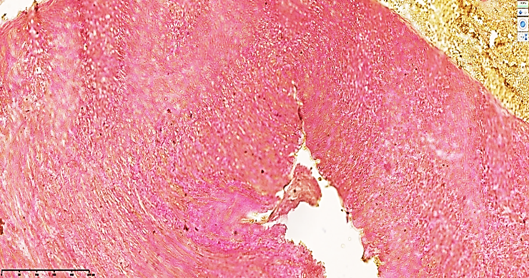
 | Figure 9. Venous vessel of the popliteal region. The vascular wall demonstrates massive accumulation of fuchsinophilic fibers. A fragment of a damaged valve with an angular configuration is identified. Wavy-oriented fuchsinophilic fibrous structures are observed. Interstitial edematous changes are visualized within the vascular stroma. Van Gieson staining. Magnification ×20×10 |
 | Figure 10. Venous vessel of the popliteal region. The vascular wall demonstrates massive accumulation of fuchsinophilic fibers. A fragment of a damaged valve with an angular configuration is identified. Wavy-oriented fuchsinophilic fibrous structures are observed. Interstitial edematous changes are visualized within the vascular stroma. Van Gieson staining. Magnification ×20×10 |
4. Discussion
- The aim of this study was to investigate histochemical alterations of the venous wall in varicose disease.The findings demonstrate that accumulation of mucopolysaccharides and plasma imbibition play a central role in the development of structural changes in the venous wall. These processes lead to disruption of extracellular matrix organization and contribute to loss of vascular elasticity.The observed increase in PAS- and Schiff-positive structures reflects metabolic disturbances and altered pH balance within the vascular wall, which promotes degradation of fibrous components. Similar findings have been reported in studies investigating extracellular matrix remodeling in chronic venous disease [6–8].Proliferation of fuchsinophilic fibers identified by Van Gieson staining indicates enhanced collagen synthesis and fibrotic transformation. These changes are consistent with previously described mechanisms of venous wall remodeling and valvular dysfunction [7,8].Endothelial damage and thrombotic deposits observed in the present study confirm the role of structural alterations in thrombogenesis. Impaired hemodynamics and turbulent blood flow further contribute to disease progression.Limitations: The study was qualitative and did not include morphometric analysis.Strengths: Detailed histochemical characterization of venous wall remodeling.Future directions: Quantitative morphometric studies are required to further elucidate pathogenetic mechanisms.
5. Conclusions
- The study demonstrated that varicose disease is associated with metabolic and structural remodeling of the venous wall, including plasma imbibition, mucoid swelling, and fibrotic transformation.These changes contribute to valvular insufficiency, impaired hemodynamics, and increased risk of thrombogenesis.Therapeutic strategies should be aimed at improving trophic support of the venous wall, limiting fibrosis, and preventing thromboembolic complications.
ACKNOWLEDGEMENTS
- The authors express gratitude to the staff of the Fergana Medical Institute of Public Health for their support in conducting this study.
Author Contributions
- All authors contributed equally to the study design, data collection, analysis, and manuscript preparation.
Conflict of Interest
- The authors declare no conflict of interest.
Funding
- The study was conducted without external financial support.