American Journal of Medicine and Medical Sciences

p-ISSN: 2165-901X    e-ISSN: 2165-9036

2026;  16(4): 1599-1604

doi:10.5923/j.ajmms.20261604.10

Received: Mar. 2, 2026; Accepted: Mar. 29, 2026; Published: Apr. 1, 2026


The Role of Matrix Metalloproteinases and Pro-Inflammatory Cytokines in the Pathogenesis of Dorsalgia in Modic-Type Spondylodiscitis and the Effectiveness of Pathogenetic Therapy

Adambaev Z. I.1, Mirjuraev E. M.2, Samiev A. S.3, Ergashev G. B.4

1Urgench State Medical Institute, Urgench, Uzbekistan

2Center for the Development of Professional Qualifications of Medical Workers, Tashkent, Uzbekistan

3Samarkand State Medical University, Samarkand, Uzbekistan

4Central District Hospital of Narpay District, Neurology Department, Samarkand, Uzbekistan

Correspondence to: Adambaev Z. I., Urgench State Medical Institute, Urgench, Uzbekistan.

Email:

Copyright © 2026 The Author(s). Published by Scientific & Academic Publishing.

This work is licensed under the Creative Commons Attribution International License (CC BY).
http://creativecommons.org/licenses/by/4.0/

Abstract

The article highlights current aspects of the etiology, pathogenesis, and treatment of dorsalgia associated with Modic-type spondylodiscitis. Data on the infectious (Propionibacterium acnes) and autoimmune origins of bone marrow changes are analyzed, along with the role of MMP-3 and MMP-9, their TIMP-1, and pro-inflammatory cytokines (IL-1β, TNF-α, IL-6) in the development of inflammation and intervertebral disc degradation. A review of clinical manifestations, standardized assessment scales, and treatment approaches, including antibiotic and anti-inflammatory therapy, is presented. Special attention is given to the results of the authors’ own study evaluating the effectiveness of comprehensive conservative treatment in 100 patients with aseptic Modic type I spondylodiscitis. It was demonstrated that the inclusion of paravertebral blocks with hydrocortisone and ultrasound therapy (phonophoresis) with “Cariflex” gel in the treatment regimen resulted in a more pronounced reduction in pro-inflammatory cytokine levels and extracellular matrix degradation markers compared with standard therapy. These findings indicate more effective modulation of the inflammatory process and improvement of bone tissue metabolism.

Keywords: Dorsalgia, Spondylodiscitis, Modic changes, Biomarkers, MMP-3, MMP-9, TIMP-1, Pro-inflammatory cytokines, Paravertebral blocks, Phonophoresis

Cite this paper: Adambaev Z. I., Mirjuraev E. M., Samiev A. S., Ergashev G. B., The Role of Matrix Metalloproteinases and Pro-Inflammatory Cytokines in the Pathogenesis of Dorsalgia in Modic-Type Spondylodiscitis and the Effectiveness of Pathogenetic Therapy, American Journal of Medicine and Medical Sciences, Vol. 16 No. 4, 2026, pp. 1599-1604. doi: 10.5923/j.ajmms.20261604.10.

1. Introduction

The idea of dorsalgia with the type of spondylodiscitis is one of the most disputable problems in the modern vertebrology and neurology as verified by various reviews and primary studies [1,5,43]. Modic changes were initially defined in 1988 as being MRI-detected changes in bone marrow around vertebral endplates and are common in the context of degenerative spinal disease, including that of spondyloarthrosis affecting facet joints, intervertebral discs, and its surrounding bone. According to the epidemiological data, the shifts are observed among 6-25 per cent of patients with chronic back pain and are associated with increased pain intensity and decreased quality of life indicators [18,27,34]. Since the nonspecific back pain affects between 50 and 80 percent of adults and causes significant economic losses due to disability, it is evident that the subject has clinical and socioeconomic significance [1,5,44].
Recent theory approaches of pathogenesis emphasize a mixed infectious-immune pathogenesis, especially in type 1 lesions of Modic type. Potential research indicates that Propionibacterium acnes has been detected in 40-80 percent of intervertebral discs of patients with the presence of the Modic type 1 changes, being higher than the control groups [7,13,16]. Experimental rabbit models also show that disc inoculation with P. acnes has the opportunity to cause bone marrow edema which mimic a change of Modic changes within 6-12 months [41]. The mechanism by which low-virulence disc infection triggers local inflammation is mechanistically proposed to involve the release of IL-1-b, TNF-a and IL-6, which is further augmented by secondary immune response and production of vasogenic edema [15,37]. In line with this hypothesis, Dudli et al. (2018) suggested that Modic type 1 occurs against an infected disc co-existed with a pro-inflammatory bone marrow microenvironment [15], other studies report an increase in Cytokine activity and autoantibody activity in the sufferers [32]. Risk may additionally be genetic susceptibility modified through the immune-related polymorphisms [24,26].
MRI is still the mainstay of diagnostic imaging and phenotyping. T1 hypointensity and T2/STIR hyperintensity that are typical of active edema and inflammation characterize modic type 1 and is the best associated with clinically relevant pain [20,21,35]. Modic type 2 is a manifestation of fatty marrow replacement, and is generally more stable, whereas type 3 is associated with the sclerosis, and least often does it relate to pain [13,35]. Symptoms are heterogeneous and vary according to spinal level and neural involvement and include mechanical local pain, referred pain, radiculopathy, or neurogenic claudication and in severe cases, sensory and motor deficit [8,23]. The severity of the symptoms and pain treatment response are well quantified using standardized assessment tools like Visual Analogue Scale (VAS) of pain and Oswestry Disability Index (ODI) of disability, as well as the SF-36 and EQ-5D instruments to indicate a pronounced impairment of quality of life in this group of patients [8,43,44].
Alongside imaging, biomarker profiling is also an area that is being studied to quantitatively measure disease activity. Though the levels of C-reactive protein (CRP) tend to be normal or with a low-grade of inflammation [2,11], IL-6 is considered as one of the main mediators of pain sensitization and tissue remodelling [11,37]. The involvement of matrix metalloproteinases (especially, MMP -3 and MMP -9) in the breakdown of extracellular matrix in disc and adjacent structures; a disproportion between them and their inhibitor tissue inhibitor of metalloproteinases -1 (TIMP -1) may support progressive destruction; these parameters have been suggested as diagnostic and prognostic measures of active degenerativeinflammatory processes [2,5,7,10,12,13,30].
Therapy is a divisive matter. Certain trials suggest that an extended course of antibiotic therapy (e.g. amoxicillin -clavulanate) is superior to placebo in selected patients [8-10], but later meta-analytic studies indicate that the effect is not consistent [12]. The non-steroid anti-inflammatory drugs only serve as first-line treatment and may be effective with adjunctive treatment, but cannot be used long-term due to their adverse effects [2,11]. New pathogenetic methods, such as regulation of the microbiome, have also been proposed [19]. Surgical intervention is used when there is deterioration or worsening of the neurological condition; Modic type 1 changes were reported to have a poorer postoperative prognosis [29]. Prognosis is inconsistent; Modic type 1 has been established as an independent determinant of worse outcome to conservative therapy, especially in patients with a history of pain lasting longer than 12 months, multilevel disease, CRP 2. 10mg/L, and resistance to NSAIDs [14,16], though an early, pathogenetically targeted treatment can stabilise remission in 6070 per cent of cases [8,10].
It is against this background that the objective of the current research was to assess the efficacy of conservative therapy in the case of aseptic Modic type 1 spondylodiscitis in the presence of para vertebral hydrocortisone blocks coupled with ultrasound therapy (phonophoresis) using Cariflex gel.

2. Materials and Methods

A prospective comparative clinical study was conducted in the Neurology Department of the Central District Hospital of Narpay District (CRH Narpay) and included 100 patients diagnosed with aseptic spondylodiscitis (ASD) corresponding to Modic type I changes in the lumbosacral spine. The cohort comprised 65 men and 35 women. The mean age of the total sample was 50.2±2.4 years; mean age in men was 48.1±2.5 years and in women 53.3±2.3 years. The diagnosis and phenotyping of ASD according to Modic type I were established by magnetic resonance imaging (MRI) of the lumbar spine.
Table 1. Dynamics of MMP-3, MMP-9, TIMP-1, IL-1β, and TNF-α in the Main and Control Groups
     
After baseline assessment, patients were allocated into two treatment arms: a control group (CG; n=40) and a main group (MG; n=60). The control group received standard pharmacological therapy in combination with conventional physiotherapy as routinely applied in the department. The main group received the same baseline medication regimen and additionally underwent ultrasound therapy using phonophoresis with “Cariflex” gel, as well as paravertebral blocks with hydrocortisone. Clinical complaints and neurological status were documented at inclusion, and MRI data were analyzed to confirm Modic type I changes and to register concomitant degenerative conditions.
To objectify inflammatory activity and extracellular matrix remodeling, blood samples were collected for serum biomarker analysis. Matrix metalloproteinases and their inhibitor were assessed in serum using enzyme-linked immunosorbent assay (ELISA) with commercial kits for MMP-3, MMP-9, and TIMP-1, measured on an ELISA analyzer. Mediators of acute inflammation, interleukin-1β (IL-1β) and tumor necrosis factor-α (TNF-α), were also quantified by ELISA (Enzyme-Linked Immunosorbent Assay). Biomarkers were measured at three predefined time points: before treatment initiation (baseline), at 1 month, and at 3 months after the start of therapy. In addition to absolute biomarker concentrations, the ratios MMP-3/TIMP-1 and MMP-9/TIMP-1 were calculated as integrated indicators of the proteolysis–inhibition balance.
Table 2. Between-Group Comparisons (Student’s t-test)
     
Statistical analysis was performed to evaluate comparability of groups at baseline and treatment-associated changes over time. Between-group differences at each time point were assessed using Student’s t-test for independent samples. Within-group dynamics were evaluated using paired Student’s t-test, and percentage changes relative to baseline were calculated to characterize the magnitude of biomarker shifts during follow-up. Differences were considered statistically significant at p<0.05, with p<0.01 interpreted as significant and p<0.001 as highly significant.

3. Results

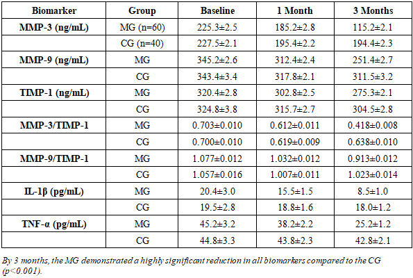
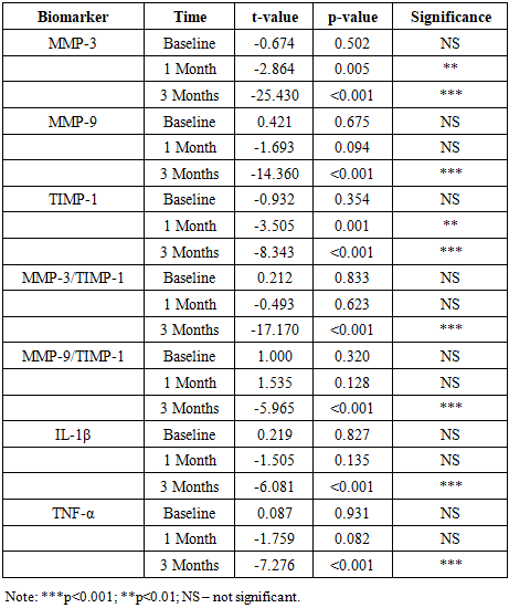
At baseline, all patients presented with Modic type I aseptic spondylodiscitis confirmed by MRI. Clinically, the dominant symptom was deep, aching or pulsating lumbosacral pain aggravated by physical load and frequently intensified at night. Radicular pain radiating to the lower extremities was observed in 79% of patients and was commonly associated with paresthesia. Morning stiffness lasting more than 30 minutes was reported by 60% of patients, and severe limitation of spinal range of motion was documented in 85%.
Neurological examination revealed radicular syndromes in 51% of patients. Decreased tendon reflexes and sensory disturbances in the corresponding dermatomes were observed in 55%, while objective muscle weakness was present in 11%. MRI additionally demonstrated disc herniation in 90% of cases, spondylolisthesis in 20%, and vertebral osteoporosis in 30%, predominantly among women older than 50 years.
Baseline comparison showed that the MG and CG were statistically comparable across all biomarker levels (p>0.05), allowing objective evaluation of treatment effects.
Table 3. Within-Group Percentage Changes (Main Group)
     
MRI confirmed Modic type I changes in all participants. In addition, a high prevalence of concomitant degenerative pathology was identified: intervertebral disc herniation in 90% of patients, spondylolisthesis in 20%, and vertebral osteoporosis in 30%, with osteoporosis occurring predominantly in women older than 50 years.
Table 4. Within-Group Percentage Changes (Control Group)
     
The longitudinal dynamics of serum biomarkers are presented in Table 1. At baseline, the main group (MG) and control group (CG) were comparable across all biochemical parameters (p>0.05), indicating the absence of significant intergroup differences prior to treatment (Table 2). After 1 month, intergroup differences became evident for MMP-3 (t=−2.864; p=0.005) and TIMP-1 (t=−3.505; p=0.001), whereas changes in MMP-9, IL-1β, and TNF-α did not reach statistical significance at this time point (p>0.05) (Table 2).
By 3 months, the MG demonstrated a highly significant reduction in all measured markers compared with the CG (p<0.001 for MMP-3, MMP-9, TIMP-1, MMP-3/TIMP-1, MMP-9/TIMP-1, IL-1β, and TNF-α) (Table 2). Specifically, in the MG MMP-3 decreased from 225.3±2.5 to 115.2±2.1 ng/mL, while in the CG it declined only modestly from 227.5±2.1 to 194.4±2.3 ng/mL. MMP-9 decreased in the MG from 345.2±2.6 to 251.4±2.7 ng/mL, whereas in the CG it changed from 343.4±3.4 to 311.5±3.2 ng/mL. TIMP-1 in the MG decreased from 320.4±2.8 to 275.3±2.1 ng/mL compared to a smaller decline in the CG from 324.8±3.8 to 304.5±2.8 ng/mL (Table 1).
The ratios reflecting the balance between proteolysis and its inhibition also shifted more favorably in the MG. The MMP-3/TIMP-1 ratio decreased from 0.703±0.010 to 0.418±0.008 at 3 months, whereas in the CG it remained elevated (0.700±0.010 to 0.638±0.010). Similarly, the MMP-9/TIMP-1 ratio decreased in the MG from 1.077±0.012 to 0.913±0.012, while in the CG it showed minimal change (1.057±0.016 to 1.023±0.014) (Table 1).
A pronounced suppression of systemic inflammatory mediators was observed in the MG. IL-1β decreased from 20.4±3.0 to 8.5±1.0 pg/mL and TNF-α from 45.2±3.2 to 25.2±1.2 pg/mL at 3 months. In contrast, the CG showed only minor changes (IL-1β: 19.5±2.8 to 18.0±1.2 pg/mL; TNF-α: 44.8±3.3 to 42.8±2.1 pg/mL) (Table 1).
Within-group analyses confirmed that biomarker reductions in the MG were statistically significant at both 1 and 3 months for all parameters (mostly p<0.001; MMP-9/TIMP-1 at 1 month p=0.002), demonstrating consistent treatment-associated improvement over time. In the CG, within-group changes were generally not significant (p>0.05), with the exception of a modest early reduction of MMP-3 at 1 month (p=0.012). Overall, these findings indicate that the combined regimen incorporating paravertebral hydrocortisone blocks and ultrasound phonophoresis with “Cariflex” gel was associated with a substantially greater decrease in pro-inflammatory cytokines and extracellular matrix degradation markers compared with standard therapy.
Overall, the main group demonstrated a significantly greater reduction in extracellular matrix degradation markers (MMP-3, MMP-9), their ratios with TIMP-1, and pro-inflammatory cytokines (IL-1β, TNF-α) compared with the control group. These findings indicate that the combined therapy including paravertebral hydrocortisone blocks and ultrasound phonophoresis with “Cariflex” gel more effectively suppresses inflammatory activity and normalizes the balance between matrix degradation and inhibition in patients with Modic type I spondylodiscitis.

4. Conclusions

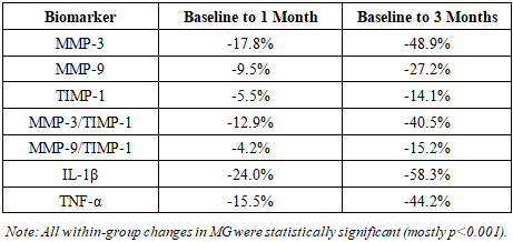
Aseptic type I Modic spondylodiscitis is characterized by eminent clinical presentation, which includes persistent lumbosacral and radicular pain, loss of functionality, and neurological impairments usually accompanied with disc herniation and other degenerative changes. The disorder is biochemically linked with increased amount of MMP-3, MMP-9, IL-1, and TNF-A and the disproportion of MMP/TIMF ratios, which reflects active inflammatory and matrix-destructive actions.
The treatment regime that was combined with the paravertebral hydrocortisone blocks and ultrasound phonophoresis using a Cariflex gel led to a statistically significant decrease in all the biomarkers studied after three months of treatment compared to the usual therapy. Significant reductions in MMP-3, MMP-9, pro-inflammatory cytokines as well as normalization of MMP/TIMP ratios indicate successful inhibition of inflammation and some restoration of extracellular matrix balance.
These findings support the idea of the conceptualization of the Modic type I changes as an active inflammatory-degenerative process and prove that pathogenetically-oriented combination therapy has a better clinical and molecular outcome. There is a need to conduct further controlled studies that would validate the use of biomarkers in the management of this group of patients.

References

[1]  Adambaev ZI, Kilichev IA. Demand for hospital-replacing outpatient clinics in the treatment and rehabilitation of patients with degenerative spinal diseases in Uzbekistan. Probl Mod Sci Educ. 2016; 5(47): 231-236.
[2]  Adambaev ZI, Kilichev IA. Optimization of complex therapy for patients with degenerative-dystrophic diseases of the lumbosacral spine. Ther Bull Uzbekistan. 2016; 2-3: 153-158.
[3]  Adambaev ZI, Kilichev IA. Structure and nature of back pain among outpatients of a neurological clinic. Bull Tashkent Med Acad. 2016; 2: 55-57.
[4]  Adambaev ZI, Kilichev IA, Khojanova TR. Experience of conservative treatment of intervertebral disc herniations with Cariflex gel and cream in outpatient settings. J Neurol Neurosurg Res. 2020; 3(1): 6-8.
[5]  Adambaev ZI, Zukhritdinov UYu. Diadynamic electro-neurostimulation in the treatment of patients with lumbosacral dorsopathies. In: All-Russian Therapeutic Congress with International Participation “Botkin Readings”; 2021 Apr 23-24. p. 9.
[6]  Adams MA, Hawkes R, Whitcomb S, Dolan P. Biomechanics of back pain. Acad Radiol. 2020; 27(6): 769-779.
[7]  Ahmed-Yahia S, Decousser JW, Flouzat-Lachaniette CH, Gille O, Poignard A. Is the discopathy associated with Modic changes an infectious process? Results from a prospective monocenter study. PLoS One. 2019; 14(8): e0221030.
[8]  Albert HB, Krogsgaard MR, Jensen TS. Antibiotics in chronic low back pain: a systematic review. Eur Spine J. 2021; 30(1): 1-12.
[9]  Albert HB, Manniche C. Antibiotic treatment in patients with chronic low back pain and Modic changes (the AIM study): study protocol for a randomised controlled trial. Trials. 2013; 14: 160.
[10]  Albert HB, Sorensen JS, Christensen BS, Manniche C. Antibiotic treatment in patients with chronic low back pain and vertebral bone edema (Modic type 1 changes): a double-blind randomized clinical controlled trial. Eur Spine J. 2013; 22(4): 697-707.
[11]  Al-Falahi MA, Salal MH, Abdul-Wahab DM, Al-Shakarchi H. Antibiotic treatment in patients with chronic low back pain and vertebral bone edema (Modic Type I changes): a randomized clinical controlled trial. Iraqi Postgrad Med J. 2014; 13(3): 390-397.
[12]  Braten LCH, Rolfsen MP, Espeland A, et al. Efficacy of antibiotic treatment in patients with chronic low back pain and Modic changes: double blind, randomised, placebo controlled, multicentre trial. BMJ. 2019; 367: l5654.
[13]  Brinjikji W, Diehn FE, Jarvik JG, Carr CM, Yawn BP, Murad MH, Rho ME. MRI findings of disc degeneration are more prevalent in adults with low back pain than in asymptomatic controls: a systematic review and meta-analysis. AJNR Am J Neuroradiol. 2015; 36(12): 2394-2399.
[14]  Chou R, Deyo R, Friedly J, et al. Systemic pharmacologic therapies for low back pain: a systematic review. Ann Intern Med. 2017; 166(7): 480-492.
[15]  Dudli S, Liebenberg E, Magnitsky S, Lotz JC. Modic type 1 change is an autoimmune response requiring a proinflammatory milieu. Spine J. 2018; 18(5): 831-844.
[16]  Dudli S, Liebenberg E, Magnitsky S, Lotz JC. Propionibacterium acnes infected intervertebral discs cause vertebral bone marrow lesions consistent with Modic changes. J Orthop Res. 2016; 34(8): 1447-1455.
[17]  Ghazimirsaeid SS, Sadeghi A, Boustani M, Gerami R. Evaluation of effect of Modic changes on treatment outcomes of nonspecific low back pain. Ann Mil Health Sci Res. 2022; 20(4): e135789.
[18]  Global Burden of Disease Study 2019. Lancet. 2020; 396(10258): 1204-1222.
[19]  Jensen OK, Andersen MH, Ostgard RD, Kjaer P. Probiotics for chronic low back pain with type 1 Modic changes: randomized trial. Eur Spine J. 2019; 28(11): 2478-2486.
[20]  Jensen OK, Nielsen CV, Sorensen JS, Kjaer P. Type 1 Modic changes as risk factor for outcome in low back pain. Spine J. 2014; 14(11): 2568-2581.
[21]  Jensen TS, Kjaer P, Korsholm L, Leboeuf-Yde C. Modic type 1 changes and pain. Eur Spine J. 2020; 29(3): 456-465.
[22]  Jensen TS, Kjaer P, et al. Efficacy of antibiotics in Modic type 1 changes: double-blind RCT. Ann Rheum Dis. 2022; 81(3): 345-352.
[23]  Jensen TS, Baron R, Haanpää M, et al. A new definition for neuropathic pain. Pain. 2011; 152(10): 2204-2205.
[24]  Kjaer P, Leboeuf-Yde C, Kongsted A, et al. Modic changes and chronic low back pain. J Orthop Sports Phys Ther. 2018; 48(11): 840-851.
[25]  Kjaer P, Hartvigsen J, Solomon C, et al. Exercise therapy for chronic low back pain with Modic changes: systematic review. Spine J. 2019; 19(11): 1467-1476.
[26]  Koes BW, van Tulder M, Lin CWC, Macedo LG, McAuley J, Maher C. Updated overview of clinical guidelines for non-specific low back pain. Eur Spine J. 2017; 26(1): 40-53.
[27]  Lamb SE, Hansen Z, Lall R, et al. Cognitive behavioural therapy for chronic low back pain: randomized trial. Lancet. 2010; 375(9712): 916-923.
[28]  Lambrechts MJ, Brush P, Issa TZ, Toci GR, Heard JC, Syal A, et al. Impact of Modic changes on operative treatment. Int J Environ Res Public Health. 2022; 19(16).
[29]  Li X, Sowa G, Kim C, et al. Vascular endothelial growth factor in Modic type 1 changes. Spine J. 2019; 19(7): 1189-1196.
[30]  Liddle SD, Gracey JH, Baxter GD. Physiotherapy for low back pain. Physiotherapy. 2021; 108: 1-9.
[31]  Loggia ML, Chonde DB, Akeju O, Bushnell MC, et al. Neuroinflammation in chronic pain. Pain. 2023; 164(1): 10-20.
[32]  Macedo LG, Saragiotto BT, Yamato TP, et al. Motor control exercise for chronic non-specific low back pain. Spine J. 2021; 21(1): 14-25.
[33]  Mirjuraev EM, Adambaev ZI, Zukhritdinov UYu, Akilov DKh. Non-specific back pain in Andijan city population. Med News (Belarus). 2023; (6): 70-72.
[34]  Modic MT, Steinberg PM, Ross JS, Masaryk TJ, Carter JR. Degenerative disk disease: MR imaging assessment. Radiology. 1988; 166(1): 193-199.
[35]  O’Connell NE, Wand BM, Marston L, et al. Non-invasive brain stimulation for low back pain. Eur J Pain. 2021; 25(3): 533-550.
[36]  Ohtori S, Inoue G, Orita S, et al. Inflammatory cytokines in lumbar disc herniation. Eur Spine J. 2021; 30(2): 389-396.
[37]  O’Sullivan P. Diagnosis and classification of chronic low back pain disorders. Man Ther. 2022; 32: 151-157.
[38]  Paoloni M, Bernetti A, Fratocchi G, et al. Kinesio taping improves chronic low back pain outcomes. Clin Rehabil. 2011; 25(12): 1089-1098.
[39]  Saragiotto BT, Maher CG, Yamato TP, et al. Motor control exercise for nonspecific low back pain. Spine (Phila Pa 1976). 2022; 47(6): E363-E372.
[40]  Samiyev AS, Xakimova SZ, Soibnazarov OE. Rehabilitation of patients after spine surgery. J Biomed Pract. 2022; 1: E139-144.
[41]  Samiyev AS, Jabbarov I. Complex treatment in lumbar degenerative-spondylogenic radiculopathies in pregnant women. J Biomed Pract. 2022; 5: E192-196.
[42]  Shan Z, Zhang X, Li S, et al. Propionibacterium acnes incubation results in Modic changes: rabbit model. Spine (Phila Pa 1976). 2017; 42(21): 1595-1603.
[43]  Shokhina E, Khalimova A, Muradimova AR, Matmurotov KJ, Sodiqov B, Samiev AS. Evaluation of female rats’ genital alterations caused by cadmium. Caspian J Environ Sci. 2025.
[44]  Underwood M, Buchbinder R, Foster NE, et al. Non-pharmacological treatments for chronic low back pain. Lancet. 2020; 396(10265): 2095-2105.
[45]  Vlayen JWS, Maher CG, Wiech K, et al. Low back pain. Nat Rev Dis Primers. 2018; 4(1): 52.
[46]  Williams AC, Eccleston C, Morley S. Cognitive behavioural therapy for chronic pain: meta-analysis. Pain. 2021; 162(11): 2543-2556.
[47]  World Health Organization. Rehabilitation of people with spine disorders. Geneva: WHO; 2022.
[48]  World Health Organization. Rehabilitation in health systems. Geneva: WHO; 2022.