-
Paper Information
- Next Paper
- Paper Submission
-
Journal Information
- About This Journal
- Editorial Board
- Current Issue
- Archive
- Author Guidelines
- Contact Us
American Journal of Medicine and Medical Sciences
p-ISSN: 2165-901X e-ISSN: 2165-9036
2026; 16(4): 1555-1557
doi:10.5923/j.ajmms.20261604.01
Received: Jan. 26, 2026; Accepted: Feb. 22, 2026; Published: Apr. 1, 2026

Structural Changes of the Intervertebral Disc in Hernias in Young Patients
 Abstract
 Reference
 Full-Text PDF
 Full-text HTML
Azimov Ulugbek Mekhritdinovich, Navruzov Rustam Rashidovich
Bukhara State Medical Institute, Bukhara City, Republic of Uzbekistan
Copyright © 2026 The Author(s). Published by Scientific & Academic Publishing.
This work is licensed under the Creative Commons Attribution International License (CC BY).
http://creativecommons.org/licenses/by/4.0/

Background. Intervertebral disc herniation in young patients represents a distinct clinical and morphological entity compared to age-related degenerative disc disease. Mechanical overload and repetitive microtrauma are considered major contributing factors. However, structural characteristics of early disc degeneration in young individuals remain insufficiently investigated. The aim of the current study was to evaluate morphological and morphometric alterations of intervertebral discs in young patients with herniation. Materials and Methods. A case-control morphological study was conducted on intervertebral disc specimens obtained from young patients diagnosed with lumbar or cervical disc herniation. Histological examination included assessment of the nucleus pulposus, annulus fibrosus, cartilaginous endplates, and paradiscal tissues. Morphometric parameters were quantified using digital image analysis software. Statistical analysis was performed using SPSS version 26.0. Differences between groups were evaluated using Student’s t-test. A p-value < 0.05 was considered statistically significant. Results. Young patients demonstrated significant thinning of the annulus fibrosus (−21%), reduction in nucleus pulposus area (−17%), decreased water content (−13%), and increased collagen fiber density (+25%) compared to controls (p < 0.050). Degenerative index scores increased more than threefold. Reactive angiogenesis in paradiscal tissues increased by 18%. The most pronounced alterations were observed in the lumbar region. Conclusion. Intervertebral disc herniation in young patients develops primarily under mechanical stress conditions, accompanied by early structural and biochemical alterations rather than advanced age-related degeneration. These findings support the concept that early degenerative-dystrophic changes form the morphological substrate for herniation and pain syndrome in young individuals.
Keywords: Intervertebral disc, Disc herniation, Morphology, Nucleus pulposus, Annulus fibrosus
Cite this paper: Azimov Ulugbek Mekhritdinovich, Navruzov Rustam Rashidovich, Structural Changes of the Intervertebral Disc in Hernias in Young Patients, American Journal of Medicine and Medical Sciences, Vol. 16 No. 4, 2026, pp. 1555-1557. doi: 10.5923/j.ajmms.20261604.01.
1. Introduction
- Intervertebral disc herniation represents one of the most frequent causes of chronic back pain and radiculopathy worldwide, significantly affecting quality of life and working capacity across different age groups. Although degenerative disc disease is traditionally regarded as an age-related process associated with progressive structural deterioration, disc herniation is increasingly diagnosed in young adults and even adolescents. This trend has been attributed to modern lifestyle factors, including sedentary behavior, prolonged static loading, insufficient physical conditioning, and repetitive biomechanical stress. In young individuals, mechanical overload, axial compression, rotational strain, and cumulative microtrauma appear to play a predominant role in disc pathology, often preceding overt age-related degeneration [1,2].The intervertebral disc is a complex fibrocartilaginous structure composed of three anatomically and functionally distinct components: the gelatinous nucleus pulposus, the surrounding annulus fibrosus, and the superior and inferior cartilaginous endplates. The nucleus pulposus is rich in proteoglycans and water, providing resistance to compressive forces, whereas the annulus fibrosus, formed by concentric lamellae of collagen fibers, ensures tensile strength and containment of the nucleus. Cartilaginous endplates regulate nutrient diffusion and metabolic exchange between the disc and adjacent vertebral bodies. The structural and biochemical integrity of these components is essential for maintaining biomechanical stability, load distribution, and flexibility of the spinal column [3,4,5].Early pathological changes in the intervertebral disc may involve disruption of collagen fiber orientation, reduction in proteoglycan concentration, decreased hydration of the nucleus pulposus, and impairment of endplate permeability. Such alterations can compromise the disc’s mechanical resilience and increase susceptibility to annular fissures and protrusion of nuclear material. Importantly, in young patients, these changes may develop independently of advanced age-related degeneration and instead reflect adaptive or maladaptive responses to excessive biomechanical stress. Progressive fibrosis, microstructural disorganization, and localized inflammatory reactions may further accelerate structural weakening and predispose to herniation [6,7,8].Despite the clinical significance of disc herniation in young patients, detailed morphological and morphometric characterization of early degenerative-dystrophic alterations remains insufficiently explored. Most available studies focus predominantly on advanced degenerative stages observed in older populations, while the structural substrate of herniation in young individuals has not been fully elucidated. A better understanding of early morphological changes may provide insight into the pathogenesis of disc pathology, improve diagnostic accuracy, and support the development of targeted preventive and therapeutic strategies [9]. Therefore, the aim of the current study was to investigate morphological and morphometric changes of intervertebral discs in young patients with diagnosed hernias.
2. Materials and Methods
- Study Design. A controlled morphological and morphometric study was performed.Study Population. Intervertebral disc specimens were obtained from young patients (mean age 32.4 ± 6.8 years) undergoing surgical treatment for lumbar or cervical disc herniation. The control group consisted of age-matched individuals without degenerative spinal pathology.Histological Analysis. Tissue samples were fixed in formalin, embedded in paraffin, sectioned, and stained using hematoxylin-eosin and Masson's trichrome techniques. Structural assessment included evaluation of collagen fiber organization, fissures of the annulus fibrosus, hydration status of the nucleus pulposus, and thickness of hyaline endplates.Morphometric Assessment. Digital image analysis was used to measure:• Thickness of the annulus fibrosus• Area of the nucleus pulposus• Collagen fiber density• Endplate thickness• Vascular bed area in paradiscal tissuesStatistical Analysis. Statistical analysis was performed using SPSS 26.0. Data are presented as mean ± standard deviation (SD). Intergroup comparisons were conducted using Student’s t-test. Statistical significance was set at p < 0.050.
3. Results
- Morphological examination demonstrated flattening of intervertebral discs, reduced elasticity, and decreased intervertebral space height. The annulus fibrosus showed collagen fiber stratification, focal tears, and disorganization. Microfractures with penetration of nucleus pulposus fragments into annular layers were observed.Nucleus Pulposus. The nucleus pulposus retained relatively high hydration (up to 85%) in general; however, herniated regions exhibited partial dehydration and mucoid degeneration. Early chondroid transformation was identified in several specimens.
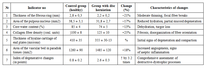 | Table 1. Morphological and morphometric changes of the intervertebral disc in young patients with hernias |
4. Discussion
- The current study demonstrated that intervertebral disc herniation in young patients is associated with early degenerative-dystrophic alterations rather than advanced age-related degeneration. The observed thinning of the annulus fibrosus and increased collagen density indicate compensatory fibrosis secondary to mechanical overload.Reduced hydration of the nucleus pulposus reflects impaired proteoglycan metabolism and decreased tissue elasticity. These changes increase susceptibility to rupture under axial stress.Reactive angiogenesis in paradiscal tissues suggests chronic microtrauma-induced inflammatory responses. The findings support the hypothesis that herniation in young individuals results from a combination of biomechanical stress and early biochemical alterations.
5. Conclusions
- Intervertebral disc herniation in young patients develops predominantly due to mechanical overload and repetitive microtrauma. Early morphological alterations include thinning of the annulus fibrosus, partial dehydration of the nucleus pulposus, fibrosis, and reactive angiogenesis. These structural changes constitute the morphological substrate of pain syndrome and neurological manifestations in young individuals. Early detection and targeted preventive strategies may reduce progression of degenerative-dystrophic processes.