-
Paper Information
- Next Paper
- Previous Paper
- Paper Submission
-
Journal Information
- About This Journal
- Editorial Board
- Current Issue
- Archive
- Author Guidelines
- Contact Us
American Journal of Medicine and Medical Sciences
p-ISSN: 2165-901X e-ISSN: 2165-9036
2026; 16(3): 1059-1064
doi:10.5923/j.ajmms.20261603.44
Received: Jan. 7, 2026; Accepted: Feb. 1, 2026; Published: Mar. 7, 2026

Cerebrospinal Fluid Flow and Spinal Cord Motion in Cervicothoracic Spinal Dysraphism: A Systematic Review
 Abstract
 Reference
 Full-Text PDF
 Full-text HTML
Ahmediyev M. M.1, Arziqulov J. M.1, Ahmediyev T. M.2, Tulayev N. B.1, Ashrapov J. R.1, Yugai I. A.1, Jumayev O. B.2
1Department Pediatric Neurosurgery, Republican Scientific Practical Medical Center of Neurosurgery, Tashkent, Uzbekistan
2Department of Traumatology, Orthopedics, Military Field Surgery and Neurosurgery, Tashkent, Uzbekistan
Correspondence to: Ahmediyev M. M., Department Pediatric Neurosurgery, Republican Scientific Practical Medical Center of Neurosurgery, Tashkent, Uzbekistan.
| Email: |  |
Copyright © 2026 The Author(s). Published by Scientific & Academic Publishing.
This work is licensed under the Creative Commons Attribution International License (CC BY).
http://creativecommons.org/licenses/by/4.0/

Cerebrospinal fluid (CSF) circulation plays a critical role in maintaining normal spinal cord physiology. Disturbances of CSF flow may occur in a wide range of congenital and acquired spinal pathologies, including spinal dysraphism, diastematomyelia, tethered cord syndrome, and syringomyelia. Traditional radiological assessment relies primarily on morphological evaluation of the subarachnoid spaces and spinal cord position; however, these static features often fail to correlate with clinical manifestations, particularly in pediatric patients. Advances in magnetic resonance imaging (MRI), especially phase-contrast and cine MRI techniques, have enabled noninvasive evaluation of CSF flow dynamics and spinal cord motion synchronized with the cardiac cycle. The present systematic review summarizes current evidence on MRI-based assessment of CSF flow and spinal cord motion in children with cervicothoracic spinal dysraphism, with particular emphasis on diastematomyelia. The review highlights the diagnostic value, clinical implications, and limitations of functional MRI techniques and discusses their role in improving risk stratification and surgical decision-making in pediatric neurosurgery.
Keywords: Cerebrospinal fluid flow, Spinal dysraphism, Diastematomyelia, Pediatric neurosurgery, Phase-contrast MRI
Cite this paper: Ahmediyev M. M., Arziqulov J. M., Ahmediyev T. M., Tulayev N. B., Ashrapov J. R., Yugai I. A., Jumayev O. B., Cerebrospinal Fluid Flow and Spinal Cord Motion in Cervicothoracic Spinal Dysraphism: A Systematic Review, American Journal of Medicine and Medical Sciences, Vol. 16 No. 3, 2026, pp. 1059-1064. doi: 10.5923/j.ajmms.20261603.44.
Article Outline
1. Introduction
- Spinal dysraphism encompasses a heterogeneous group of congenital malformations resulting from incomplete closure of the neural tube and associated mesenchymal structures during early embryogenesis [12,10]. In the pediatric population, cervicothoracic forms of spinal dysraphism are less common than lumbosacral variants; however, they are associated with a disproportionately higher risk of neurological deterioration due to the high density of ascending and descending neural pathways within this region [11,4].Alterations in cerebrospinal fluid (CSF) circulation are increasingly recognized as a key pathophysiological mechanism contributing to neurological dysfunction in spinal dysraphism. Obstructive lesions, abnormal CSF spaces, formation of closed compartments, and the development of pathological cavities such as cysts and syrinxes may significantly impair normal CSF dynamics [1,8]. Nevertheless, conventional magnetic resonance imaging (MRI) assessment remains largely limited to static morphological features, which may not adequately reflect functional disturbances of CSF flow or spinal cord mobility, particularly in pediatric patients [2,3].Recent advances in MRI technology have enabled direct visualization and quantitative assessment of pulsatile CSF flow and spinal cord motion using phase-contrast and cine MRI techniques. These methods provide novel insights into the biomechanical behavior of the spinal cord and surrounding CSF spaces, offering opportunities for improved understanding of disease mechanisms, early detection of functional impairment, and optimization of surgical strategies in children with congenital spinal disorders [5,13].The aim of the present systematic review is to analyze current evidence on MRI-based assessment of CSF flow and spinal cord motion in pediatric cervicothoracic spinal dysraphism, with particular emphasis on diastematomyelia and split cord malformations, in order to clarify their diagnostic value and clinical relevance for pediatric neurosurgical practice.
2. Methods
- Study designThis systematic review was conducted in accordance with the PRISMA 2020 (Preferred Reporting Items for Systematic Reviews and Meta-Analyses) guidelines.Search strategyA comprehensive literature search was performed in PubMed/MEDLINE, Scopus, and Web of Science databases for studies published between 1995 and 2024. The search terms included combinations of the following keywords: cerebrospinal fluid flow, CSF dynamics, phase-contrast MRI, cine MRI, spinal cord motion, spinal dysraphism, diastematomyelia, split cord malformation, and pediatric.Eligibility criteriaInclusion criteria were: original clinical studies or systematic reviews; pediatric population (0–18 years); evaluation of CSF flow and/or spinal cord motion using MRI-based techniques; inclusion of patients with spinal dysraphism.Exclusion criteria were: isolated case reports without functional analysis; experimental studies without clinical correlation; studies not reporting CSF flow or spinal cord motion parameters.Study selectionAfter removal of duplicates, titles and abstracts were screened independently. Full-text articles were reviewed to determine final eligibility. Discrepancies were resolved by consensus.
3. Results
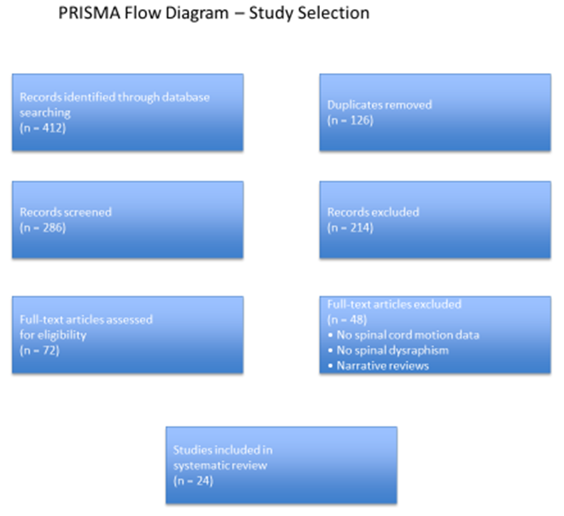
- Study selection (PRISMA)The initial search identified 412 publications. After removal of duplicates, 286 articles remained. Screening of titles and abstracts resulted in exclusion of 214 studies. Full-text assessment was performed for 72 articles, of which 48 were excluded due to insufficient functional data or lack of pediatric dysraphism cases. A total of 24 studies were included in the final analysis.
 | Figure 1 |
 | Table 1. Pediatric studies on CSF flow and spinal cord motion |
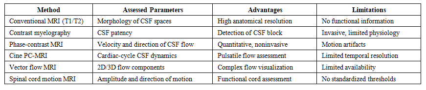 | Table 2. Methods for Assessment of CSF Flow: Advantages and Limitations |
4. Discussion
- The findings of the present review support the concept that spinal dysraphism represents a dynamic disorder in which neurological impairment may arise from disturbed CSF biomechanics rather than from static compression alone. Multiple studies have demonstrated that abnormalities in CSF flow and spinal cord motion may precede clinical deterioration, particularly in pediatric patients with cervicothoracic dysraphism and diastematomyelia [4,6,17,22–24]. These observations highlight the limitations of morphology-based assessment and emphasize the need for functional MRI techniques in pediatric neurosurgical decision-making.Limitations of morphological imaging aloneConventional MRI remains indispensable for anatomical characterization of spinal dysraphism. However, reliance on morphological criteria alone may underestimate the extent of functional compromise. Numerous studies included in this review demonstrated discordance between static imaging findings and neurological status, reinforcing the concept that functional impairment may precede or occur independently of overt structural abnormalities.This discrepancy is particularly evident in cervicothoracic dysraphism, where small changes in biomechanics can have significant neurological consequences.Clinical value of functional CSF imagingFunctional MRI techniques offer several potential advantages in the evaluation of pediatric spinal dysraphism. By directly visualizing CSF dynamics and spinal cord motion, these methods provide insights into pathophysiological mechanisms that cannot be inferred from static images alone.From a clinical perspective, functional imaging may: aid in identifying children at risk of neurological deterioration, support differentiation between clinically relevant and incidental anatomical findings, assist in surgical planning by localizing functionally significant obstruction or tethering, provide objective markers for postoperative assessment.Implications for surgical decision-makingIn diastematomyelia, particularly Type I lesions, surgical intervention often involves removal of the septum and reconstruction of the dural sac. Restoration of normal CSF flow is a primary goal of surgery, yet traditional decision-making has relied largely on morphological considerations.The evidence summarized in this review suggests that functional assessment of CSF dynamics may refine surgical indications and help tailor operative strategies. For example, demonstration of significant flow asymmetry or spinal cord motion restriction at the cervicothoracic level may support earlier intervention, even in the absence of severe clinical symptoms.Pediatric-specific considerationsChildren differ fundamentally from adults in terms of spinal compliance, CSF volume distribution, and neurodevelopmental plasticity. These factors must be taken into account when interpreting functional MRI findings. The absence of standardized pediatric reference values for CSF flow parameters represents a major limitation and highlights the need for age-specific normative data.Clinical implications and future directionsThe integration of functional MRI into routine evaluation of pediatric spinal dysraphism has the potential to transform clinical practice. Future research should focus on: establishing standardized imaging protocols, de-fining quantitative thresholds predictive of neurological deterioration, correlating functional imaging findings with long-term clinical outcomes, incorporating CSF dynamics into prognostic indices and treatment algorithms.Multicenter prospective studies will be essential to validate the clinical utility of these approaches.
5. Conclusions
- Functional MRI assessment of cerebrospinal fluid flow and spinal cord motion provides critical insights into the pathophysiology of pediatric cervicothoracic spinal dysraphism. The evidence reviewed suggests that disturbances in CSF dynamics and spinal cord mobility are common in conditions such as diastematomyelia and may precede overt neurological deterioration.Incorporation of functional imaging into diagnostic and surgical decision-making frameworks holds promise for earlier risk stratification, more precise operative planning, and improved neurological outcomes in children with spinal dysraphism. Further standardization and high-quality clinical studies are required to fully realize the potential of these techniques in pediatric neurosurgery.
ACKNOWLEDGEMENTS
- The authors acknowledge all contributors whose work supported the preparation of this systematic review.
DISCLOSURE
- The authors declare no conflicts of interest related to this study.