American Journal of Medicine and Medical Sciences

p-ISSN: 2165-901X    e-ISSN: 2165-9036

2026;  16(2): 811-816

doi:10.5923/j.ajmms.20261602.88

Received: Jan. 25, 2026; Accepted: Feb. 20, 2026; Published: Feb. 27, 2026


Perforations and Other Injuries of the Duodenum: Current Approaches to Diagnosis and Therapy

Urokov Sh. T.1, Abidov U. O.1, Masharipov Kh. K.2

1Bukhara State Medical Institute named after Abu Ali ibn Sina, Bukhara, Uzbekistan

2Bukhara Branch of the Republican Scientific Center for Emergency Medical Care Uzbekistan, Bukhara, Uzbekistan

Copyright © 2026 The Author(s). Published by Scientific & Academic Publishing.

This work is licensed under the Creative Commons Attribution International License (CC BY).
http://creativecommons.org/licenses/by/4.0/

Abstract

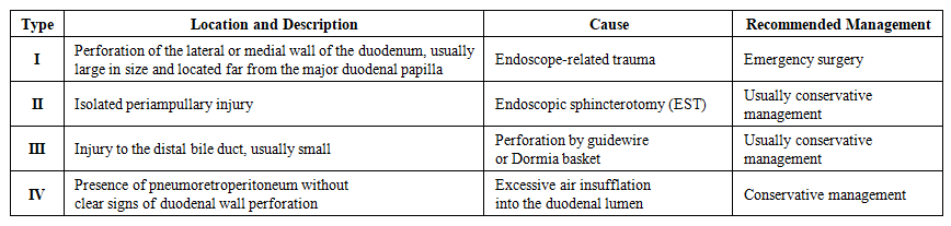
This review presents current aspects of diagnosis and treatment of peptic ulcer perforations and other duodenal integrity disorders, including retroperitoneal posterior wall injuries. It outlines etiological factors, classifications (including Stapfer), clinical features, imaging approaches, and treatment strategies. Special emphasis is placed on minimally invasive techniques - Over-the-Scope Clip (OTSC) and Endoscopic Vacuum Therapy (EVT) - which show high efficacy in selected cases and can reduce complication and mortality rates. The need for further research and broader implementation of these technologies in local clinical practice is highlighted.

Keywords: Ulcer perforation, Injury, Duodenum, Retroduodenal injury, OTSC, Endoscopic vacuum therapy

Cite this paper: Urokov Sh. T., Abidov U. O., Masharipov Kh. K., Perforations and Other Injuries of the Duodenum: Current Approaches to Diagnosis and Therapy, American Journal of Medicine and Medical Sciences, Vol. 16 No. 2, 2026, pp. 811-816. doi: 10.5923/j.ajmms.20261602.88.

1. Introduction

This review presents up-to-date data on the diagnosis and treatment of peptic ulcer perforations and other conditions associated with disruption of the integrity of the duodenum (DU). Etiological factors, classifications of injuries, clinical manifestations, and diagnostic approaches are examined. Particular attention is paid to minimally invasive methods such as OVESCO (OTSC) placement and endoscopic vacuum therapy (EVT). The indications, technical aspects, and outcomes of these technologies are discussed both in comparison with and in combination with conventional surgery [3,4,5,8,15,19,20,21,22,23,24,25].
Disruption of duodenal integrity is one of the key causes of emergency abdominal surgery and is associated with high mortality, especially in cases of delayed diagnosis and sepsis. Among the causes of duodenal wall disruption, perforations of ulcer etiology account for the majority, whereas trauma, anastomotic suture failure, and iatrogenic endoscopic injuries constitute the remaining proportion. Injuries of the posterior duodenal wall should be highlighted separately. With delayed diagnosis, mortality in these conditions may reach 40–70%, particularly with the development of peritonitis, retroperitoneal phlegmon, and sepsis [10,11,14].
It is necessary to point out the existing challenges in early diagnosis and effective treatment of posterior duodenal wall injuries. The relevance and necessity of applying modern technologies in cases of posterior duodenal wall disruption are emphasized by a number of authors [1,2,7].
The contemporary diagnostic and therapeutic approach relies on early imaging (primarily CT) and a personalized choice among surgical, endoscopic, and combined strategies. Major guidelines (WSES) emphasize the need for rapid stabilization, antibiotic therapy, and early source control, including laparoscopic repair or alternative methods in critically ill and high-risk patients [4,5,15].
Disruption of the posterior wall of the duodenum represents a particularly rare and extremely serious pathology requiring immediate attention due to the high likelihood of complications and a significant risk of mortality. These injuries are often accompanied by atypical clinical presentations, which substantially complicates diagnosis. The main problem lies in the penetration of gastrointestinal contents into the retroperitoneal space, creating conditions for severe inflammatory processes such as phlegmon, abscess formation, and sepsis, as well as multiple organ failure [9,17,18].
Diagnostics and Imaging. In cases of retroduodenal injuries, traditional examination methods such as diagnostic laparotomy and laparoscopy have limited effectiveness due to difficulties in accessing the retroperitoneal space, the inability to obtain sufficient diagnostic information, and the risk of developing various infectious complications.
Therefore, this pathological condition requires the development of effective approaches aimed at reducing potential complications and improving treatment outcomes.
There are also alternative approaches that lie between conservative and surgical management. These include stent placement, the use of biodegradable biological materials to close the defect at the site of injury, and the application of mesenchymal stem cells to accelerate healing processes. However, these methods are currently applied only at the experimental level and have not completed the full cycle of clinical trials; indications for their use, as well as their applicability across different patient populations, remain insufficiently defined [27].
In some cases, conventional surgical techniques prove to be ineffective, which may necessitate repeated surgical interventions and, as a consequence, lead to increased mortality or reduced quality of life for patients.
The diagnosis of retroduodenal perforations requires an integrated approach using modern imaging modalities. Contrast-enhanced computed tomography (CT) is considered the gold standard, as it allows detection of signs of perforation such as free gas or fluid in the retroperitoneal space.
Contrast-enhanced CT is recommended as the method of choice for confirming perforation, localizing the defect, and assessing the extent of spread, including retroperitoneal involvement. Plain radiography for detection of “free gas” has low sensitivity; water-soluble contrast duodenography and/or duodenoscopy are used selectively, particularly when small or retroperitoneal defects or post-procedural complications are suspected.
Magnetic resonance imaging (MRI) may be useful for detailed evaluation of soft tissues and assessment of the extent of the inflammatory process.
Ultrasonography (US), especially when combined with Doppler imaging, helps identify fluid collections in the abdominal cavity, although it has limited diagnostic value for retroperitoneal processes.
Laparoscopy serves as both a diagnostic and therapeutic modality, allowing visualization of the affected area and performance of necessary interventions.
Clinical Presentation. Sudden onset of pain and signs of peritonitis are typical in cases of “free” perforation; in retroperitoneal rupture, the clinical picture is often more subtle and may include lumbar pain, subcutaneous emphysema, and fever.
In recent years, an increase in the prevalence of chronic diseases has been observed. The growing number of patients with peptic ulcer disease, chronic inflammatory gastrointestinal disorders, and the use of gastrotoxic medications (e.g., NSAIDs, corticosteroids) contributes to an increased risk of perforation. In addition, patients with comorbid conditions such as diabetes mellitus, liver cirrhosis, and immunodeficiency states have a higher risk of complications.
Atypical perforation, including retroperforation of a posterior duodenal wall ulcer, represents a rare but clinically significant problem that requires special attention in modern medicine. Its relevance is determined by several factors: high mortality rates, and the fact that delayed diagnosis and inadequate treatment lead to rapid progression of severe complications, including phlegmon, sepsis, and multiple organ failure. According to published data, mortality in advanced cases reaches 40–70%.
Perforation of the posterior wall of the duodenum results in leakage of gastric contents rich in enzymes and acids into the retroperitoneal space. This triggers a rapidly progressing inflammatory process that may advance to retroperitoneal phlegmon, sepsis, and multiple organ failure.
Diagnostic difficulties are primarily related to the atypical clinical presentation, which is associated with the confinement of gastrointestinal contents within the retroperitoneal space, hindering early detection. Standard diagnostic methods, such as plain radiography, are often of limited value, necessitating the use of advanced imaging techniques.
The limitations of traditional treatment methods are related to the difficulty of accessing the retroperitoneal space and the high risk of infectious complications, which significantly complicate surgical management. This underscores the need to develop new, more effective, and safer therapeutic approaches.
Thus, conditions associated with disruption of duodenal integrity—particularly perforations and injuries of the posterior wall of the duodenum—remain among the most complex and pressing problems in modern surgery, requiring innovative solutions and a multidisciplinary approach.
Treatment. Alongside traditional conservative and surgical methods for managing conditions associated with disruption of duodenal integrity, modern minimally invasive technologies have been actively developed and introduced in recent years. These include endoscopic defect closure using OTSC systems, the use of endoscopic vacuum therapy (EVT) for anastomotic leakage and perforations, placement of temporary covered self-expanding stents, and the application of biodegradable materials for defect closure. These approaches make it possible to minimize surgical trauma, reduce the length of hospital stay, and lower the risk of postoperative complications. However, they require advanced technical resources and highly experienced specialists, and their effectiveness and indications within domestic healthcare settings have not yet been sufficiently studied [12,13].
At the present stage, the treatment strategy for peptic ulcer perforations and disruptions of duodenal integrity is determined by the location of the defect, the time elapsed since injury, the severity of peritonitis, the patient’s general condition, and the technical capabilities of the healthcare facility.
General principles of diagnosis and treatment have been developed in accordance with established guidelines (WSES, 2020; SAGES, 2019; ESGE, 2020). The core principles of management include immediate stabilization—correction of fluid and electrolyte disturbances and normalization of hemodynamics; broad-spectrum antibacterial therapy; antisecretory therapy; and early source control.
Surgical Methods.
1. Laparoscopic closure with Graham omentoplasty is considered the “gold standard” for small defects (<10–12 mm) in hemodynamically stable patients.
2. Resection or duodenoplasty is indicated for larger defects.
3. Retroduodenal injuries are surgically challenging due to limited access and often require a combined approach, including open surgical debridement and drainage of the retroperitoneal space.
4. Endoscopic and minimally invasive technologies recommended by the ESGE (2020) include:
- OTSC (OVESCO) is used for acute perforations up to 20 mm in size when detected early.
- Endoscopic vacuum therapy (EVT) is indicated for anastomotic leaks, suture-line failure, and selected perforations; reported clinical success rates range from 80% to 100% in observational series.
- Covered self-expanding stents are applied in cases of extended defects and when primary closure is not feasible.
Table 1 summarizes the key approaches to the management of duodenal perforations, outlining their main indications, advantages, and limitations.
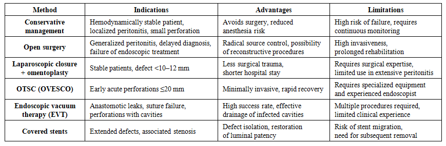
Table 1. Comparative Characteristics of Treatment Methods for Duodenal Perforations
Conservative management is applied in strictly selected patients with a stable clinical condition, limited (localized) peritonitis, and a small perforation size. The main advantage of this approach is the possibility of avoiding surgery and the associated risks of anesthesia and surgical trauma. However, this strategy is associated with a high risk of failure and requires continuous clinical and instrumental monitoring, since any deterioration in the patient’s condition necessitates immediate surgical intervention.
Open surgery remains the treatment of choice in cases of generalized peritonitis, delayed diagnosis of perforation, or failure of endoscopic techniques. It provides radical source control and allows for definitive defect repair. The disadvantages include high invasiveness, prolonged postoperative rehabilitation, and more pronounced postoperative pain.
Laparoscopic closure with omentoplasty is suitable for hemodynamically stable patients with small defects (<10–12 mm). Compared with open surgery, this approach is less traumatic, promotes faster recovery, and shortens hospital stay. Its limitations include the need for a highly experienced surgeon and technical difficulties in the presence of extensive peritonitis, where laparoscopic access may be challenging.
Placement of covered self-expanding stents is justified in cases of extended defects and strictures, allowing isolation of the defect and restoration of intestinal lumen patency. Limitations include the risk of stent migration, the need for subsequent removal, and potential complications associated with prolonged intraluminal placement.
Endoscopic defect closure using the OTSC (OVESCO) system is indicated for acute perforations up to 10 mm in size detected at an early stage, particularly in patients with a high anesthetic risk. Systematic reviews demonstrate high technical and clinical success rates in perforations and leaks (approximately 75–90%), reducing the need for “rescue” surgery in selected patients. This method is minimally invasive and ensures rapid recovery; however, it requires specialized equipment and an experienced endoscopist [28,29].
Endoscopic vacuum therapy (EVT) is used for anastomotic leaks, suture-line failure, and perforations with cavity formation. It provides a high success rate and effective decontamination of infected cavities but requires multiple procedures (up to 5–6 sponge exchanges) and currently has limited experience in widespread clinical practice. Adequate visualization of the defect margins, cavity sanitation, and decompression are essential. For periampullary/ERCP-related injuries, the choice in favor of endoscopic closure is determined by the Stapfer classification and the patient’s clinical stability [22,23,30]. Management strategies depending on the cause of ERCP-related injury and localization are presented in Table 2.
Table 2. Classification of Iatrogenic Duodenal Injuries According to Stapfer and Recommended Management Strategy
In recent years, the use of vacuum-assisted closure (VAC) therapy and sponge drains has been actively investigated in the treatment of complex infectious processes and wounds, especially in hard-to-access areas. VAC systems facilitate removal of exudate and enhance tissue healing by creating negative pressure, thereby improving drainage, reducing inflammation, and lowering the risk of abscess formation. Sponge drains, in turn, enable effective local drainage, particularly in prolonged inflammatory processes within the retroperitoneal space.
Relevance of VAC systems and sponge drainage in retroduodenal perforations.
The incorporation of VAC systems and sponge drainage into a comprehensive treatment strategy for posterior duodenal wall integrity disruption may represent an important element in improving outcomes for patients with this pathology. These methods allow for:
• Improved drainage of the retroperitoneal space, minimizing the risk of fluid accumulation and ongoing inflammation.
• Reduction in the incidence of infectious complications.
• Shorter recovery time due to accelerated tissue healing.
Thus, the choice of treatment method is determined by the clinical scenario, the technical capabilities of the healthcare facility, and the experience of the specialists involved. Contemporary strategies increasingly favor a combination of approaches with an emphasis on minimally invasive technologies, which allows for improved outcomes and a reduced rate of complications.
Technologies such as vacuum-assisted closure therapy, endoscopic techniques, and modern drainage approaches demonstrate significant potential for improving treatment outcomes. However, their application remains insufficiently studied and requires further clinical investigation.
Accordingly, research aimed at developing a comprehensive approach to the diagnosis and management of retroduodenal injuries and duodenal perforations using VAC therapy and sponge drainage is timely and of high clinical relevance.
Endoscopic Vacuum Therapy (EVT) creates negative pressure within the area of the defect or cavity, ensuring active drainage of infected contents and stimulation of granulation tissue formation. Retrospective series and systematic reviews demonstrate successful use of EVT in leaks and perforations of the duodenum and small intestine, including retroperitoneal processes. This method is particularly useful in the presence of drainable cavities and anastomotic leaks, as well as in situations where safe laparotomy is not feasible.
Combined use of EVT and OTSC techniques. In cases of small defects, a combination of OTSC and EVT may be applied: the clip is used to achieve luminal defect closure, while the sponge-based drainage system provides control of the residual cavity or phlegmon.
Iatrogenic perforations after ERCP. Systematic reviews confirm that for Stapfer type I injuries (free perforation of the duodenal wall), early surgical intervention more often yields better outcomes. In contrast, for Stapfer types II–IV (periampullary and/or retroperitoneal injuries), conservative and/or endoscopic strategies are preferred, including nasobiliary or nasogastric drainage, stenting, clipping, and EVT, provided that the patient is hemodynamically stable and adequate drainage is ensured. ESGE guidelines recommend confirming suspected perforation with CT and considering endoscopic closure depending on defect size and the experience of the center.
Unresolved issues in treatment. A substantial body of scientific research by international authors has been devoted to the diagnosis and management of conditions associated with disruption of duodenal integrity. In particular, numerous studies address duodenal trauma, ulcer-related perforations, anastomotic suture failure, and iatrogenic perforations. In most cases, these studies are based on traditional surgical approaches.
However, analysis of contemporary literature indicates that pathologies involving disruption of the retroperitoneal segments of the duodenum remain insufficiently studied, with several controversial and unresolved aspects. This is primarily related to the limited possibilities for early and accurate diagnosis of retroperitoneally localized posterior duodenal wall injuries, as well as to the relatively low effectiveness of treatment.
In recent years, international literature has increasingly highlighted the prospects of managing duodenal trauma, perforations, and iatrogenic injuries using modern minimally invasive technologies. In particular, new methods such as mechanical clipping with the OVESCO (OTSC) system and endoscopic vacuum therapy (EVT) have been introduced. Nevertheless, the effectiveness of these technologies, their indications, and clinical outcomes within domestic healthcare systems have not yet been sufficiently evaluated or systematically studied.
At the same time, in national clinical practice, experience with modern endoscopic techniques for treating conditions associated with disruption of duodenal integrity remains extremely limited, and the number of studies by domestic authors in this field is minimal. Although some local reports note the shortcomings of traditional methods and the high rates of complications and mortality, scientifically substantiated practical recommendations for the implementation of modern minimally invasive approaches are virtually absent.
Therefore, the application of endoscopic and minimally invasive technologies appears highly relevant. This will help address existing gaps in the literature and establish a scientific basis for the introduction of new approaches into national clinical practice.

2. Conclusions

Contemporary approaches to the diagnosis and management of duodenal perforations include early imaging (contrast-enhanced CT), adequate stabilization, and a targeted choice of treatment strategy based on the type of injury, defect size, and the patient’s clinical condition. Minimally invasive methods—OTSC and EVT—have demonstrated high clinical effectiveness, particularly in patients with high operative risk. Their wider implementation in clinical practice has the potential to reduce the number of re-laparotomies, shorten hospital stay, and decrease mortality rates.

References

[1]  Abakumov, M.M., Chirkov, R.N. Injuries of the Duodenum. Moscow: Binom Publishing House; 2014. 152 p.
[2]  Agarwal, N. Triple tube drainage for difficult gastroduodenal perforations: a prospective study / N. Agarwal, N.K. Malviya, N. Gupta [et al.] // World J. Gastrointestinal Surg. - 2017. - Vol. 9, № 1. - P. 19-24.
[3]  Anton, J.J. Management of duodenal trauma / J.J. Anton // Trauma Acute Care Surg. - 2019. - Vol. 87, № 3. - P. 739-740.
[4]  Balasubramanian, G. Isolated rupture of duodenum following blunt trauma abdomen: report of a case of avulsion of pylorus / G. Balasubramanian, C. Vijayakumar, I. Anbarasu [et al.] // I.S.J. - 2017. - Vol. 4, № 8. - P. 2845-2847.
[5]  Bartell N, et al. Clinical efficacy of the OTSC device: systematic review. Clin Endosc. 2020. PMC
[6]  Chevallay M, et al. Outcome of EVT for duodenal leaks. Endosc Int Open. 2022/2023. PMCPubMed
[7]  Garcia, A.F. Management of duodenal trauma: a retrospective review from the Panamerican Trauma Society / A.F. Garcia, P. Ferrada, J. Duchesne [et al.] // Journal of Trauma and Acute Care Surgery. - 2019. - Vol. 86, № 3. - P. 392-396.
[8]  Imaging blunt pancreatic and duodenal trauma [Electronic resource] / S. Shah [et al.] // Applied Radiology. – 2016. – Vol. 45 (11). – P. 22-28. – Mode of access: http://appliedradiology. com/articles/imaging-blunt-pancreatic and-duodenal -trauma.
[9]  Ioskevich, N.N. Duodenal injuries: issues of diagnosis and surgical treatment. Journal of Grodno State Medical University (Journal GrSMU) [Internet]. November 15, 2018 [cited May 11, 2025]; 16(5): 615–621. Available from: http://journal-grsmu.by/index.php/ojs/article/view/2331/.
[10]  Irarrazaval Mainguyague, M.J. Blunt abdominal trauma due to handlebar injury / M.J. Irarrazaval Mainguyague, J. Saez Binelli, С. Kychenthal Loyola [et al.] // Rev. Chil. Pediatr. - 2020. - Vol. 91, № 5. - P. 754-760.
[11]  Kong, V. Duodenal trauma / V. Kong, J.L. Bruce // Scandinavian Journal of Surgery. - 2019. - Vol. 108, № 2. - Р. 1-5.
[12]  Korita, V.R., Mikhetkina, S.I., Sidorenko, M.G. Duodenal injuries. Far Eastern Medical Journal. 2012; 1: 37–39.
[13]  Krasilnikov, D.M., Minnullin, M.M., Nikolaev, Ya.Yu. Surgical tactics in closed duodenal injuries. Practical Medicine. 2013; 2(67): 42–45.
[14]  Kumarl, A. Complete Isolated Duodenal Transection Due to Blunt Abdominal Trauma: A Case Report / A. Kumarl, V. Bhasker, M.C. Misra [et al.] // I.J.S.R. - 2017. - V. 6, № 4. - P. 1447-1448.
[15]  Louro, J. Multiple isolated small bowel perforations following blunt abdominal trauma / J. Louro, M. Albano, L. Reis [et al.] // Int. J. Surg. Case Rep. - 2018. - Vol. 53, № 1. - P. 46-49.
[16]  Minnullin, M.M., Krasilnikov, M.D., Tolstikov, P.P. Duodenal injuries: diagnosis and surgical treatment. Bulletin of Contemporary Clinical Medicine. 2015; 8(Suppl. 1): 81–85.
[17]  Mizrahi, H. Laparoscopic treatment of duodenal injury caused by gunshot / H. Mizrahi, N. Geron, A. Nicola // Injury. - 2014. - Vol. 45, № 5. - P. 916-917
[18]  Monino L., Moreels T.G., 2023. Endoscopic Vacuum Therapy of Upper Gastrointestinal Anastomotic Leaks.
[19]  Monino, L., & Moreels, T. G. (2023). Endoscopic Vacuum Therapy of Upper Gastrointestinal Anastomotic Leaks: How to Deal with the Challenges (with Video). Life, 13(6), 1412. https://doi.org/10.3390/life13061412.
[20]  Paspatis GA, et al. ESGE Guideline: Diagnosis and management of iatrogenic endoscopic perforations (update 2020). Endoscopy. 2020. esge.com+1PubMed
[21]  Samara AA, et al. Post-ERCP retroperitoneal perforation: surgical vs non-operative initial management. Ann Gastroenterol. 2021. annalsgastro.gr
[22]  Seyfried, F. Successful treatment of a gastric leak after bariatric surgery using endoluminal vacuum therapy / F. Seyfried, S. Reimer, A.D. Miras // Endoscopy. - 2013. - Vol. 45, № 2. - P. 267-268.
[23]  Shah, S. Imaging blunt pancreatic and duodenal trauma / S. Shah, M.D. Faisal Khosa, S. Rai [et al.] // Applied Radiology. - 2016. - Vol. 45, № 11. - P. 22-28.
[24]  Smallwood, N.R. The use of endoluminal vacuum (E-Vac) therapy in the management of upper gastrointestinal leaks and perforations / N.R. Smallwood, J.W. Fleshman, S.G. Leeds [et al.] // Surg. Endosc. - 2016. - Vol. 30, № 6. - P. 2473-2480.
[25]  Soreide K., Thorsen K., Harrison E.M. Perforated peptic ulcer // Lancet, 2015.
[26]  Sukhodolya, A.I., Petrushenko, V.V., Kozak, I.O. Surgical treatment of duodenal injuries. Surgery. 2014; 7: 17–20.
[27]  Topchiashvili, Z.A., Geynits, A.V., Mustafaev, R.D., et al. Surgical treatment of traumatic injuries of the duodenum. Bulletin of the REAVIZ Medical Institute. 2013; 3: 34.
[28]  Wang ZZ, et al. Effectiveness and safety of OTSC in closing GI defects. World J Gastroenterol. 2021. WJGnet
[29]  Wei JJ, et al. OTSC for perforated peptic ulcer (серия случаев). Surg Endosc. 2019. PubMed.
[30]  Wichmann D, et al. Endoscopic negative pressure therapy for duodenal/jejunal perforations. Front Surg. 2023. Frontiers.