-
Paper Information
- Next Paper
- Previous Paper
- Paper Submission
-
Journal Information
- About This Journal
- Editorial Board
- Current Issue
- Archive
- Author Guidelines
- Contact Us
American Journal of Medicine and Medical Sciences
p-ISSN: 2165-901X e-ISSN: 2165-9036
2026; 16(2): 803-806
doi:10.5923/j.ajmms.20261602.86
Received: Feb. 2, 2026; Accepted: Feb. 23, 2026; Published: Feb. 27, 2026

Morphological Evaluation of Acute Oral Poisoning
 Abstract
 Reference
 Full-Text PDF
 Full-text HTML
Khayot Khamidullaevich Yakubov, Gulbakhor Bakhshillaevna Juraeva, Tulkin Karimovich Nasirov
Tashkent State Medical University, Tashkent, Uzbekistan
Copyright © 2026 The Author(s). Published by Scientific & Academic Publishing.
This work is licensed under the Creative Commons Attribution International License (CC BY).
http://creativecommons.org/licenses/by/4.0/

Acute oral poisoning remains a significant cause of mortality, posing a complex challenge for clinical toxicology and forensic medicine. The morphological evaluation of internal organ damage in these conditions is crucial for objectively establishing thanatogenesis, clarifying the mechanisms of toxic action, and improving the reliability of expert conclusions. The aim of this study was a comprehensive morphological assessment of changes in internal organs during acute oral poisoning of various etiologies from the perspective of their diagnostic and forensic significance. The study material consisted of autopsy and histological results from individuals who died of acute oral poisoning with phenobarbital, acetic acid, and ethyl alcohol. Autopsies were performed within 24 hours of death. Macroscopic examination was supplemented by histological analysis with hematoxylin-eosin (H&E) staining. The study established that the morphological pattern of acute oral poisoning is systemic, characterized by combined damage to vital organs. The brain exhibited signs of severe edema, venous congestion, and perivascular hemorrhages, reflecting hypoxic-toxic effects. In the lungs, edema, capillary congestion, and alveolar hemorrhages predominated, indicating impaired microcirculation and respiratory function. The liver was characterized by dystrophic changes in hepatocytes, centrilobular necrosis, and signs of fatty infiltration, pointing to a pronounced cytolytic syndrome. The kidneys revealed proximal tubular epithelial necrosis and interstitial edema, corresponding to the development of acute renal failure. Dystrophic changes in cardiomyocytes and microcirculatory disorders were noted in the myocardium. Thus, morphological evaluation in acute oral poisoning allows for objective confirmation of systemic toxic damage, clarifies the mechanism of death, and significantly increases the informativeness of forensic diagnostics, especially when combined with clinico-toxicological and laboratory data.
Keywords: Forensic toxicology, Morphological pattern, Acute poisoning, Ethyl alcohol, Phenobarbital
Cite this paper: Khayot Khamidullaevich Yakubov, Gulbakhor Bakhshillaevna Juraeva, Tulkin Karimovich Nasirov, Morphological Evaluation of Acute Oral Poisoning, American Journal of Medicine and Medical Sciences, Vol. 16 No. 2, 2026, pp. 803-806. doi: 10.5923/j.ajmms.20261602.86.
1. Introduction
- Acute oral poisoning continues to hold a leading position in the structure of exogenous mortality and remains an urgent medico-social and forensic problem. According to national and international studies, intoxications with medications, alcohol, and industrial or household chemicals are consistently among the main causes of sudden and violent death, particularly among individuals of working age. Despite the advancement of analytical toxicology and the introduction of high-sensitivity laboratory methods, establishing the immediate mechanism of death remains difficult in a significant number of cases. Frequently, a low-toxicity chemical substance becomes a potent poison as a result of metabolic transformations within the organism itself [1,2]. Forensic diagnosis of acute poisoning is complicated by the lack of clinical history in most cases, the absence of specific morphological signs, and the vast variety of combined poisons, along with the limitations of existing methods for detecting toxins in biological media [3,4]. Literature data indicate that the concentration of a toxicant in biological fluids does not always correlate with the clinical severity of intoxication or the lethal outcome. This is due to individual reactivity, comorbidities, metabolic rate, and the specific toxicodynamics of the substance. In this regard, the morphological assessment of organ and tissue damage takes on special significance, allowing for an objective judgment of the depth and systemic nature of toxic damage [5,6,7]. Several authors emphasize that morphological signs of a hypoxic, cytotoxic, and hemodynamic nature reflect the final links of thanatogenesis in acute intoxications [8,9,10].Current publications note insufficient unification of morphological criteria for diagnosing death from acute oral poisoning and fragmented data on pathomorphological changes across different groups of toxicants. This leads to variability in forensic conclusions and reduces their evidentiary value [11,12,13]. Thus, the in-depth study of morphological changes in acute oral poisoning based on the analysis of modern literature and practical forensic material is an urgent direction aimed at improving the diagnosis of causes of death and forming scientifically grounded criteria for evaluating lethal outcomes.Study Objective The objective of this study is to identify and scientifically substantiate morphological diagnostic criteria and evaluate the mechanism of death in acute oral poisonings to enhance the accuracy of forensic medical expert conclusions.
2. Materials and Methods
- The research material comprised the medical histories and forensic medical expert reports of 78 victims of acute poisoning, supplemented by personal observations (forensic examinations) of 32 cadavers of individuals who died from acute oral poisoning with phenobarbital, acetic acid, and ethyl alcohol. Autopsy examinations were performed no later than 24 hours after the onset of death. Following the macroscopic description of internal organs and the systematic entry of all identified changes into a computerized database, histological examinations were conducted. For histological analysis, tissue samples were fixed in 10% formalin and embedded in paraffin blocks. Histological sections were subsequently stained with hematoxylin and eosin (H&E).
3. Results and Discussion
- The study was conducted at the Republican Scientific and Practical Center of Forensic Medicine and its branches between 2021 and 2025. A total of 110 cases were analyzed, including clinical, laboratory, toxicological, and morphological data.
|
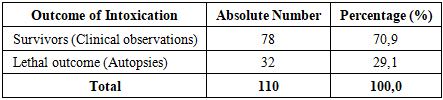
 | Figure 1. Myocardium represented by bundles of cardiomyocytes arranged unevenly, with pronounced signs of acute toxic-hypoxic damage. Stained with H&E, mag. 10x20 |
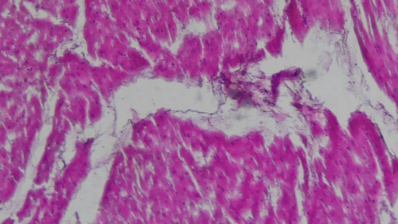
 | Figure 2. Hepatocytes are predominantly enlarged, with pale, granular cytoplasm showing pronounced signs of acute toxic dystrophy. Stained with H&E, mag. 10x20 |
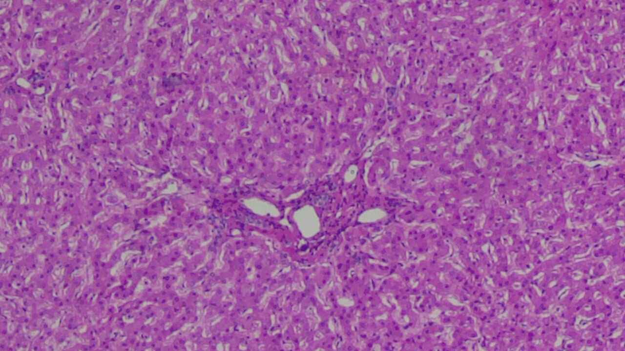
 | Figure 3. Glomeruli are generally preserved in configuration; however, some exhibit congestion of the capillary loops, and in certain areas, their moderate collapse. The lumina of the tubules are unevenly dilated and partially filled with proteinaceous masses, consistent with the pattern of acute tubular necrosis. Stained with H&E, mag. 10x20 |
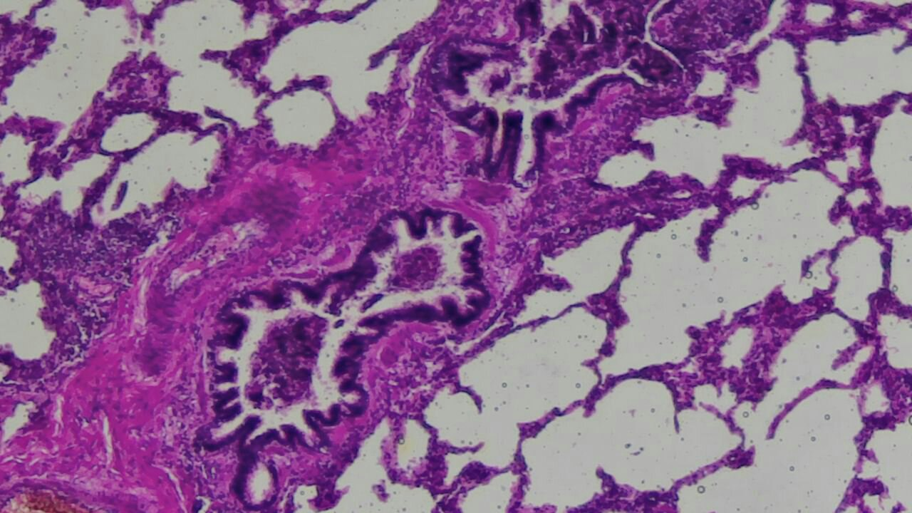 | Figure 4. Lung tissue with pronounced signs of acute toxic-hypoxic injury and microcirculatory disorders. Alveoli are unevenly dilated, in some areas sharply overdistended, with focal areas of alveolar collapse. Stained with H&E, mag. 10x20 |
4. Conclusions
- The study demonstrated that acute oral poisonings are accompanied by the development of pronounced and systemic morphological changes in the internal organs, reflecting complex toxic-hypoxic damage to the organism. The identified pathomorphological changes are consistent and result from the direct toxic action of poisons, microcirculatory disturbances, tissue hypoxia, and metabolic disorders. The findings confirm the high diagnostic value of morphological examination in the forensic medical evaluation of acute oral poisoning. A comprehensive interpretation of morphological changes in combination with toxicological and clinical laboratory data allows for a more accurate determination of the mechanism of death, objectifies expert findings, and increases the reliability of forensic medical conclusions. The results of the study can be used to improve the practice of forensic medical examination and to develop unified morphological criteria for the diagnosis of lethal poisonings.