American Journal of Medicine and Medical Sciences

p-ISSN: 2165-901X    e-ISSN: 2165-9036

2026;  16(2): 774-779

doi:10.5923/j.ajmms.20261602.80

Received: Feb. 3, 2026; Accepted: Feb. 23, 2026; Published: Feb. 27, 2026


Cognitive Disorders in Post-Traumatic Encephalopathies and Increase the Effectiveness of Treatment

Hikmatova Sh. Sh., Haydarov N. Q., Abdullaeva M. B., Sobirova S. K., Aduqodirov E. I.

Tashkent State Medical University, Tashkent, Uzbekistan

Copyright © 2026 The Author(s). Published by Scientific & Academic Publishing.

This work is licensed under the Creative Commons Attribution International License (CC BY).
http://creativecommons.org/licenses/by/4.0/

Abstract

Post-traumatic encephalopathy (PTE) is a chronic progressive condition that develops after traumatic brain injury and is characterized by cognitive, morphological, and functional impairments of the central nervous system. The present study aimed to evaluate cognitive disorders in patients with PTE and assess the effectiveness of комплекс therapy. A total of 100 patients were examined using clinical, neuropsychological (MMSE), neuroimaging (MRI), and functional (EEG) methods before and after treatment. Prior to therapy, most patients demonstrated pre-dementia cognitive impairment (MMSE 24–25 points), along with diffuse cerebral atrophy, gliosis, leukoaraiosis, and pathological EEG changes. After комплекс treatment, MMSE scores improved to 26–27 points, accompanied by stabilization of MRI findings and normalization of EEG parameters. The results confirm the interrelationship between structural, functional, and cognitive disturbances in PTE and demonstrate the clinical effectiveness of timely comprehensive therapeutic intervention.

Keywords: Post-traumatic encephalopathy, Traumatic brain injury, Cognitive impairment, MMSE, MRI, EEG, Neurodegeneration, Комплекс therapy, Cerebral atrophy

Cite this paper: Hikmatova Sh. Sh., Haydarov N. Q., Abdullaeva M. B., Sobirova S. K., Aduqodirov E. I., Cognitive Disorders in Post-Traumatic Encephalopathies and Increase the Effectiveness of Treatment, American Journal of Medicine and Medical Sciences, Vol. 16 No. 2, 2026, pp. 774-779. doi: 10.5923/j.ajmms.20261602.80.

1. Introduction

Encephalopathy is a brain injury that leads to degeneration of nerve tissue and is characterized by impaired cerebral function. [1] Pathological impairment of cerebral function occurs mainly through two mechanisms:
Ischemia occurs as a result of insufficient oxygen reaching the cells of the brain due to damage to the blood vessels of the brain. This can be caused by arterial hypertension, diabetes, atherosclerosis, thrombosis, etc. [2]
Intoxication is poisoning by products that are harmful for metabolism to brain cells. This condition often occurs as a result of long-term alcohol consumption, liver diseases, kidney diseases, etc. [3]
Encephalopathy is most common in people over 50 years of age. However, the disease is now becoming more common in younger people. It can even occur in newborns (perinatal encephalopathy), caused by hypoxia during pregnancy. [4]
Another type of encephalopathy that is currently becoming increasingly serious and has serious consequences is post-traumatic encephalopathy. Post-traumatic encephalopathy (PTE) is a group of mental and neurological disorders that include atrophic, dystrophic, and degenerative changes in the layers of the brain that develop over one or more years after a traumatic brain injury. [5] The symptoms and degree of damage to the PTE depend on the patient's age, the severity of the traumatic brain injury, the effect of medical treatment, changes in the blood vessels of the brain, and the effects of chronic and alcoholic intoxication. [10-11]
Symptoms of posttraumatic encephalopathy: memory loss, weakness, headache, dizziness, ringing in the ears, depressed mood, speech disorders, tremors, urinary disorders. [6,7]
80% of injuries in neurotraumatology are mild traumatic brain injuries, which occur in an average of 120 to 300 people per 1,000,000 population in different countries. [12-14] In addition, due to modern urbanization and the increase in the number of vehicles on city roads, traumatic brain injuries, as well as post-traumatic encephalopathy, which occurs after injuries, are becoming the most urgent problem. [15] Complications arising from various traumatic brain injuries worsen the quality of life of patients and make their social adaptation difficult. [9] The fact that traumatic brain injuries are more common, especially in young people, causes many social problems. [14-16] Therefore, timely diagnosis of such patients and improvement of treatment tactics have become an urgent task.
The purpose of the study: to evaluate the development of post-traumatic encephalopathies and to develop effective diagnostic and therapeutic measures.

2. Research Material and Methods

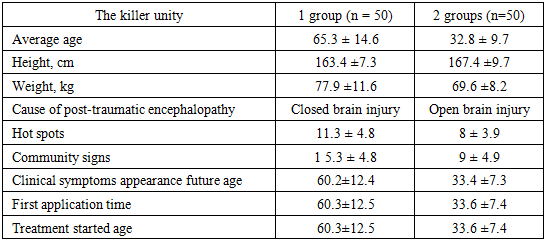
100 patients with post-traumatic encephalopathy participated in our clinical study, their average age was 23-80 years. The differences in age and gender of the patients are described in Table 1. All the patients included in our study were divided into two groups: 50 patients received conventional treatment and 50 patients received complex treatment.
Table 1. Indicators of patients by age and gender
     
Table 1 mainly Men 40%, women 60% organization to men relatively women 1.5 times how many organization will come 70% of patients big aged and elderly The patients were men. 50.8±19.05 years old on average, women and died at the average age of 55.5 ±13.03 years.
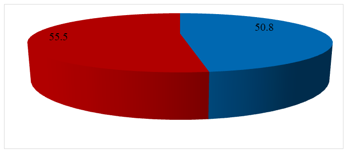
Figure 1. Average age of patients by gender
In table 2 to be described See, Taken to the information according to the study included 1st and 2nd group patients between young, clinical symptomatology weight and of the disease come exit characteristics according to noticeable differences exists.
Table 2. Clinical characteristics of research groups
     
The mean age of patients in group 1 was 65.3 ± 14.6 years, which was significantly higher than that of patients in group 2 (32.8 ± 9.7 years). This indicates that in group 1 the disease mainly manifests itself in old age and the proportion of age-related neurodegenerative and vascular changes is higher.
Analysis of anthropometric parameters showed that the average height of patients in group 1 was slightly lower (163.4 ± 7.3 cm) and higher weight (77.9 ± 11.6 kg). In group 2, the height was relatively higher (167.4 ± 9.7 cm) and lower weight (69.6 ± 8.2 kg), which is explained by the difference in age structure.
As a result of the analysis of the causes of posttraumatic encephalopathy, it was found that in group 1, closed brain injury prevails, and in group 2, open brain injury is more common. This case illustrates the differences between the mechanism of injury and its clinical consequences.
Indicators reflecting the severity of clinical symptoms also differed between groups. In particular, focal neurological signs were 11.3 ± 4.8 points in group 1, and 8.0 ± 3.9 points in group 2. Total scores were also significantly higher in group 1 (15.3 ± 4.8) compared to 9.0 ± 4.9 points in group 2. This situation indicates a wider and more severe course of the pathological process in group 1.
The analysis of the age of onset of clinical symptoms, time of first presentation, and age at initiation of treatment also revealed significant differences between the groups. In group 1, these indicators were on average around 60 years old, while in group 2 they were between 33–34 years old. This is characterized by an earlier onset of the disease in group 2 and a relatively earlier request for medical care.
All patients underwent standard clinical laboratory, neurological and instrumental examinations, including history taking, physical examination, clinical and biochemical blood tests, electroencephalography, duplex scanning of the common carotid arteries, and MRI of the brain. In addition, neuropsychological tests were performed to assess the cognitive functions of all patients, using the MMSE, modified Rankin scale, Barthel scale, as well as the Revermid index and NISS scale to assess the neurological and cognitive status of patients. In addition, serum levels of (NGF), homocysteine, VEGF (vascular endothelial growth factor), HIF1 alpha (hypoxia-induced factor) were measured. Brain biorhythms were assessed by EEG.
We studied 100 patients aged 45 to 65 years with posttraumatic encephalopathy due to open and closed head injuries, with a mean age of 59.7±5.6 years, of whom 43 were men and 32 were women. The control group consisted of 20 healthy individuals of similar age.

3. Research Results

The cognitive function of 100 patients with post-traumatic encephalopathy included in the study was assessed using the MMSE scale. According to the results, the majority of patients had MMSE scores in the range of 24–26.
The most common MMSE scores during the follow-up period were 24 and 25 points, indicating that a large proportion of patients had predementia cognitive impairment. Although scores of up to 26 points were observed in some patients, results that fully corresponded to normal cognitive function (28–30 points) were almost nonexistent.
MMSE scale to the interpretation suitable:
• 24-27 points - preliminary cognitive disruptions,
• 20-23 points - mild dementia,
• 28-30 points - cognitive violations no.
This from the criteria come out without, in the study participation did of patients in most cases cognitive violations clinical in terms of formed, however still dementia to the level insufficient situation that It was determined. This situation post-traumatic in encephalopathy cognitive functions decrease chronic and progressive to the feature owner that it is confirms.
Also, the accumulation of MMSE scores in a relatively narrow range (mainly 24–25) indicates the presence of a uniform clinical profile of cognitive deficits, namely, moderate impairments in memory, attention, calculation, and executive functions predominate. According to the results of the analysis of 100 patients with post-traumatic encephalopathy, the majority of patients had MMSE scores in the range of 24–26 points, which indicates the predominance of pre-dementia cognitive impairment. The identified changes confirm the presence of a persistent decline in cognitive functions in post-traumatic encephalopathy and justify the need for early neuropsychological diagnosis, dynamic monitoring, and cognitive rehabilitation measures in these patients.
After a course of treatment, cognitive function was reassessed in 100 patients with post-traumatic encephalopathy. The results showed that MMSE scores were mostly in the range of 25–27.
The most common values after treatment were 26 and 27 points. This indicates a positive trend compared to the pre-treatment values of around 24–25 points. Although in some patients MMSE scores remained at 25 points, in most cases an improvement in cognitive functions was noted.
MMSE scale to the interpretation suitable:
• 28-30 points - cognitive violations no,
• 24-27 points - preliminary cognitive disruptions,
• 20-23 points - mild dementia.
When analyzed based on these criteria, pre-dementia cognitive impairments remained in the majority of patients after treatment, but their severity significantly decreased. In particular, a steady trend of improvement was observed in the indicators of attention, short-term memory, calculation, and orientation functions.
The shift of MMSE scores to the upper limit (26–27 points) indicates that the treatment measures have a positive effect on cognitive functions, and the neuropsychological deficit is reversible. This confirms the effectiveness of comprehensive and timely therapy in post-traumatic encephalopathy. The results of the assessment of 100 patients with post-traumatic encephalopathy after treatment showed an increase in MMSE scale indicators to the range of 25–27 points. This confirms that as a result of treatment, cognitive functions have significantly improved, the risk of dementia has decreased, and there is a possibility of functional recovery of cognitive disorders. The results obtained substantiate the important clinical significance of the MMSE scale in assessing the effectiveness of treatment in patients with post-traumatic encephalopathy.
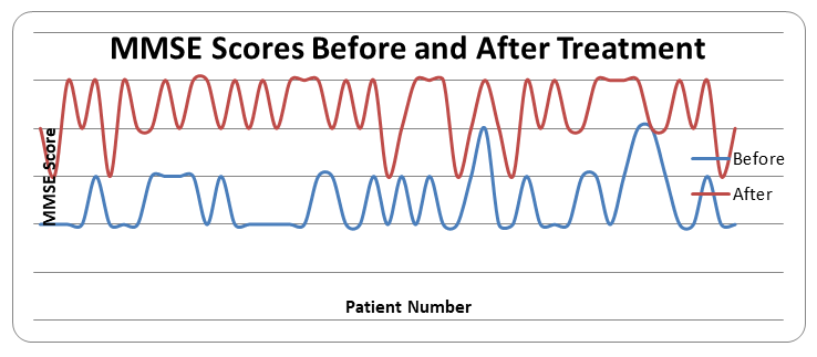
Figure 2. MMSE scores before and after treatment
Analysis of the chart data showed that the cognitive function status of patients with posttraumatic encephalopathy before treatment was mainly in the range of 24–25 points, which indicates the predominance of predementia cognitive impairment. In the pretreatment period, most patients had moderate impairments in memory, attention, calculation, and executive functions.
After the course of treatment, a significant improvement in MMSE scores was noted. As shown in the diagram, the scores after treatment mainly shifted to the range of 26–27, which confirms the stable positive dynamics of cognitive functions. In particular, in the majority of patients, an increase in MMSE scores by 1–3 points was observed, which is a clinically significant change.
At the same time, even after treatment, MMSE scores remained at 25 points in a certain part of patients, which indicates the chronic nature of cognitive impairment in post-traumatic encephalopathy. However, the analysis of the overall dynamics showed that the tendency for regression and stabilization prevailed, rather than worsening cognitive deficits.
A visually clear representation of the difference between the indicators before and after the treatment in the diagram proves that the used therapy methods had a positive effect on neurocognitive functions.
All patients in our study underwent brain MRI before and after treatment.
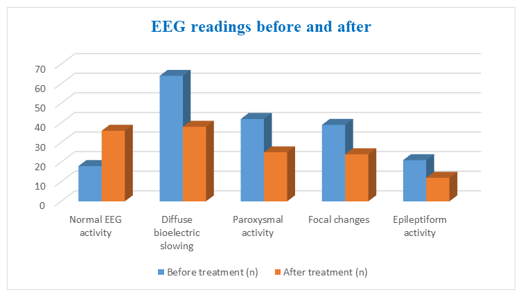
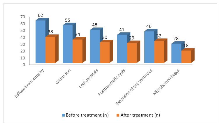
Figure 3. MRI findings before and after treatment
The data in the diagram reflect the pre- and post-treatment MRI brain parameters in 100 patients with post-traumatic encephalopathy. According to the results of the analysis, all assessed morphological changes were present with a high frequency before treatment, which indicates that post-traumatic damage has a chronic and multifactorial pathogenesis.
The most common MRI change was diffuse cerebral atrophy, which was detected in 62 patients before treatment. After the course of treatment, this figure decreased to 38, which indicates a slowdown in the progression of neurodegenerative processes in the brain tissue. Analysis of the diagram shows that after complex treatment in patients with post-traumatic encephalopathy, a tendency to decrease and stabilize was observed in most of the changes in the brain MRI. The results obtained confirm the slowdown in the progression of neurodegenerative and post-traumatic morphological changes and justify the important diagnostic value of brain MRI in assessing the effectiveness of treatment.
Foci of gliosis were observed in 55 patients before treatment, and in 34 patients after treatment. This is explained by partial regression or stabilization of post-traumatic inflammation and ischemic changes.
The leukoaraiosis index was also characterized by positive dynamics, detected in 48 patients before treatment and in 30 patients after treatment. This indicates a decrease in microcirculatory disorders in the white matter of the brain.
Structural changes such as post-traumatic cysts and ventricular dilation also showed a tendency to decrease after treatment. In particular, post-traumatic cysts decreased from 41 to 29 patients, and ventricular dilation from 46 to 32 patients. Microhemorrhages were the least common change, recorded in 28 patients before treatment. After treatment, this indicator decreased to 18 patients, indicating stabilization of the vascular walls and improvement of microcirculation. In general, the diagram clearly shows that the pre-treatment indicators are superior and that all parameters decrease after treatment, which clearly indicates a positive effect of therapy on neuroimaging results.
All our patients underwent EEG before and after treatment.
Figure 4. EEG readings before and after treatment
The diagram data reflect the electroencephalography (EEG) parameters of the brain in 100 patients with post-traumatic encephalopathy before and after treatment. According to the results of the analysis, pathological EEG changes prevailed in the majority of patients before treatment, which indicates a significant deterioration in the functional state of the brain.
Before treatment, diffuse bioelectric slowing was the most common EEG change, recorded in 64 patients. After the course of treatment, this indicator was found to have decreased to 38, indicating stabilization of brain excitability and neuronal activity.
Paroxysmal activity was observed in 42 patients before treatment and in 25 patients after treatment. This situation indicates a decrease in the frequency of paroxysmal reactions and a decrease in the risk of epileptogenic activity.
Focal changes also showed a significant reduction trend after treatment: this indicator, which was detected in 39 patients before treatment, was observed in 24 patients after treatment. This situation indicates that functional recovery processes are activated against the background of local brain injuries.
Epileptiform activity is the least common but clinically significant EEG change. It was observed in 21 patients before treatment and in 12 patients after treatment, indicating a decrease in the susceptibility to epilepsy.
At the same time, the proportion of patients with normal EEG activity increased from 18 before treatment to 36 after treatment. This confirms the restoration of brain bioelectric activity and the high functional effectiveness of the applied therapy. Analysis of the diagram shows that after complex treatment in patients with post-traumatic encephalopathy, stable positive dynamics of brain EEG parameters were observed. The decrease in pathological bioelectric changes and an increase in the proportion of normal EEG activity indicate an improvement in the functional state of the brain. The results obtained justify the fact that the electroencephalography method is an important functional diagnostic tool in assessing the effectiveness of treatment in post-traumatic encephalopathy.
In order to comprehensively assess the morphological, functional, and cognitive state of the central nervous system in patients with posttraumatic encephalopathy, brain MRI, electroencephalography (EEG), and MMSE scores were analyzed before and after treatment.
Analysis of MRI data showed that before treatment, a large proportion of patients had a high frequency of morphological changes such as diffuse brain atrophy, foci of gliosis, leukoaraiosis, post-traumatic cysts, and ventricular enlargement. After the course of treatment, most of these changes showed a tendency to decrease and stabilize, which indicates a slowdown in the progression of neurodegenerative processes.
Analysis of EEG indicators showed that diffuse slowing of brain bioelectric activity, paroxysmal activity, focal changes and epileptiform activity predominated before treatment. After treatment, a significant decrease in the frequency of pathological EEG changes and an increase in the percentage of normal bioelectrical activity were noted. This situation confirms the improvement of the functional state of the brain and the stabilization of the activity of neuronal networks.
The results of the MMSE scale assessment showed that most patients had pre-dementia cognitive impairment (24–25 points) before treatment. After the treatment course, an increase in MMSE scores to 26–27 points was noted, indicating an improvement in cognitive functions, in particular, memory, attention, calculation, and executive functions.
Integral analysis showed that the reduction and stabilization of morphological changes on MRI were closely related to the normalization of bioelectrical activity on EEG and an increase in MMSE indicators. That is, the regression of pathological changes in brain structures occurred simultaneously with functional recovery and improvement of cognitive status.
These data show that morphological, functional and cognitive impairments in posttraumatic encephalopathy are interconnected and that complex treatment has a positive effect on these systems at the same time.

4. Conclusions

The development of SBE in patients with SCD significantly worsens the quality of life of patients. Patients develop SBE, which causes complications in many other systems. Post-traumatic encephalopathy is also characterized by complications involving the nervous system, including the peripheral nervous system. Neurological dysfunctions observed in post-traumatic encephalopathy lead to a sharp decrease in the quality of life of patients.
Seizure syndrome lasting more than three months, a significant increase in cerebral hypertension during physical activity was observed.
Deep reflex assessment revealed asymmetric reduction of various reflexes, and in some patients complete loss of reflexes was observed. It is noteworthy that in a large proportion of patients, seizures and cognitive impairment were detected and corrected.
Overall, these results highlight the complexity of neurologic manifestations and severe clinical manifestations in patients with posttraumatic encephalopathy, emphasizing the importance of comprehensive evaluation and management strategies to address cerebral complications in this patient population.

References

[1]  Bazarian, J. J., Blyth, B., & Cimpello, L. (2006). Bench to bedside: Evidence for brain injury after concussion. The Lancet Neurology, 5(1), 67–75.
[2]  Matmurodov, R., Abduqodirov, E., Kalanov, A., Babayeva, F., & Jamalova, S. (2025). Myofascial pain syndromes in parkinsonism. Movement Disorders, 4(S977), 156.
[3]  Matmurodov, R., Jumanazarova, S., Abduqodirov, E., & Abdullayeva, M. (2025). Analysis of statistical data on patients with Parkinson’s disease. Movement Disorders (MDS Journal), 4(1), S801.
[4]  Gardner, R. C., & Yaffe, K. (2015). Epidemiology of mild traumatic brain injury and neurodegenerative disease. Alzheimer’s Research & Therapy, 7(1), 30.
[5]  Graham, D. I., & Sharp, D. J. (2019). Understanding neurodegeneration after traumatic brain injury. Brain, 142(4), 881–882.
[6]  Iverson, G. L. (2005). Outcome from mild traumatic brain injury. Current Opinion in Psychiatry, 18(3), 301–317.
[7]  Johnson, V. E., Stewart, W., & Smith, D. H. (2013). Axonal pathology in traumatic brain injury. Experimental Neurology, 246, 35–43.
[8]  Levin, H. S., Diaz-Arrastia, R. R., & Shum, D. (2014). Cognitive rehabilitation in traumatic brain injury. The Lancet Neurology, 13(3), 315–326.
[9]  Maas, A. I. R., Stocchetti, N., & Bullock, R. (2008). Moderate and severe traumatic brain injury in adults. The Lancet Neurology, 7(8), 728–741.
[10]  McInnes, K., Friesen, C. L., MacKenzie, D. E., Westwood, D. A., & Boe, S. G. (2017). Mild traumatic brain injury (mTBI) and chronic cognitive impairment: A scoping review. PLoS ONE, 12(4), e0174847.
[11]  Stuss, D. T., & Levine, B. (2002). Adult clinical neuropsychology: Lessons from studies of the frontal lobes. Annual Review of Psychology, 53, 401–433.
[12]  Wilson, B. A., Gracey, F., Evans, J. J., & Bateman, A. (2009). Neuropsychological rehabilitation: Theory, models, therapy and outcome. Cambridge University Press.
[13]  Abduqodirov, E., Matmurodov, R., Muminov, B., Daminova, H., Khalimova, K., Naimov, O., & Juraev, R. (2023). Aspects of early detection of Parkinson’s disease in ambulatory settings. Movement Disorders, 38(478), 203.
[14]  Abduqodirov, E., Matmurodov, R., Juraev, R., Naimov, O., Muminov, B., Khalimova, K., & Daminova, H. (2023). Glial neurotrophiz factor as a differential marker of Parkinson’s disease and vascular parkinsonism. Movement Disorders, 38(289), 120.
[15]  Abduqodirov, E., Muminov, B., Matmurodov, R., & Khalimova, K. (2023). Extent of cognitive impairment in Parkinson’s disease with COVID-19. Movement Disorders, 38(332), 141.
[16]  Abdukodirov, E. I., Naimov, O. Y., Matmurodov, R. J., Khalimova, H. M., & Muminov, B. A. (2023). Dynamic assessment of levels of depression and anxiety impairment in Parkinson's disease. Movement Disorders, 38.