-
Paper Information
- Next Paper
- Previous Paper
- Paper Submission
-
Journal Information
- About This Journal
- Editorial Board
- Current Issue
- Archive
- Author Guidelines
- Contact Us
American Journal of Medicine and Medical Sciences
p-ISSN: 2165-901X e-ISSN: 2165-9036
2026; 16(2): 767-770
doi:10.5923/j.ajmms.20261602.78
Received: Jan. 24, 2026; Accepted: Feb. 17, 2026; Published: Feb. 26, 2026

Mineral Status of Blood in Patients with Computer Visual Syndrome Depending on Their Severity
 Abstract
 Reference
 Full-Text PDF
 Full-text HTML
Mamatkhujaev Minkhojiddin Sadirdinkhoji ugli1, Karimova Muyassar Khamitovna2, Mamatkhujaeva Gulnarahan Najmidinovna1
1Andijan State Medical Institute, Andijan, Uzbekistan
2Republican Specialized Scientific and Practical Medical Center for Eye Microsurgery, Tashkent, Uzbekistan
Copyright © 2026 The Author(s). Published by Scientific & Academic Publishing.
This work is licensed under the Creative Commons Attribution International License (CC BY).
http://creativecommons.org/licenses/by/4.0/

The article presents an assessment of the state of blood mineral status and an analysis of its relationship with the severity of computer vision syndrome (CVS) in computer users. 160 patients with clinical manifestations of CVS and 40 healthy individuals from the control group were examined. Depending on the severity of the syndrome, patients were divided into subclinical, mild, moderate, and severe CVS groups. The severity of symptoms was assessed using the author's questionnaire CVS-44, comprehensive ophthalmological examination, and the author's clinical and functional classification of COPS. The concentrations of zinc, selenium, copper, iron, magnesium, calcium, and phosphorus were determined in blood serum. It has been established that the progression of CVS is accompanied by a dose-dependent decrease in the levels of zinc, selenium, iron, magnesium, and calcium against a background of a relative increase in copper and phosphorus. In moderate and severe CVS, a number of indicators reached threshold or subnormal values. Significant correlations have been identified between the severity of CVS and blood mineral status indicators, which confirms their pathogenetic significance and the expediency of considering them in the diagnosis and prevention of CVS.
Keywords: Computer vision syndrome, Mineral status, Micro and macro elements, Computer users
Cite this paper: Mamatkhujaev Minkhojiddin Sadirdinkhoji ugli, Karimova Muyassar Khamitovna, Mamatkhujaeva Gulnarahan Najmidinovna, Mineral Status of Blood in Patients with Computer Visual Syndrome Depending on Their Severity, American Journal of Medicine and Medical Sciences, Vol. 16 No. 2, 2026, pp. 767-770. doi: 10.5923/j.ajmms.20261602.78.
1. Introduction
- In the context of global digitalization and the widespread introduction of information technologies, computer vision syndrome (CVS) is considered one of the most common forms of functional visual pathology in people of working age. According to epidemiological studies, CVS symptoms are detected in 60-90% of digital device users, while the severity of clinical manifestations directly correlates with the duration and intensity of visual load [1,5].According to modern concepts, CVS is a multifactorial condition, the pathogenesis of which includes accommodation disorders, tear film instability, decreased blinking frequency, development of asthenopic disorders, and autonomic nervous system dysfunction [3]. In recent years, special attention has been paid to the role of systemic metabolic mechanisms capable of reducing the adaptive capabilities of the visual analyzer during chronic visual strain [4].Microelements play a key role in ensuring antioxidant protection, energy metabolism, neuromuscular transmission, and the regulation of vascular tone. Zinc and selenium participate in the functioning of antioxidant system enzymes, magnesium and calcium in the regulation of neuromuscular excitability and accommodation processes, iron in tissue respiration, and copper in oxidative stress and inflammation reactions [2]. Disruption of microelement homeostasis can contribute to increased oxidative stress, vegetative dysfunctions, and functional decompensation of the visual system.Despite the existence of studies dedicated to the role of microelements in various ophthalmological and neurosensory disorders, data on the nature of changes in the blood's microelement status in patients with CVS, depending on the severity of the disease, remain limited and contradictory [6]. The lack of systematized information on this issue makes it difficult to develop differentiated diagnostic and preventive approaches.In this regard, studying the microelement status of the blood in patients with computer vision syndrome, taking into account the severity of clinical manifestations, is a pressing scientific and practical task aimed at deepening understanding of the pathogenesis of computer vision syndrome and substantiating comprehensive therapeutic and preventive measures.Purpose of the study: The purpose of this study was to assess the state of blood microelement status and to identify its relationship with the severity of computer vision syndrome in computer users.
2. Material and Methods
- The object of the study was 160 computer users with clinical signs of computer vision syndrome. The control group consisted of 40 healthy individuals comparable in gender and age, regularly using computers, but without clinical manifestations of CVS.The research protocol was in accordance with the principles of the Helsinki Declaration, and all the examined individuals signed informed voluntary consent to participate in the study.The severity of computer vision syndrome symptoms was assessed using the author's COVS-44 questionnaire, the results of which, in combination with ophthalmological examination data, were used to distribute patients according to the degrees of CVS according to the developed author's clinical and functional classification.Depending on the severity of CVS, the examined patients were divided into four clinical groups (each with 40 people): subclinical CVS, mild, moderate, and severe CVS.Ophthalmological examination included visometry, refractometry, biomicroscopy of the anterior segment of the eye, ophthalmoscopy, as well as assessment of accommodative and binocular functions, the state of tear production, and the stability of the tear film.To assess the microelement status, a biochemical study of blood serum was conducted to determine the concentrations of zinc, selenium, copper, iron, magnesium, calcium, and phosphorus. Venous blood collection was performed in the morning on an empty stomach. The analyses were conducted using standard certified laboratory methods.Statistical processing of the data was carried out using methods of variation statistics. The results are presented as the mean and the error of the mean (M ± m). For intergroup comparisons, Student's criterion was used for independent samples, and correlation analysis was performed using Pearson and Spearman coefficients. Differences at p < 0.05 were considered statistically significant.
3. The Results and Discussion
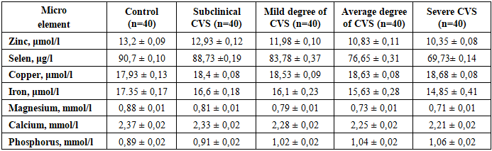
- Analysis of mineral element concentrations in blood serum revealed a clear correlation between impaired mineral homeostasis and an increase in the severity of computer vision syndrome (CVS). In the control group, all the studied indicators were within the reference values. As CVS progressed from the subclinical form to the severe stage, statistically significant and unidirectional changes in most of the studied elements were noted (Table 1).
|
4. Conclusions
- The conducted research showed that in computer vision syndrome, changes occur in the mineral composition of the blood, the severity of which increases with the severity of the disease. Even in the early stages of CVS, deviations from the control values are noted, and in moderate and severe cases, they become most pronounced.For zinc, selenium, magnesium, iron, and calcium, a gradual decrease in concentrations is characteristic, while the levels of copper and phosphorus, on the contrary, increase. The most noticeable changes were identified for selenium and zinc, which allows us to consider them as the most sensitive indicators related to the degree of CVS.Overall, computer vision syndrome is accompanied by the formation of a systemic mineral imbalance, which confirms the presence of a metabolic component in the development and progression of the disease. Considering the mineral status of the blood can supplement the clinical assessment of the severity of computer vision syndrome.