American Journal of Medicine and Medical Sciences

p-ISSN: 2165-901X    e-ISSN: 2165-9036

2026;  16(2): 719-727

doi:10.5923/j.ajmms.20261602.69

Received: Jan. 2, 2026; Accepted: Jan. 22, 2026; Published: Feb. 26, 2026


Erosive and Ulcerative Gastroduodenal Bleeding in Patients with Comorbid Cardiovascular Pathology

Rasul I. Rakhimov1, Sherzod Sh. Zhanibekov2, Bobir I. Shukurov2, 3, Karimali S. Elmurodov3

1City Clinical Hospital No. 7, Tashkent, Uzbekistan

2Tashkent State Medical University, Tashkent, Uzbekistan

3Republican Research Center of Emergency Medicine, Tashkent, Uzbekistan

Correspondence to: Bobir I. Shukurov, Tashkent State Medical University, Tashkent, Uzbekistan.

Email:

Copyright © 2026 The Author(s). Published by Scientific & Academic Publishing.

This work is licensed under the Creative Commons Attribution International License (CC BY).
http://creativecommons.org/licenses/by/4.0/

Abstract

This review is devoted to the problem of erosive and ulcerative gastroduodenal bleeding (EUGDB) in patients with comorbid cardiovascular pathology. EUGDB remains one of the most significant causes of emergency hospitalization, accounting for up to 90% of all gastrointestinal bleeding cases and being characterized by high recurrence rates and mortality. Particular attention is paid to the relationship between cardiovascular diseases, polypharmacy, and age-related changes with the risk of ulcerative lesions and their complications. Data are presented on the influence of antiplatelet agents, anticoagulants, nonsteroidal anti-inflammatory drugs, and Helicobacter pylori infection on the pathogenesis of EUGDB. Current evidence is summarized regarding the development of bleeding in patients with ischemic heart disease (IHD), chronic heart failure (CHF), and acute ischemic stroke (AIS), as well as their impact on prognosis and treatment outcomes. The importance of a multidisciplinary approach to diagnosis and therapy is emphasized, including prophylactic use of proton pump inhibitors, H. pylori eradication, and individualized selection of anticoagulant therapy. Special attention is given to new anticoagulants, particularly factor XI inhibitors, which demonstrate a promising efficacy and safety profile.

Keywords: Erosive and ulcerative gastroduodenal bleeding, Cardiovascular diseases, Comorbidity, Anticoagulant therapy, Helicobacter pylori, Ischemic heart disease, Acute ischemic stroke

Cite this paper: Rasul I. Rakhimov, Sherzod Sh. Zhanibekov, Bobir I. Shukurov, Karimali S. Elmurodov, Erosive and Ulcerative Gastroduodenal Bleeding in Patients with Comorbid Cardiovascular Pathology, American Journal of Medicine and Medical Sciences, Vol. 16 No. 2, 2026, pp. 719-727. doi: 10.5923/j.ajmms.20261602.69.

1. Introduction

Erosive and ulcerative gastroduodenal bleeding (EUGDB) remains one of the most serious complications of many gastrointestinal disorders, accounting for 80–90% of all gastrointestinal bleeding (GIB) cases [1]. These bleedings are characterized by sudden onset, rapid progression, and high mortality rates [2]. In the United States, more than 320,000 patients are hospitalized annually due to EUGDB, with mortality rates ranging from 2% to 15%, and the economic burden associated with this type of bleeding exceeding 2.5 billion USD [3,4,5]. In Europe, between 48 and 147 individuals per 100,000 population are admitted to hospitals with gastroduodenal hemorrhage. Mortality directly attributed to bleeding or decompensation of comorbid conditions may reach 10% [6]. Despite substantial advances in surgery and anesthesiology, mortality from acute gastrointestinal bleeding remains at 5–14%, rising to 30–40% in cases of rebleeding, with no clear trend toward improvement [7]. Among patients with EUGDB, death directly related to blood loss occurs in approximately 18%, whereas the remaining fatal outcomes are associated with complications of underlying comorbidities [8].
In recent years, the rising incidence of erosive and ulcerative gastroduodenal bleeding (EUGDB) in patients with cardiovascular diseases and other somatic disorders has acquired an epidemic character. According to various authors, the frequency of acute erosive and ulcerative lesions of the gastric and duodenal mucosa (GDM) in patients with therapeutic pathology reaches 80–90%, while EUGDB attributable to these lesions accounts for 45–55% of cases [9]. For example, analysis of 6,900 autopsies of patients who died from myocardial infarction and stroke in multidisciplinary hospitals demonstrated a nearly one-third increase in the detection rate of erosive and ulcerative GDM lesions over the past decade. Among patients with cardiovascular diseases, acute ulcers and erosions constitute 60–70% of all GDM mucosal lesions, whereas chronic peptic ulcers are diagnosed in 30–40% of cases. Acute ulcers predominate in individuals with acute cerebrovascular accidents and myocardial infarction, which is associated with stress-induced hypersecretory responses and impaired mucosal microcirculation, whereas chronic ulcers are more commonly observed in long-standing ischemic heart disease and prolonged use of antiplatelet agents [10].
EUGDB of varying severity developed in 67% of deceased patients with ischemic heart disease (IHD) and in 55% of those with stroke; however, their contribution to mechanisms of fatal outcome differed. The incidence of fatal bleeding (EUGDB as the direct cause of death) was 3% in myocardial infarction, 2% in chronic forms of IHD accompanied by chronic heart failure (CHF), and 1% in stroke. At the same time, the frequency of acute erosive and ulcerative lesions of the GDM in deceased patients with myocardial infarction reaches 18%, in CHF – 10%, in stroke – 14%, and may rise to 21% when IHD is combined with cerebrovascular disease [11].
The challenge of timely diagnosis and treatment of erosive and ulcerative GDM lesions – and consequently, prevention of EUGDB – is exacerbated by the fact that in 30–90% of patients with chronic somatic pathology, gastropathies are asymptomatic; in 46–58% of cases, typical clinical manifestations are absent; and in 25–42% of patients, symptoms of the underlying disease predominate. As a result, mucosal lesions of the GDM often remain undiagnosed until the development of life-threatening bleeding. In this patient population, EUGDB is the first clinical manifestation of gastroduodenal mucosal injury in 25–55% of cases, while in most instances this severe complication is identified for the first time at autopsy [8].
On the other hand, several risk factors are becoming increasingly prevalent. Population aging has led to a higher incidence of cardiovascular diseases and other comorbid conditions, thereby increasing mortality risk in patients with EUGDB. In addition, the growing use of low-dose aspirin, NSAIDs, antiplatelet agents, and anticoagulants further contributes to this risk [12,13]. In this context, it is important to clarify that within the framework of this review, the term “cardiovascular diseases” encompasses both chronic forms of ischemic heart disease (stable angina, chronic heart failure) and acute vascular events – acute myocardial infarction and AIS. These conditions share common pathogenetic mechanisms, the use of antiplatelet and anticoagulant therapy, as well as similar risk factors for the development of erosive and ulcerative gastroduodenal lesions and bleeding.

2. The Relationship Between Comorbid Cardiovascular Pathology and Erosive–Ulcerative Gastroduodenal Bleeding

The term “comorbidity” was first introduced in 1970 by the American epidemiologist A.R. Feinstein, who defined it as “any distinct additional clinical entity that has existed, exists, or may occur during the clinical course of a patient’s index disease” [14,15]. Comorbidity refers to the presence of two or more chronic diseases in a single patient, which may be etiopathogenetically interconnected or may simply coexist temporally, regardless of the level of activity of each condition. This concept implies that comorbid states arise due to shared or unified pathogenetic mechanisms.
A comprehensive understanding of the relationship between comorbid pathology and EUGDB is a crucial component in improving outcomes of this severe urgent condition. It is well established that various chronic diseases significantly increase the risk of gastrointestinal bleeding, particularly EUGDB. For example, conditions such as liver cirrhosis and peptic ulcer disease show a strong association with the risk of gastrointestinal hemorrhage [16]. In addition, the elderly population faces additional risks due to factors such as polypharmacy and the long-term management of chronic diseases, especially when over-the-counter medications such as NSAIDs are used, which can exacerbate gastrointestinal complications [17].
NSAID-associated ulcer bleeding accounts for more than 20,000 deaths annually in the United Kingdom and over 16,500 deaths in the United States [23]. Nevertheless, many patients take antiplatelet agents for years without pronounced adverse effects. Large cohort studies – Biobank (n=213,598) and ESTHER (n=7,737) – have confirmed that low-dose aspirin is an independent risk factor for peptic ulcer disease. Use of aspirin for less than one year was associated with a higher risk of ulcer formation compared with long-term use (more than one year); however, even during prolonged administration, aspirin maintains a statistically significant association with peptic ulcers [19].
The combination of aspirin with proton pump inhibitors (PPIs) significantly reduces the risk of peptic ulcer disease and its complications. Moreover, this approach improves treatment tolerability and enhances patient adherence. In clinical practice, when IHD coexists with peptic ulcer disease, clopidogrel should be preferred as an antiplatelet agent due to its lower rate of gastrotoxic complications compared with aspirin. When dual antiplatelet therapy (aspirin + clopidogrel) is required, concomitant use of PPIs (omeprazole, esomeprazole) is mandatory. The use of ticlopidine and ticagrelor requires caution in patients with active ulcer disease, particularly in the presence of H. pylori infection, which should be eradicated prior to initiating therapy. The ESC (2020) and ACC/AHA (2019) guidelines emphasize that the choice of antiplatelet agent should take into account the risk of gastrointestinal bleeding, and in patients at high risk, clopidogrel monotherapy with PPI protection is preferred [18].
Antiplatelet agents are not the only cause of ulcer formation and gastrointestinal bleeding. In a study involving 1,000 patients with stable IHD, erosive and ulcerative lesions of the gastric mucosa were observed even during PPI therapy [19]. Possible contributing factors include reduced mucosal blood supply due to atherosclerosis, adverse effects of PPIs, and H. pylori infection. According to REGATA-1 data, H. pylori infection was identified in 87.5% of patients with severe gastrointestinal bleeding [20].
The impact of H. pylori on the pathogenesis of IHD remains a subject of ongoing investigation. Patients with H. pylori infection were more frequently diagnosed with coronary stenosis (7.6% vs. 2.9%, p=0.01) [18]. Proposed mechanisms of this association include systemic inflammation, vascular endothelial injury, oxidative stress, dyslipidemia, and enhanced platelet aggregation [21,22,24]. H. pylori eradication therapy has been shown to reduce the frequency of angina episodes and improve exercise tolerance [18,22].
Patients with IHD who are receiving antiplatelet therapy should undergo regular screening for H. pylori infection followed by eradication, if detected. Long-term PPI use is recommended to reduce the likelihood of ulcer formation and bleeding [19,20].
Analysis of the pathogenetic role of comorbid conditions allows for more accurate identification of high-risk groups among patients with EUGDB. This, in turn, enables the implementation of preventive strategies such as the use of proton pump inhibitors, correction of metabolic disturbances, and rational prescribing of medications, taking into account their adverse effects on the gastrointestinal mucosa (Table 1).
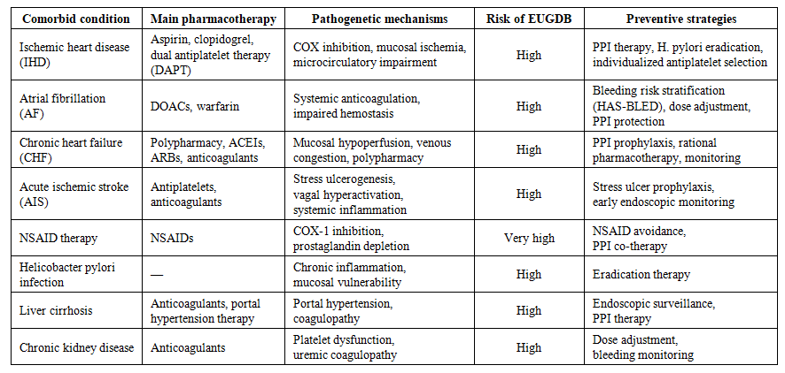
Table 1. Comorbid cardiovascular and systemic conditions, pharmacological therapy, and risk of erosive and ulcerative gastroduodenal bleeding (EUGDB)
Peptic ulcer disease is one of the most common gastrointestinal disorders, affecting 11–14% of men and 8–11% of women during their lifetime [23]. Studies demonstrate a clear association between peptic ulcer disease and IHD. A case–control analysis confirmed an increased risk of ulcer development in patients with IHD [24]. Both conditions share common risk factors, including smoking, unbalanced nutrition, and other unfavorable influences, which further contribute to their progression.
The development of peptic ulcers in patients with IHD is often associated with the use of antiplatelet agents, which constitute a standard component of cardiovascular therapy. Long-term aspirin use, as demonstrated in multiple studies, increases the likelihood of upper gastrointestinal complications by 2–4 times [24,25,26]. However, the actual number of such cases is even higher, as up to two-thirds of endoscopically confirmed ulcerative lesions are asymptomatic [27].
Assessing bleeding risk in patients with cardiovascular disease presents a significant challenge due to the complex clinical picture and the multifactorial nature of cardiac pathology. These patients – particularly those who develop heart failure (HF) – frequently have concomitant conditions such as renal dysfunction, hepatic impairment, and frailty, each of which independently increases bleeding risk [28]. Moreover, the use of multiple medications, including anticoagulants, antiplatelet agents, and heart failure therapies such as angiotensin-converting enzyme inhibitors (ACEIs) or angiotensin receptor blockers (ARBs), further complicates risk assessment by increasing the likelihood of drug interactions and adverse effects [29]. To optimize clinical decision-making during anticoagulant therapy, several bleeding risk assessment tools have been developed, the most widely used of which is the HAS-BLED score (Hypertension, Abnormal renal/liver function, Stroke, Bleeding history or predisposition, Labile INR, Elderly, Drugs/alcohol) [30].
Several clinical trials and observational studies have evaluated the efficacy and safety of anticoagulants in patients with IHD. The WARCEF trial (Warfarin Versus Aspirin in Reduced Heart Ejection Fraction) compared warfarin and aspirin in patients with IHD, reduced ejection fraction (HFrEF), and sinus rhythm [31]. Although warfarin did not demonstrate a significant reduction in the primary outcome of ischemic stroke, it was associated with a lower rate of the composite outcome of ischemic stroke or all-cause mortality compared with aspirin. However, the use of warfarin was accompanied by a higher risk of major bleeding, underscoring the need for careful bleeding risk assessment before initiating anticoagulant therapy in patients with IHD.
In patients with atrial fibrillation (AF), the ARISTOTLE trial (Apixaban for Reduction in Stroke and Other Thromboembolic Events in Atrial Fibrillation) confirmed the efficacy and safety of the direct oral anticoagulant (DOAC) apixaban compared with warfarin in reducing the risk of stroke or systemic embolism [32]. Subgroup analyses of AF patients demonstrated consistent advantages of apixaban in preventing stroke while simultaneously lowering the risk of major bleeding, supporting the use of DOACs as the preferred anticoagulants in this population.
Balancing the risks of thromboembolism and bleeding poses a significant challenge in the management of patients with IHD. Decisions regarding the initiation of anticoagulant therapy in such patients require an individualized approach that takes into account both thrombosis and bleeding risks, as well as patient preferences [33]. Anticoagulant therapy effectively reduces the risk of thromboembolic events and is recommended for selected categories of patients with IHD in accordance with current clinical guidelines. However, its benefits must be weighed against the risk of bleeding, which may lead to serious consequences, including worsening of coronary and heart failure symptoms, the need for hospitalization, and increased mortality [34,35].
GIB in patients with AIS is of particular clinical interest. In a retrospective study of 6,853 AIS patients conducted in Canada, O’Donnell et al. reported GIB in 1.5% of hospitalized individuals [36]. In contrast, a study by Hsu et al. involving patients of Asian origin demonstrated that the incidence of GIB in AIS reached 7.8% [37]. Similar findings were presented by Rumalla and Mittal [38], who noted that patients of Asian descent remained at a higher risk of developing GIB after AIS even after adjusting for comorbid factors. This may be attributed to the higher prevalence of H. pylori infection in Asian populations [36]. An interesting observation was made by Chen et al. [39], who reported that gastroduodenal ulcers developed in 44% of all patients admitted to neurological intensive care units with a diagnosis of AIS.
GIB most frequently develops within the first week after the onset of AIS [40]. The source of bleeding may originate from either the upper or lower gastrointestinal tract. In a study by Ogata et al. involving Japanese patients with AIS, upper gastrointestinal bleeding accounted for 50.4% of cases. The most common causes were peptic ulcer disease, malignant neoplasms, reflux esophagitis, Mallory–Weiss syndrome, and esophageal varices. In 25.8% of cases, the bleeding originated from the lower gastrointestinal tract, including pseudomembranous colitis, ischemic colitis, angiodysplasia, polyps, and diverticula. However, in 23.6% of patients, endoscopic and colonoscopic evaluation failed to identify a definitive bleeding source [40].
Several pathophysiological mechanisms have been proposed to explain the association between GIB and AIS. Studies have shown that stroke induces hyperactivation of the vagus nerve, leading to increased gastric acid and pepsin secretion, mucosal injury, and the development of stress ulcers [37,41]. The review by Camara-Lemarroy et al. [42] emphasizes the significant contribution of systemic inflammation, oxidative stress, nitric oxide pathway inhibition, and the use of antiplatelet agents in ulcerogenesis, thereby increasing the risk of post-stroke GIB. Similar mechanisms were described by Fu [43], who noted activation of noradrenergic neurons and impaired central nervous system regulation of gastrointestinal blood flow, contributing to mucosal injury. Collectively, these mechanisms explain the heightened risk of GIB in severe ischemic strokes.
The autonomic nervous system pathway, which runs from the hypothalamus through the midbrain to the spinal cord, may explain the connection between ischemia of the posterior circulation and GIB [44]. Schaller et al. proposed that the release of catecholamines after AIS causes vasospasm, leading to ischemia and impaired trophism of the gastric mucosa, and consequently, GIB [45]. Izumiyama and Kogure also identified reduced gastric mucosal blood flow in patients with ischemic stroke, which may contribute to ulcer formation [46].
Patients with ischemic stroke who developed GIB were predominantly older [36,40,43,44,47] and male [44], and were also more likely to have impaired daily activities before the onset of bleeding [40]. It was found that a history of peptic ulcer disease significantly increases the risk of GIB in the context of AIS. Notably, gastroduodenal bleeding occurred even in patients with a history of ulcers who were on prophylactic proton pump inhibitor (PPI) therapy [40]. Moreover, GIB more frequently developed in patients with a high National Institutes of Health Stroke Scale (NIHSS) score upon admission, indicating a correlation between stroke severity, the extent of neurological deficit, and an increased risk of hemorrhagic complications [40,43]. Hypertension and previous episodes of GIB were also associated with an increased risk of recurrence [44].
Fu [43] identified additional independent risk factors for GIB in patients with acute cerebral infarction, including a low Glasgow Coma Scale (GCS) score, presence of infection, and infarction within the posterior circulation [48]. Among infections associated with GIB, the most common were pneumonia and urinary tract infections (UTIs) [49]. Patients with stroke who developed GIB also demonstrated leukocytosis, a higher frequency of carotid artery stenosis, and elevated fasting glucose levels compared with those without GIB [43]. Ischemia in the middle cerebral artery territory and anterior circulation infarction were likewise associated with an increased risk of GIB, whereas partial anterior circulation infarct (PACI) showed no such association [44]. A history of malignancy and coagulopathies is also considered a significant risk factor for the development of GIB in AIS patients [36,40,44]. Furthermore, atrial fibrillation was markedly more common in patients with GIB compared with those without bleeding [36,37,38,40,43]. Additional factors – such as cardioembolic stroke, concomitant ischemic heart disease, prior stroke, and a family history of stroke – were also more frequently observed in patients with GIB relative to those without hemorrhagic complications [40,49].
One explanation for the higher prevalence of atrial fibrillation, carotid artery stenosis, and cardioembolic stroke in patients with GIB is that these factors are commonly associated with more severe strokes, which in turn increase the risk of GIB [50]. Similarly, impaired consciousness, reflected by a low Glasgow Coma Scale (GCS) score, also indicates severe stroke [37,43].
Infections, sepsis, and leukocytosis are associated with an increased risk of GIB in AIS [43]. One possible explanation is that septic states lead to visceral hypoperfusion and ulcerative mucosal injury due to the release of pro-inflammatory cytokines [51].
Renal and hepatic dysfunction are also important risk factors for bleeding from both the upper and lower gastrointestinal tract in patients with AIS [52]. Liver cirrhosis results in the formation of esophageal and gastric varices due to portal hypertension, as well as coagulopathy associated with hepatic dysfunction. These conditions can contribute to the development of GIB after AIS [44]. Similarly, renal insufficiency leads to platelet dysfunction, which increases bleeding tendency [53].
In a larger cohort of AIS patients, Rumalla and Mittal identified additional risk factors for GIB that had not been described previously [38]. They noted that patients with fluid and electrolyte disturbances, paralysis, alcohol abuse, and iron-deficiency anemia had a higher likelihood of developing GIB following AIS.
According to the study by Wijdicks et al. [54], long-term use of NSAIDs and aspirin also increased the risk of GIB in the setting of AIS. An association was likewise observed between pre-stroke steroid use and the risk of GIB; however, this relationship lost statistical significance after adjustment for confounding factors [40]. In contrast, statin therapy prior to stroke was associated with a reduced risk of hemorrhagic complications [40].
A decreased risk of GIB has also been observed in patients with dyslipidemia [40]. This phenomenon may be explained by the fact that H. pylori eradication is often followed by the development of dyslipidemia, manifested by elevated serum cholesterol or triglycerides [55]. At the same time, peptic ulcer disease associated with H. pylori infection is known to increase the risk of GIB in AIS patients [40].
In their study of an Asian population, Hsu et al. demonstrated that patients with multiple risk factors had a significantly higher incidence of hemorrhagic complications [37]. This suggests that bleeding risk correlates directly with the number of predisposing factors. Based on this observation, in 2014 Ji et al., using data from the China National Stroke Registry, developed the 18-point AIS-GIB score. The scale includes independent predictors of GIB such as age, sex, hypertension, liver cirrhosis, peptic ulcer disease, history of GIB, dependence on assistance before stroke, NIHSS and GCS scores at admission, and stroke subtype. This tool is used to predict in-hospital GIB risk and to identify the most vulnerable patients. The higher the score, the greater the likelihood of GIB [44].
The development of GIB in patients with AIS leads to increased rates of complications, higher mortality, and poorer overall prognosis. According to Ogata et al. [40] and Fu et al. [43], GIB is associated with worsening neurological status, increased in-hospital mortality, prolonged hospitalization, unfavorable functional outcomes, higher disability at three months, and increased one-year mortality. A likely mechanism behind the progressive deterioration in neurological status is the need to discontinue antithrombotic therapy to treat and prevent bleeding, which results in a prothrombotic state [56]. This may lead to recurrent strokes, further neurological decline, and adverse clinical outcomes.
O’Donnell et al. found that GIB was associated with a threefold increase in the risk of death or severe neurological impairment at discharge, while six-month mortality increased by 1.5-fold [36]. Moreover, GIB, along with infection, a high modified Rankin Scale score (≥4), and ischemic heart disease, was identified as an independent predictor of mortality during the first year after stroke [43]. Chou et al. demonstrated that the presence of GIB in patients with a first-ever ischemic stroke was associated with increased three-year mortality [47].
Du et al. demonstrated that GIB is associated with approximately a 1.5-fold increase in the risk of recurrent stroke at three, six, and twelve months after AIS [49]. This association may be partly explained by reduced use of antithrombotic therapy in patients with GIB; however, this hypothesis appears unlikely, as the relationship persisted even after adjustment for antithrombotic medication use. The association also remained significant after controlling for other factors, including age, sex, NIHSS score at admission, hypertension, hyperlipidemia, diabetes mellitus, IHD, prior transient ischemic attack or stroke, family history of stroke, atrial fibrillation, smoking, and alcohol abuse.
Several hypotheses have been proposed to explain the more severe course and high mortality observed in AIS complicated by major bleeding. First, the hemodynamic impact of substantial blood loss is likely an important contributor to cerebral ischemia [57]. Second, discontinuation of antithrombotic therapy in the setting of bleeding may increase the risk of myocardial infarction, venous thromboembolism, and ischemic stroke [58]. Third, bleeding may itself induce a prothrombotic state through activation of platelets and the coagulation cascade, thereby increasing the likelihood of thrombotic complications [58]. Finally, blood transfusion has been associated with increased mortality, as shown in several studies, including clinical trials [51,59].
Numerous studies indicate a bidirectional relationship between GIB and various in-hospital complications, such as pneumonia, deep vein thrombosis (DVT), pulmonary embolism (PE), urinary tract infection (UTI), septicemia, and acute kidney injury [36,60]. Management of GIB often requires discontinuation of anticoagulants, which contributes to a prothrombotic state and increases the likelihood of DVT and PE. Conversely, treatment of DVT and PE with anticoagulant therapy may increase the risk of developing GIB [38].
To minimize bleeding risk while optimizing thromboprophylaxis in patients with IHD and AIS, several strategies are recommended. First, careful assessment of bleeding risk using validated tools such as the HAS-BLED score allows identification of patients at increased risk of bleeding-related complications [61]. Second, regular monitoring is required to detect early signs of bleeding. This includes assessment of clinical status, laboratory parameters, and patient adherence, followed by appropriate adjustment of anticoagulant therapy when necessary.
In addition, the selection of anticoagulants and their dosing regimens should be individualized based on patient-specific factors such as renal function, hepatic function, and concomitant medications [62]. Direct oral anticoagulants (DOACs) offer several advantages over warfarin due to their predictable pharmacokinetics, fewer drug–drug interactions, and lower risk of intracranial hemorrhage [63]. However, careful consideration of dose adjustments and monitoring requirements is essential, particularly in patients with renal impairment or other comorbid conditions. It is also important to discuss lifestyle modifications with patients, including dietary adjustments to minimize interactions with anticoagulants and the avoidance of activities that may increase bleeding risk. Finally, patient education and counseling on recognizing signs and symptoms of bleeding, maintaining adherence to therapy, and the importance of regular medical follow-up are key components of bleeding risk management in individuals receiving anticoagulant therapy [64].
New anticoagulants offer improved therapeutic approaches for patients with cardiovascular diseases (CVD), providing alternatives with potentially more favorable efficacy and safety profiles compared with traditional agents. These drugs target specific components of the coagulation cascade and possess advantages such as rapid onset of action, predictable pharmacokinetics, and reduced monitoring requirements, allowing them to overcome several limitations of current therapy. Among the most promising classes of novel anticoagulants for the treatment of heart failure (HF) are direct oral anticoagulants (DOACs). This class includes dabigatran, rivaroxaban, apixaban, and edoxaban, which selectively inhibit key factors of the coagulation cascade – such as thrombin or factor Xa – thereby preventing thrombus formation [65,66]. Clinical studies evaluating the efficacy and safety of DOACs have demonstrated comparable or superior effectiveness in reducing the risk of stroke and systemic embolism compared with warfarin, as well as a lower risk of intracranial hemorrhage [67].
Moreover, new anticoagulants currently under investigation, such as factor XI inhibitors, offer additional therapeutic opportunities for patients with CVD [68]. These agents have the potential to provide effective thromboprophylaxis while minimizing bleeding risk – an especially important consideration for high-risk patient groups in whom traditional anticoagulants may be poorly tolerated or contraindicated [65,66,67].

3. Conclusions

Gastrointestinal bleeding represents a serious clinical challenge, particularly when it is associated with comorbid conditions that complicate both diagnosis and treatment. Diseases such as cardiovascular disorders, hepatic dysfunction, and coagulopathies frequently coexist with gastrointestinal pathology, creating a complex interplay that can hinder effective management. The presence of these comorbid conditions may not only complicate the diagnosis of gastrointestinal bleeding but also increase the risk of severe complications, ultimately affecting patient outcomes.
Comorbidities such as diabetes mellitus, arterial hypertension, chronic kidney disease, and metabolic disturbances significantly modify the pathogenesis of ulcerative lesions. They increase the vulnerability of the gastric and duodenal mucosa to injurious factors and impair regenerative and protective processes, thereby contributing to the development of complications. The presence of multiple comorbid conditions, as well as their pharmacological treatment (for example, the use of anticoagulants, antiplatelet agents, or nonsteroidal anti-inflammatory drugs), markedly increases the risk not only of primary bleeding but also of recurrent hemorrhage.
This problem is further compounded by age-related physiological changes, as most patients with erosive and ulcerative gastroduodenal bleeding (EUGDB) are older than 60 years. These patients exhibit reduced mucosal resistance to acid and pepsin, decreased gastric motility, and other changes that heighten susceptibility to injury. It is also essential to recognize that comorbid conditions influence the clinical presentation of EUGDB, complicating diagnosis and necessitating a more detailed interdisciplinary approach to management.
Identifying and addressing these interconnected health issues is crucial for clinicians when formulating an effective treatment strategy. Given the close association between EUGDB and comorbid diseases, examining the role of comorbidity becomes particularly important for developing effective approaches to prevention, diagnosis, and treatment.

References

[1]  Ray-Offor E., Opusunju K. Re-bleed and mortality amongst patients following initial endoscopy for upper gastrointestinal bleeding: a single-center Nigeria study. Cureus. 2021; 13: e12939. doi: 10.7759/cureus.12939.
[2]  Marmo R., Soncini M., Bucci C., Occhipinti V., Pellegrini L., Zullo A., et al. Derivation and validation of Re.Co.De death score risk in patients with acute nonvariceal upper GI bleeding. Gastrointest Endosc. 2022; 96: 36-43.e8. doi: 10.1016/j.gie.2022.01.029.
[3]  Antunes C., Copelin E.L. Upper gastrointestinal bleeding. StatPearls [Internet]. Treasure Island (FL): StatPearls Publishing; 2023.
[4]  Laine L., Yang H., Chang S.C., Datto C. Trends for incidence of hospitalization and death due to GI complications in the United States from 2001 to 2009. Am J Gastroenterol. 2012; 107: 1190-1195. doi: 10.1038/ajg.2012.168.
[5]  Kim J. Management and prevention of upper GI bleeding. In: Chessman K., Richardson M., Chant C., eds. PSAP-VII Gastroenterology and Nutrition. Arlington (VA): American College of Clinical Pharmacy; 2022. p.7-26.
[6]  Romstad K.K., Aabakken L., Brenna E., Moum B., Schulz T., Ødegaard S., et al. Treatment and outcome of gastrointestinal bleeding due to peptic ulcers and erosions (BLUE study). Scand J Gastroenterol. 2022; 57(1): 8-15. doi: 10.1080/00365521.2021.2000509.
[7]  Khadjibaev A.M., Akhmedov F.Sh., Niyazov A.Kh., Mukhiddinov U.R., Aliev A.A., Makhmudov R.A. Results of surgical treatment of ulcer bleeding in patients with coronary heart disease. Hospital-Replacing Technologies: Ambulatory Surgery. 2020; (1-2): 110–116. In Russian. doi: 10.21518/1995-1477-2020-1-2-110-116.
[8]  Sung J.J.Y., Tsoi K.K.F., Ma T.K.W., Yung M.Y., Lau J.Y.W., Chiu P.W.Y., et al. Causes of mortality in patients with peptic ulcer bleeding: a prospective cohort study of 10,428 cases. Am J Gastroenterol. 2010; 105(1): 84-89. doi: 10.1038/ajg.2009.507.
[9]  Polyantsev A.A., Frolov D.V., Linchenko D.V., Karpenko S.N., Chernovolenko A.A., Dyachkova Yu.A. Prevalence of acute erosive and ulcerative gastroduodenal lesions in patients with cardiovascular diseases receiving anticoagulants and antiplatelet agents. Experimental and Clinical Gastroenterology. 2019; 169(9): 65-74. In Russian. doi: 10.31146/1682-8658-ecg-169-9-65-74.
[10]  Budnevskiy A.V., Popov A.V., Cherednikov E.F., Kuranosov A.Yu., Volkova I.V., Fursov K.O., et al. Erosive and ulcerative lesions of the stomach and duodenum in therapeutic patients: prevention of bleeding. Scientific-Medical Bulletin of the Central Black Earth Region. 2016; 65: 64-71. In Russian.
[11]  Zayrat’yants O.V., Ivanov A.N., Kryukov N.N., Sorokin A.V., Gurevich K.G., Trunov A.N., et al. Acute erosive and ulcerative gastroduodenal lesions in therapeutic and neurological patients. In: Collection of scientific works dedicated to the memory of Yu.L. Perov. Moscow: Moscow State University Publishing House; 2009. pp. 35–43. In Russian.
[12]  Laine L., Jensen D.M. Management of patients with ulcer bleeding. Am J Gastroenterol. 2012; 107: 345-360. doi: 10.1038/ajg.2011.480.
[13]  Lau J.Y., Barkun A., Fan D.M., Kuipers E.J., Yang Y.S., Chan F.K. Challenges in the management of acute peptic ulcer bleeding. Lancet. 2013; 381: 2033-2043. doi: 10.1016/S0140-6736(13)60596-6.
[14]  Feinstein A.R. Pre-therapeutic classification of comorbidity in chronic diseases. J Chronic Dis. 1970; 23(7): 455-468. doi: 10.1016/0021-9681(70)90007-6.
[15]  Vertkin A.L., Skotnikov A.S. Komorbidnost’. Lechashchiy vrach. 2013; (6): 9-66. In Russian.
[16]  Gubergrits N.B., Lukashevich G.M. Hepatogenic gastropathies and hepatogenic ulcers: an old story that remains forever new. News of Medicine and Pharmacy. 2013; (5): 51-55. In Russian.
[17]  Khoroshun M.S., Lazareva A.A. Prescription of nonsteroidal anti-inflammatory drugs: benefits and risks. University Therapeutic Bulletin. 2022; 4(1): 4-10. In Russian.
[18]  Lavie C.J., Howden C.W., Scheiman J., Tursi J. Upper gastrointestinal toxicity associated with long-term aspirin therapy: consequences and prevention. Curr Probl Cardiol. 2017; 42(5): 146-164. doi: 10.1016/j.cpcardiol.2016.11.001.
[19]  Bjarnason I., Scarpignato C., Holmgren E., Olszewski M., Rainsford K.D., Lanas A. Mechanisms of damage to the gastrointestinal tract from nonsteroidal anti-inflammatory drugs. Gastroenterology. 2018; 154(3): 500-514. doi: 10.1053/j.gastro.2017.10.049.
[20]  Komarov A.L., Shakhmatova O.O., Korobkova V.V., Kurilina E.V., Shuleshova A.G., Panchenko E.P. Condition of the gastric mucosa in patients with ischemic heart disease at high risk of gastrointestinal bleeding (REGATA-1 registry). Therapeutic Archive. 2021; 93(12): 1457-1462. In Russian. doi: 10.26442/00403660.2021.12.201018.
[21]  Kountouras J., Polyzos S.A., Katsinelos P., Zeglinas C., Vardaka E., Tziallas D., et al. Cardiocerebrovascular disease and Helicobacter pylori-related metabolic syndrome: we consider eradication therapy as a potential cardio-cerebrovascular prevention strategy. Int J Cardiol. 2017; 229: 17-18. doi: 10.1016/j.ijcard.2016.11.281.
[22]  Zuin M., Rigatelli G., Del Favero G., Carraro M., Roncon L. Coronary artery disease and Helicobacter pylori infection: should we consider eradication therapy as cardiovascular prevention strategy? Int J Cardiol. 2016; 223: 711-712. doi: 10.1016/j.ijcard.2016.08.237.
[23]  Ivashkin V.T., Maev I.V., Tsar’kov P.V., Sheptulin A.A., Trukhmanov A.S., Lapina T.L., et al. Diagnosis and treatment of peptic ulcer disease in adults (Clinical guidelines of the Russian Gastroenterological Association, Russian Society of Colorectal Surgeons, and Russian Endoscopic Society). Russian Journal of Gastroenterology, Hepatology, Coloproctology. 2020; 30(1): 49-70. In Russian. doi: 10.22416/1382-4376-2020-30-1-49-70.
[24]  Chmiela M., Gajewski A., Rudnicka K. Helicobacter pylori vs coronary heart disease – searching for connections. World J Cardiol. 2015; 7(4): 187-203. doi: 10.4330/wjc.v7.i4.187.
[25]  Simonova Zh.G., Martusevich A.K., Tarlovskaya E.I. Clinical and pathogenetic aspects of the course of ischemic heart disease in combination with peptic ulcer disease. Therapeutic Archive. 2014; 86(1): 33-36. In Russian. doi: 10.17116/terarkh201486133-36.
[26]  Wunker C. The diagnosis and treatment of peptic ulcer disease and its complications: a review. Consultant. 2018; 58(1): 10-16.
[27]  Nguyen T.N.M., Sha S., Chen L.J., Holleczek B., Brenner H., Schöttker B. Strongly increased risk of gastric and duodenal ulcers among new users of low-dose aspirin: results from two large cohorts with new-user design. Aliment Pharmacol Ther. 2022; 56(2): 251-262. doi: 10.1111/apt.17011.
[28]  Salmon T., Essa H., Tajik B., Isanejad M., Akpan A., Sankaranarayanan R. The impact of frailty and comorbidities on heart failure outcomes. Card Fail Rev. 2022; 8: e14. doi: 10.15420/cfr.2021.33.
[29]  Strauss M.H., Hall A.S., Narkiewicz K. The combination of beta-blockers and ACE inhibitors across the spectrum of cardiovascular diseases. Cardiovasc Drugs Ther. 2023; 37: 757-770. doi: 10.1007/s10557-023-07466-3.
[30]  Zhu W., He W., Guo L., Wang X., Hong K. The HAS-BLED score for predicting major bleeding risk in anticoagulated patients with atrial fibrillation: a systematic review and meta-analysis. Clin Cardiol. 2015; 38: 555-561. doi: 10.1002/clc.22415.
[31]  Di Tullio M.R., Qian M., Thompson J.L., Mann D.L., Sacco R.L., Mohr J.P., et al. Left ventricular ejection fraction and risk of stroke and cardiac events in heart failure: data from the warfarin versus aspirin in reduced ejection fraction trial. Stroke. 2016; 47: 2031-2037. doi: 10.1161/STROKEAHA.116.013073.
[32]  Lopes R.D., Alexander J.H., Al-Khatib S.M., Ansell J., Diaz R., Easton J.D., et al. Apixaban for reduction in stroke and other thromboembolic events in atrial fibrillation (ARISTOTLE) trial: design and rationale. Am Heart J. 2010; 159: 331-339. doi: 10.1016/j.ahj.2009.11.026.
[33]  Larson E.A., German D.M., Shatzel J., DeLoughery T.G. Anticoagulation in the cardiac patient: a concise review. Eur J Haematol. 2019; 102: 3-19. doi: 10.1111/ejh.13172.
[34]  Shantsila E., Kozieł M., Lip G.Y. Anticoagulation versus placebo for heart failure in sinus rhythm. Cochrane Database Syst Rev. 2021; 5: CD003336. doi: 10.1002/14651858.CD003336.pub3.
[35]  Hirsh J., Anand S.S., Halperin J.L., Fuster V. Guide to anticoagulant therapy: heparin: a statement for healthcare professionals from the American Heart Association. Circulation. 2001; 103: 2994-3018. doi: 10.1161/01.CIR.103.24.2994.
[36]  O'Donnell M.J., Kapral M.K., Fang J., Saposnik G., Eikelboom J.W., Oczkowski W., et al. Gastrointestinal bleeding after acute ischemic stroke. Neurology. 2008; 71: 650-655. doi: 10.1212/01.wnl.0000319680.04696.9d.
[37]  Hsu H.L., Lin Y.H., Huang Y.C., Weng H.H., Lee M., Huang W.Y., Lee J.D. Gastrointestinal hemorrhage after acute ischemic stroke and its risk factors in Asians. Eur Neurol. 2009; 62: 212-218. doi: 10.1159/000228250.
[38]  Rumalla K., Mittal M.K. Gastrointestinal bleeding in acute ischemic stroke: a population-based analysis of hospitalizations in the United States. J Stroke Cerebrovasc Dis. 2016; 25: 1728-1735. doi: 10.1016/j.jstrokecerebrovasdis.2016.03.012.
[39]  Chen Y.D., Li S.J., Sun F.H., Liu Y.Y., Hu W.L. Monitoring of medical complications after acute ischemic stroke in a neurological intensive care unit. Eur Neurol. 2011; 66: 204-209. doi: 10.1159/000330558.
[40]  Ogata T., Kamouchi M., Matsuo R., Hata J., Ago T., Kuroda J., et al. Gastrointestinal bleeding in acute ischemic stroke: recent trends from the Fukuoka Stroke Registry. Cerebrovasc Dis Extra. 2014; 4: 156-164. doi: 10.1159/000369119.
[41]  Chan K.H., Mann K.S., Lai E.C., Ngan J., Tuen H., Yue C.P. Factors influencing the development of gastrointestinal complications after neurosurgery: results of multivariate analysis. Neurosurgery. 1989; 25: 378-382. doi: 10.1227/00006123-198909000-00005.
[42]  Camara-Lemarroy C.R., Ibarra-Yruegas B.E., Gongora-Rivera F. Gastrointestinal complications after ischemic stroke. J Neurol Sci. 2014; 346: 20-25. doi: 10.1016/j.jns.2014.08.014.
[43]  Fu J. Factors affecting the occurrence of gastrointestinal bleeding in acute ischemic stroke patients. Medicine (Baltimore). 2019; 98: e17945. doi: 10.1097/MD.0000000000017945.
[44]  Ji R., Shen H., Pan Y., Wang P., Liu G., Wang Y., et al. Risk score to predict gastrointestinal bleeding after acute ischemic stroke. BMC Gastroenterol. 2014; 14: 130. doi: 10.1186/1471-230X-14-130.
[45]  Schaller B.J., Graf R., Jacobs A.H. Pathophysiological changes of the gastrointestinal tract in ischemic stroke. Am J Gastroenterol. 2006; 101: 1655-1665. doi: 10.1111/j.1572-0241.2006.00567.x.
[46]  Izumiyama K., Kogure K. Experimental analysis of gastric mucosal blood flow after transient forebrain ischemia. Res Exp Med (Berl). 1990; 190: 301-303. doi: 10.1007/BF02576221.
[47]  Chou Y.F., Weng W.C., Huang W.Y. Association between gastrointestinal bleeding and 3-year mortality in patients with acute, first-ever ischemic stroke. J Clin Neurosci. 2017; 44: 289-293. doi: 10.1016/j.jocn.2017.06.034.
[48]  Qiu W., Liu C., Ye J., Wang W., Luo Z., Chen H., et al. Age-to-Glasgow Coma Scale score ratio predicts gastrointestinal bleeding in patients with primary intracerebral hemorrhage. Front Neurol. 2023; 14: 1034865. doi: 10.3389/fneur.2023.1034865.
[49]  Du W., Zhao X., Wang Y., Pan Y., Wang A., Meng X., et al. Gastrointestinal bleeding during acute ischaemic stroke hospitalisation increases the risk of stroke recurrence. Stroke Vasc Neurol. 2020; 5: 116-120. doi: 10.1136/svn-2019-000288.
[50]  Nolte C.H., Rossnagel K., Jungehuelsing G.J., Reich A., Villringer A., Kessler C., et al. Gender differences in knowledge of stroke in patients with atrial fibrillation. Prev Med. 2005; 41: 226-231. doi: 10.1016/j.ypmed.2004.11.013.
[51]  Lu W.Y., Rhoney D.H., Boling W.B., Johnson J.D., Smith T.C. A review of stress ulcer prophylaxis in the neurosurgical intensive care unit. Neurosurgery. 1997; 41: 416-425. doi: 10.1097/00006123-199708000-00051.
[52]  Luo J.C., Leu H.B., Hou M.C., Huang C.C., Lin H.C., Lee F.Y., et al. Nonpeptic ulcer, nonvariceal gastrointestinal bleeding in hemodialysis patients. Am J Med. 2013; 126: 264-272. doi: 10.1016/j.amjmed.2012.07.022.
[53]  Weigert A.L., Schafer A.I. Uremic bleeding: pathogenesis and therapy. Am J Med Sci. 1998; 316: 94-104. doi: 10.1097/00000441-199808000-00002.
[54]  Wijdicks E.F., Fulgham J.R., Batts K.P. Gastrointestinal bleeding in stroke. Stroke. 1994; 25: 2146-2148. doi: 10.1161/01.STR.25.10.2146.
[55]  Papamichael K.X., Papaioannou G., Karga H., Roussos A., Mantzaris G.J. Helicobacter pylori infection and endocrine disorders: is there a link? World J Gastroenterol. 2009; 15: 2701-2707. doi: 10.3748/wjg.15.2701.
[56]  Eikelboom J.W., Mehta S.R., Anand S.S., Xie C., Fox K.A., Yusuf S. Adverse impact of bleeding on prognosis in patients with acute coronary syndromes. Circulation. 2006; 114: 774-782. doi: 10.1161/CIRCULATIONAHA.106.612812.
[57]  Kim B.S., Li B.T., Engel A., Samra J.S., Clarke S., Norton I.D., Li A.E. Diagnosis of gastrointestinal bleeding: a practical guide for clinicians. World J Gastrointest Pathophysiol. 2014; 5: 467-478. doi: 10.4291/wjgp.v5.i4.467.
[58]  Hébert P.C., Yetisir E., Martin C., Biro G., Blajchman M.A., Wells G.A., et al. Is a low transfusion threshold safe in critically ill patients with cardiovascular diseases? Crit Care Med. 2001; 29: 227-234. doi: 10.1097/00003246-200102000-00001.
[59]  Wu W.C., Rathore S.S., Wang Y., Radford M.J., Krumholz H.M. Blood transfusion in elderly patients with acute myocardial infarction. N Engl J Med. 2001; 345: 1230-1236. doi: 10.1056/NEJMoa010615.
[60]  Ji R., Wang D., Shen H., Wu G., Zhang J., Li F. Interrelationship among common medical complications after acute stroke: pneumonia plays an important role. Stroke. 2013; 44: 3436–3444.
[61]  Lip G.Y., Andreotti F., Fauchier L., Huber K., Narasimhan C., Rubboli A., et al. Bleeding risk assessment and management in atrial fibrillation patients: a position document from the European Heart Rhythm Association, endorsed by the European Society of Cardiology Working Group on Thrombosis. Europace. 2011; 13: 723–746.
[62]  Alings M. Individualising anticoagulant therapy in atrial fibrillation patients. Arrhythm Electrophysiol Rev. 2016; 5: 102–109.
[63]  Julia S., James U. Direct oral anticoagulants: a quick guide. Eur Cardiol. 2017; 12: 40–45.
[64]  Metaxas C., Albert V., Habegger S., Messerli M., Hersberger K.E., Arnet I. Patient knowledge about oral anticoagulation therapy assessed during an intermediate medication review in Swiss community pharmacies. Pharmacy (Basel). 2020; 8: 54.
[65]  Chandika P., Tennakoon P., Kim T.H., Fernando I.P.S., Oh G.W., Jeon Y.J., et al. Marine biological macromolecules and chemically modified macromolecules: potential anticoagulants. Mar Drugs. 2022; 20: 654.
[66]  Jwaid M.M., Alwan M.J., Ihsan I., Jwaid M.M., Muhsin Y.F., Al-Hussaniy H.A., Al Iraqi M.K. Novel anticoagulants in the management of atrial fibrillation: a comprehensive comparative analysis. Pharmacia. 2024; 71: 1–6.
[67]  Kumar V., Ilkhanoff L. Anticoagulants for atrial fibrillation: from warfarin and DOACs to the promise of factor XI inhibitors. Front Cardiovasc Med. 2024; 11: 1352734.
[68]  Cave B.E., Shah S.P. Turning up to eleven: factor XI inhibitors as novel agents to maximize safety and maintain efficacy in thromboembolic disease. Curr Probl Cardiol. 2021; 46: 100696.