-
Paper Information
- Next Paper
- Previous Paper
- Paper Submission
-
Journal Information
- About This Journal
- Editorial Board
- Current Issue
- Archive
- Author Guidelines
- Contact Us
American Journal of Medicine and Medical Sciences
p-ISSN: 2165-901X e-ISSN: 2165-9036
2026; 16(2): 576-580
doi:10.5923/j.ajmms.20261602.40
Received: Jan. 5, 2026; Accepted: Jan. 27, 2026; Published: Feb. 7, 2026

Morphological Evidence for a Minimally Traumatic Method of Epididymal Local Anesthesia
 Abstract
 Reference
 Full-Text PDF
 Full-text HTML
Kodirov M. D.1, Khasanova D. A.2
1PhD Student, Bukhara State Medical Institute, Bukhara, Uzbekistan
2DSc., Professor, Bukhara State Medical Institute, Bukhara, Uzbekistan
Correspondence to: Kodirov M. D., PhD Student, Bukhara State Medical Institute, Bukhara, Uzbekistan.
| Email: |  |
Copyright © 2026 The Author(s). Published by Scientific & Academic Publishing.
This work is licensed under the Creative Commons Attribution International License (CC BY).
http://creativecommons.org/licenses/by/4.0/

This article presents a morphological rationale for selecting the least traumatic method of local anesthesia for the epididymis based on a comparative evaluation of tissue reactions to different administration routes of local anesthetics. The experimental study was conducted on 90 white outbred male rats (250–300 g) randomly assigned to six groups: control; intratesticular lidocaine; lidocaine conduction anesthesia; intratesticular novocaine; novocaine conduction anesthesia; and a combined technique (intratesticular novocaine with additional lidocaine conduction block). Epididymal tissue samples were obtained at 24 hours, 7 days, and 14 days after anesthetic exposure. Morphological analysis was performed using hematoxylin and eosin staining and Van Gieson staining, with assessment of the convoluted tubule epithelium, interstitial components, microcirculatory bed, and the severity of connective tissue remodeling. The findings demonstrated that intratesticular administration of anesthetics leads to more evident degenerative alterations and fibrotic transformation in epididymal tissues, whereas conduction anesthesia with lidocaine is associated with the least structural impairment. Notably, the combined technique contributed to a reduction in the intensity of morphological abnormalities, suggesting its potential value as a functionally sparing approach, particularly relevant for interventions in subjects with reproductive system disorders.
Keywords: Local anesthesia, Intratesticular injection, Conduction anesthesia, Lidocaine, Novocaine, Morphological alterations, Histology, Fibrosis, Van Gieson staining
Cite this paper: Kodirov M. D., Khasanova D. A., Morphological Evidence for a Minimally Traumatic Method of Epididymal Local Anesthesia, American Journal of Medicine and Medical Sciences, Vol. 16 No. 2, 2026, pp. 576-580. doi: 10.5923/j.ajmms.20261602.40.
1. Introduction
- Local anesthesia is an important component of pain relief during procedures on the scrotum and male reproductive system. The choice of anesthetic administration route (intratesticular, conduction, or combined) influences not only the intensity of analgesia but also the nature of the tissue reaction. The epididymis is a functionally significant organ involved in sperm maturation and transport; therefore, even moderate morphological disturbances in its architecture can be accompanied by a decrease in reproductive potential and the development of chronic alterations [1–3].According to the literature, intratesticular lidocaine administration is widely used in surgical practice and experimental models; however, it may be accompanied by local inflammatory changes and immune activation of tissues. For example, differences in immune response have been demonstrated depending on the technique of regional anesthesia during testicular procedures, including changes in inflammatory markers and regulatory cytokines [4]. Furthermore, experimental animal studies have demonstrated that intratesticular anesthesia can influence tissue reactivity and subsequent healing processes, especially with repeated procedures [5,6]. Therefore, an assessment of the morphological consequences of this method is necessary to substantiate its safety.At the same time, conduction anesthesia (spermatic cord block) is considered a more gentle method of pain relief, minimizing the direct effect of the drug on the parenchyma and interstitial components of the organ. The efficacy and safety of the spermatic cord anesthetic block as an independent method of anesthesia for scrotal surgery has been proven in clinical settings [7], and more recent studies confirm the advantages of the spermatic cord block in reducing intraoperative adverse reactions and improving the course of interventions [8]. However, despite the clinical appeal of this approach, morphological comparison of the routes of anesthetic administration in the context of structural damage to the epididymal tissue remains limited.Morphological assessment of connective tissue remodeling and fibrotic changes is particularly important. Chronic inflammation and tissue damage in the structures of the male reproductive system are known to lead to remodeling and the development of fibrosis [9], which is considered an unfavorable factor in terms of maintaining function. Furthermore, the issue of tissue toxicity of local anesthetics and their potential damaging effects on various structures of the body is actively discussed in the scientific literature [10]. Therefore, a comparative morphological study of epididymal tissue after intratesticular and conduction administration of local anesthetics represents a relevant scientific and practical task and allows for the morphological justification of the choice of the most gentle method of local anesthesia.Purpose of the study. To conduct a comparative morphological analysis of the epididymis tissue after intratesticular, conduction and combined administration of local anesthetics with an assessment of the degree of epithelial damage, interstitial changes, vascular disorders and connective tissue restructuring to substantiate the gentlest method of local anesthesia.
2. Materials and Methods
- The study was performed as an experimental controlled randomized trial on 90 white outbred male rats weighing 250-300 g. The animals were divided into 6 groups of 15 individuals: control; intratesticular administration of 1% lidocaine (L-IT); conduction anesthesia with 1% lidocaine (L-CB); intratesticular administration of 1% novocaine (N-IT); conduction anesthesia with 1% novocaine (N-CB); combined technique (N-IT + L-CB).Epididymal tissue samples were collected 24 hours, 7 days, and 14 days after the procedure (five animals at each time point). Morphological examination was performed on 4-5 µm thick paraffin sections stained with hematoxylin and eosin to assess overall histoarchitecture and using van Gieson staining to analyze connective tissue remodeling and the degree of fibrotic changes.The condition of the convoluted tubule epithelium, interstitial tissue, and microcirculation was assessed. Morphological changes were quantified on a 0–3 point scale (edema, epithelial dystrophy/desquamation, vascular abnormalities, foci of necrosis/dystrophy) with the calculation of an integral damage index (0–12 points). Statistical analysis was performed using Student's t-test, χ², and Pearson's correlation analysis; differences were considered statistically significant at p<0.05.
3. Results and Discussion
- After intratesticular injection of lidocaine into the tissues of the epididymis, signs of severe epithelial damage and an inflammatory reaction are formed (Fig. 1).
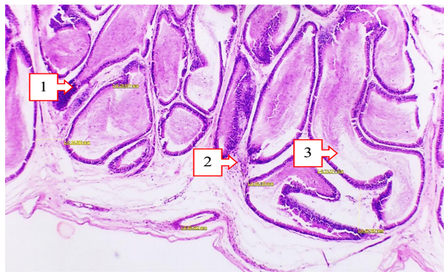 | Figure 1. Epididymis after intratesticular injection of lidocaine. Hematoxylin and eosin staining, 4x20 ml |
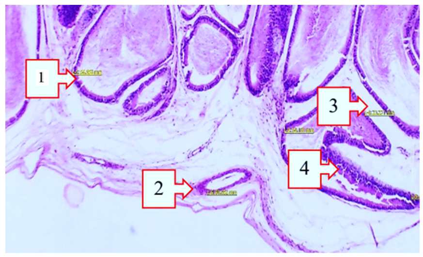 | Figure 2. Epididymis after intratesticular injection of novocaine. Hematoxylin and eosin staining, Ok. 10xOb.20 |
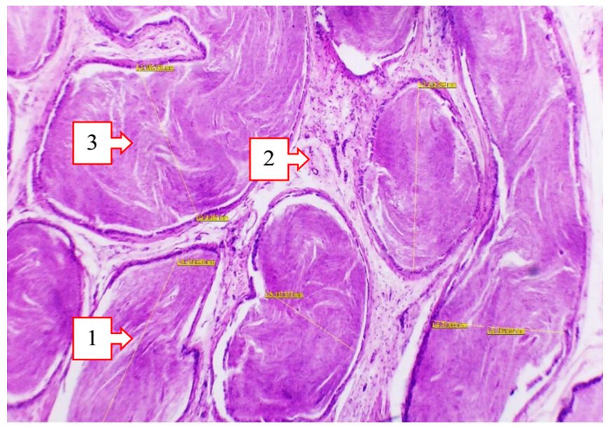 | Figure 3. Epididymis after conductive administration of lidocaine. Hematoxylin and eosin staining, 10% x 20% volume |
 | Figure 4. CD56 expression after combined use of novocaine and lidocaine. DAB, ×400 |
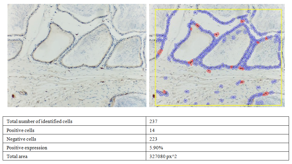
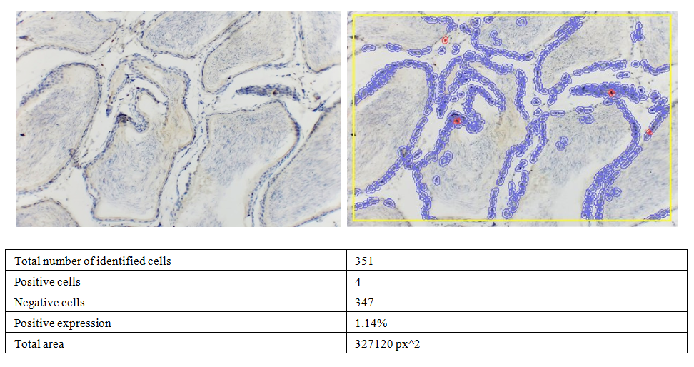 | Figure 5. CD45 expression after combined use of novocaine and lidocaine. DAB, ×400 |
4. Conclusions
- 1. Intratesticular administration of anesthetics causes the most pronounced morphological damage to epididymal tissue. This is manifested by focal epithelial desquamation, necrosis, and inflammatory infiltration, as well as a decrease in sperm count within the lumen of the testicular tubules.2. Intratesticular administration of novocaine is accompanied by the most severe stromal-vascular disorders, including hypertrophy of smooth muscle fibers, thickening of the vascular wall and narrowing of the lumen, edema and increased connective tissue remodeling.3. Conduction anesthesia with lidocaine is the most gentle method, as it ensures the preservation of the tubular structure, the absence of severe inflammation and edema, and the preserved presence of mature spermatozoa.