-
Paper Information
- Next Paper
- Previous Paper
- Paper Submission
-
Journal Information
- About This Journal
- Editorial Board
- Current Issue
- Archive
- Author Guidelines
- Contact Us
American Journal of Medicine and Medical Sciences
p-ISSN: 2165-901X e-ISSN: 2165-9036
2026; 16(2): 500-502
doi:10.5923/j.ajmms.20261602.25
Received: Jan. 6, 2026; Accepted: Jan. 22, 2026; Published: Feb. 3, 2026

Morphometric and Histological Changes in the Spleen Under the Influence of Carbon Monoxide Exposure
 Abstract
 Reference
 Full-Text PDF
 Full-text HTML
Qodirov Oybek O‘ktam ugli
PhD., Associate Professor, Department of Histology, Cytology and Embryology Bukhara State Medical Institute, Bukhara, Uzbekistan
Correspondence to: Qodirov Oybek O‘ktam ugli, PhD., Associate Professor, Department of Histology, Cytology and Embryology Bukhara State Medical Institute, Bukhara, Uzbekistan.
Copyright © 2026 The Author(s). Published by Scientific & Academic Publishing.
This work is licensed under the Creative Commons Attribution International License (CC BY).
http://creativecommons.org/licenses/by/4.0/

Morpho-functional changes in the spleen as a result of chronic carbon monoxide exposure to laboratory animals were detected under experimental conditions. As a result of chronic carbon monoxide exposure, specific changes were found in each part of the spleen, and in its wall, connective tissue growth, cell hypertrophy, signs of inflammation, and vascular congestion were observed.
Keywords: Сhronic carbon monoxide exposure, Oxidative stress, Apoptosis, Spleen, Morphological changes, Experimental study
Cite this paper: Qodirov Oybek O‘ktam ugli, Morphometric and Histological Changes in the Spleen Under the Influence of Carbon Monoxide Exposure, American Journal of Medicine and Medical Sciences, Vol. 16 No. 2, 2026, pp. 500-502. doi: 10.5923/j.ajmms.20261602.25.
Article Outline
1. Introduction
- Despite the success of carbon monoxide treatment, toxicity remains a major concern [1,2]. In carbon monoxide poisoning, damage to the spleen and spleen tissue can lead to altered or increased drug concentrations, which can increase toxicity, prolong hospital stays, and increase mortality (Department of Animal Science, Debre Berhan University, Department of Veterinary Medicine, Jimma University, Jimma, Ethiopia) [3,4].In experimental studies, the morphometric changes in the spleen and the adverse effects of carbon monoxide on the human body are very diverse, and many scientific studies are being conducted around the world to address this urgent problem [5,6,7]. This work was carried out at leading research centers and universities around the world (Western Kentucky University, Dominican University of California, Harvard University, University of Missouri-Columbia, University of Nebraska–Lincoln, Colby College (USA), Oxford University (Great Britain) [8,9,10]. In an organism poisoned by carbon monoxide, carbon and protein metabolism are disrupted, resulting in symptoms of acidosis. The balance of potassium and calcium in the blood and the functioning of the central nervous system are disrupted, and thus a person can die prematurely [11,12,13]. The harmful effects of carbon monoxide include changes in the body, including the morphological characteristics of organs, and the development of therapeutic and preventive measures to reduce the harmful effects of carbon monoxide has not lost its relevance [14,15,16].Оbjеctivе оf thе study. To identify the morphological and functional changes in spleen tissue under chronic inhalational exposure to carbon monoxide (CO), evaluate their severity and developmental stages, and scientifically substantiate the mechanism of CO influence on spleen immunological reactivity based on morphological, morphometric, histological, and biochemical parameters.
2. Mаtеriаl аnd Mеthоds оf Rеsеаrch
- The experiments were conducted on 208 white female outbred rats born in vivarium conditions. Rats aged 3, 6, 9 and 12 months were involved. The experiments followed the ethical rules for the use of animals and the requirements of the Helsinki Congress. Before the start of the experiments, all sexually mature rats were quarantined for a week and, excluding somatic or infectious diseases, they were transferred to the usual vivarium regime under the same conditions. During the experiment, the behavior and physiological state of animals in the standard and experimental groups were monitored. Rats were divided into 4 groups (n = 208): I-control group (n = 40); II-IV-groups (n = 168) experimental animals were chronically poisoned with a dose of carbon monoxide in the air of 0.01-0.05 mg / l. During the experiment, 4 3-month-old, 1 6-month-old, and 1 12-month-old rats died as a result of chronic carbon monoxide poisoning. After that, we divided 162 outbred white female rats chronically poisoned with carbon monoxide into 3 more groups. Group 2 (n = 54) rats chronically poisoned with carbon monoxide in the experiment; Group 3 (n = 54) rats chronically poisoned with carbon monoxide were administered intragastrically through a metal stomach tube for 14 days with 1 ml of infusion of the plant Gulimansar; In the 4th group (n = 54) experiment, rats chronically poisoned with carbon monoxide were administered intragastrically through a metal gastric tube for 14 days with a volume of 0.1 ml of an alcoholic solution of asparagus oil (in a ratio of 1:9). A total of 208 rats were used in the experiments, of which only 6 died during the experiments (Table 1).
|
3. Rеsults аnd Discussiоn
- As a result of chronic exposure to hydrogen sulfide, the body weight of 6-month-old laboratory animals ranged from 160 to 230 g, with a mean of 201.8 ± 7.36 g. The absolute organ weight ranged from 0.44 to 0.76 g, with a mean of 0.59 ± 0.03 g. The weight index ranged from 0.217% to 0.322%, with a mean of 0.295 ± 0.01%.Spleen length ranged from 25.4 to 33.8 mm, with a mean of 29.3 ± 0.77 mm. The growth rate was 22.2% higher compared to 6-month-old rats that consumed chemically enriched underground water. Spleen width ranged from 4.0 to 6.4 mm, with a mean of 5.16 ± 0.22 mm. The growth rate was 9.8% higher compared to 6-month-old rats that consumed chemically enriched underground water. Spleen thickness ranged from 2.0 to 3.8 mm, with a mean of 3.02 ± 0.16 mm. The growth rate was 12.0% higher compared to 3-month-old irradiated rats (Figure 1).
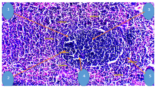 | Figure 1. Morphometry of the spleen of a 6-month-old white outbred rat after chronic hydrogen sulfide exposure. Staining: Hematoxylin-eosin. Magnification: 20×10. 1 – lymphatic nodule, 2 – periarterial region, 3 – germinal center, 4 – mantle zone, 5 – marginal zone |
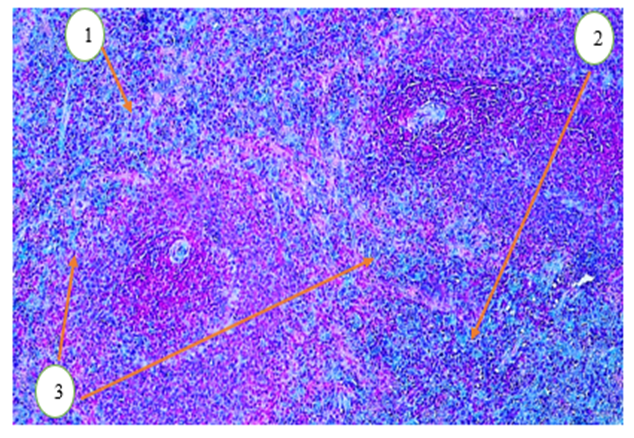 | Figure 2. Microscopic appearance of the spleen of a 6-month-old white outbred rat after chronic hydrogen sulfide exposure.Staining: Alcian blue. Magnification: 20×10. 1 – densely stained capsule zone, 2 – medullary zone, 3 – trabeculae and adipose tissue between lobules |
4. Discussion
- Histological structure: The image shows the spleen’s lymphoid tissue against a background, with germinal centers clearly visible in the central regions. These centers are associated with immune responses and serve as sites for lymphocyte development.Red and blue coloration: Red or reddish cells (stained with Alcian blue) indicate a high concentration of lymphocytes. Blue areas are associated with fibrous tissue or degenerated cells.It should be noted that the enlargement of germinal centers indicates heightened immune system activity. As a result of chronic hydrogen sulfide exposure, cellular infiltration and slight disruption of lymphoid tissue were observed.
5. Cоnclusiоns
- Chronic exposure to chemical agents led to the development of fibrotic regions in the spleen tissue, which were stained blue.Morphometric parameters of the spleen were determined in 3-, 9-, and 12-month-old white laboratory rats. It was found that the spleen dimensions in 9- to 12-month-old rats were larger than those in 6-month-old outbred rats, particularly evident in the size of the peripheral layer and the blood vessels.Chronic exposure to carbon monoxide (CO) gas resulted in hypertrophy of spleen cells, interstitial edema in the intercellular spaces, and the development of congestion in small blood vessels.To address the morphological changes caused by chronic CO exposure, a decoction of Gulmansasar (herbal plant) was administered. Histological examination of spleen tissue from treated outbred rats revealed positive effects, including a reduction in intercellular edema, decreased inflammatory elements, and improvement in dystrophic changes in the tissue.Histological methods for analyzing the spleen under chronic CO exposure are widely used for diagnosing and differentially diagnosing spleen tissue diseases of various etiologies, as they allow evaluation of potential morphofunctional alterations.