American Journal of Medicine and Medical Sciences

p-ISSN: 2165-901X    e-ISSN: 2165-9036

2026;  16(2): 436-439

doi:10.5923/j.ajmms.20261602.11

Received: Jan. 2, 2026; Accepted: Jan. 22, 2026; Published: Feb. 3, 2026


Morphological Aspects of Early Carcinogenesis of the Stomach and the Role of Helicobacter Pylori Strains

Ismailova J. A. , Rakhimova Sh. Sh. , Pulatov S. S.

Republican Specialized Scientific Practical Medical Center of Therapy and Medical Rehabilitation, Uzbekistan

Copyright © 2026 The Author(s). Published by Scientific & Academic Publishing.

This work is licensed under the Creative Commons Attribution International License (CC BY).
http://creativecommons.org/licenses/by/4.0/

Abstract

With the aim of early diagnosis of stomach cancer, the authors studied the significance of morphological studies and pathogenic strains of H. pylori in 100 patients with gastric diseases infected with H. pylori and in a control group of 20 individuals. The results of the study showed that a mild degree of gastric mucosal damage by H. pylori in chronic gastritis is associated with low intensity of inflammatory changes, and the CagA gene can be considered a marker gene indicating the pathogenicity status of H. pylori.

Keywords: H. pylori, Chronic gastritis, Endoscopic studies, Gastric mucosa, ureC and CagA genes, Biopsy, Molecular genetic studies, OLGA and OLGIM systems, Atrophy, Intestinal metaplasia

Cite this paper: Ismailova J. A. , Rakhimova Sh. Sh. , Pulatov S. S. , Morphological Aspects of Early Carcinogenesis of the Stomach and the Role of Helicobacter Pylori Strains, American Journal of Medicine and Medical Sciences, Vol. 16 No. 2, 2026, pp. 436-439. doi: 10.5923/j.ajmms.20261602.11.

1. Introduction

In 2022, 20 million new cancer cases were registered worldwide, and 9.7 million deaths were recorded. 53.3 million cancer patients survived 5 years after diagnosis [3,14,16]. Approximately 1 in 5 people will develop cancer during their lifetime. According to IARC, it is 12 times more common in underdeveloped and developing countries than in developed countries [17]. Several factors contribute to the development of cancer, one of the main factors of which is Helicobacter pylori (H. pylori). Studies have shown that aggressive strains of H. pylori are known to cause severe forms of the disease [1,8,15]. In addition, molecular genetic factors of a person have been confirmed to play an important role. Gastric cancer ranks 2nd in the structure of oncological diseases in our country, with an incidence of 6.8 per 100,000 population (2022). In our country, gastric cancer, regardless of etiopathogenetic factors, is treated mainly on the basis of oncological principles, and unlike other types of cancer, the 5-year survival rate does not exceed 50% even in early stages [7]. The high aggressiveness, moderate sensitivity to radiation and chemotherapy indicate the need to focus on the molecular genetic factors of this disease and develop personalized treatment methods in the future [9,12]. To date, gastric cancer and precancerous lesions in our country have not been sequenced or genotyped for exome, labeled, expression-based marker genes, marker polymorphisms, region-specific H. pylori strains, and disease-associated polymorphisms [2,13]. Therefore, the percentage of patients diagnosed at an early stage of cancer remains quite low. Of course, morphological examinations play a key role in the early diagnosis of gastric cancer and help to identify precancerous changes in the gastric mucosa due to H. pylori infection [5,10]. Numerous literature reviews have shown that the risk of gastric cancer is higher in patients with advanced age, gender (more men than women), atrophic gastritis, intestinal metaplasia, and gastric dysplasia than in patients with other precancerous diseases (for example, gastric polyposis), and aggressive strains of H. pylori are involved as their “conductors” [4,6,11]. This, in turn, requires a serious and individual approach to precancerous diseases.
The aim of the study is to study the importance of morphological examinations and pathogenic strains of H. pylori in the early diagnosis of stomach cancer.
The object of the study was 100 patients with gastric diseases infected with H. pylori, who underwent molecular genetic testing and morphological examination of targeted biopsy samples to identify pathological changes in the gastric mucosa.

2. Methods of the Study

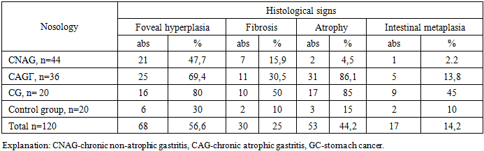
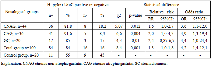
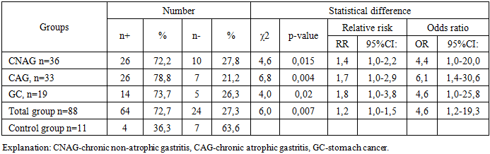
All patients were divided into three study groups according to the nosology of the pathological process, and their results were studied comparatively: 80 patients with chronic gastritis (CG), and 20 patients with gastric cancer (GC) and a control group of 20 healthy people.
All patients underwent gastroscopy and a five-point gastric biopsy. Based on the studied material, a morphological study of biopsies was carried out to identify preneoplastic changes (fibrosis, atrophy, intestinal metaplasia (IM), dysplasia) in patients with gastric diseases infected with H. pylori (Table 1).
Table 1. Morphological incidence of gastric diseases infected with H. pylori (based on the study of biopsies taken from the gastric mucosa), n=120
     
The table shows that, depending on the nosological form of gastric diseases infected with H. pylori, the morphological appearance differs in the variability of gradations. The results obtained indicate that the intensity of morphological signs depends on the nosological forms of gastric diseases infected with H. pylori. In general, gastric diseases infected with H. pylori were morphologically manifested by atrophy of the gastric mucosa - 44.2%, fibrosis - 25%, and intestinal metaplasia of varying degrees - 14.2%. The analysis of morphological studies, which included qualitative and quantitative indicators, shows that diseases infected with H. pylori are characterized by variable changes in the gastric mucosa. In all studied nosological groups, morphological changes according to the OLGA system were zero or I degree: 27.2% and 15.1%, respectively. Intestinal metaplasia was observed in only 7.3% of cases in all groups according to the OLGA system. When analyzed using the OLGIM system, the same morphological changes showed a different rate. Intestinal metaplasia of stages I and II was detected in 17.6% and 10.8% of cases, respectively. The study showed that irreversible stages III and IV of intestinal metaplasia were detected in 11.2% and 5.6% of patients, respectively. CG and GC were accompanied by varying degrees of activity of the local inflammatory process. The general interpretation of the results obtained showed the feasibility of using the above systems, since it is morphological analysis from the point of view of detecting atrophy and intestinal metaplasia that can be the most basic platform for creating a further strategy of therapeutic measures. Therefore, it is a predictive risk factor for the development of neoplastic changes, detected in 17.6% of patients with stage II and III according to the OLGA system and in 17.7% of patients with stage III and IV according to the OLGIM system. If this indicator was not observed in our studies in CNAG, then in CAG it is very rare and in GC it has the most accurate indicator.
The ureC (glm) gene of H. pylori bacteria was amplified using the Real time PCR method in the feces of all 120 people in the study group. In order to determine the pathogenic status of the identified H. pylori bacteria, PCR amplification of the EPIYA motif of the CagA gene was performed.
Analysis of the obtained PCR results showed that 95 (79.1%) of the 120 people examined had H. pylori ureC gene amplification, while 25 (20.8%) patients did not have the ureC gene. The detection rate of H. pylori in all nosological groups was 1.5 times higher than in the control group (84% and 55% of cases), and it was confirmed that H. pylori bacteria are a risk factor for the development of these diseases (χ2=8.4; p=0.001). In a control group of 20 healthy individuals, 11 (55%) had the ureC gene detected, while 9 (45%) did not (Table 2).
Table 2. Statistical analysis of the H. pylori ureC gene in gastric diseases
     
CNAG, САG ва GC H. pylori was confirmed to be a significant risk factor for the development of diseases (p=0.001).
In order to determine the pathogenic status of H. pylori and to study its association with the disease, the CagA gene was obtained and the association between the H. pylori CagA gene and the development of diseases was studied.
The results showed that the CagA gene was detected in 69 (78.4%) of 84 patients, while it was not detected in 19 (21.5%). Of the 11 control subjects, the CagA gene was detected in 6 (54.5%) of 11 subjects, while it was not detected in 5 (45.4%) (see Table 3). Analysis of the CagA gene by nosological groups showed that the percentage of detection of the pathogenic CagA gene increased with the development of the disease. Analysis of the study results showed that, unlike healthy controls in the control group, the CagA gene was detected 1.5 times more often in patients with H. pylori-associated gastric diseases (78.4% / 54.5%, respectively).
Table 3. Association of the CagA gene with H. pylori–associated gastric diseases
     
In healthy people, the incidence of ureC gene-positive patients with no CagA gene was 2.8 times lower than in patients with nosocomial disease (21.5% versus 45.4%). According to the data obtained, the CagA gene, which determines the pathogenicity of H. pylori, had an aggravating effect on the course of gastric diseases associated with all its forms. The CagA gene significantly increased the risk of developing the disease by 2.9-6.2 times. Analysis of the data obtained indicates the role of the CagA gene as a factor in the development of severe forms of gastric diseases, which was confirmed by a 2.9-fold increase in the risk of developing CABG in patients with a positive CagA gene (χ2=8.4; p=0.002; OR=2.9; 95%Cl 1.4-6.5) and a 6.2-fold increase in the risk of gastric cancer compared to the control group (χ2=8.6; p=0.001).
Of the 88 patients with the H. pylori ureC gene, 19 (21.5%) did not detect the CagA gene. Perhaps the H.pylori bacteria present in these patients lack the CagA gene or have strains that contain motifs other than the A motif (B, C, D, E). Another hypothesis is that these isolates may have the CagA gene, but there may be a nucleotide mutation at the primer binding site. Analysis of the association of the CagA gene with nosological groups indicates its leading role in the development of severe forms of the disease. It is clearly seen that the CagA gene is a risk factor for the development of severe forms of gastrointestinal diseases. In terms of diagnostic efficiency (AUC-classifier), the CagA gene showed an average indicator (61.8-66.8 %), in all diseases. This means that the CagA gene can be used as a marker gene in the diagnosis of pathogenic or non-pathogenic strains of H. pylori.

3. Conclusions

1) In chronic gastritis, the weak level of infection of the gastric mucosa with H. pylori is associated with a low intensity of inflammatory changes.
2) The results of the study showed that the CagA gene is a risk factor for the development of gastrointestinal diseases and that this gene can be used as a marker gene for the pathogenic status of H. pylori.

References

[1]  Bordin, D. S., Livzan, M. A., Mozgovoi, S. I., & Gaus, O. V. (2025). Autoimmune gastritis and Helicobacter pylori infection: molecular mechanisms of relationship. International Journal of Molecular Sciences, 26(16), 7737.
[2]  Chiang, T. H., Hsu, Y. N., Chen, M. H., Chen, Y. R., Cheng, H. C., Chen, M. J., ... & Chen, C. S. (2025). A rural-to-center artificial intelligence model for diagnosing Helicobacter pylori infection and premalignant gastric conditions using endoscopy images captured in routine practice. Endoscopy.
[3]  Coelho MCF, Ribeiro HG, Gomes CGO, Marinho FP, Barbosa AJA, Coelho lgv. Нelicobacter pylori chronic gastritis on patients with premalignant conditions: olga and olgim evaluation and serum biomarkers performance. Arq Gastroenterol. 2021 Jan-Mar; 58(1): 39-47. doi: 10.1590/S0004-2803.202100000-08. PMID: 33909795.
[4]  Ding, Lijia, et al. "Risk factors analysis for pathological upgrade after endoscopic submucosal dissection in patients with gastric intraepithelial neoplasia: a single-center retrospective study." Therapeutic Advances in Gastroenterology 18 (2025): 17562848251397847.
[5]  Fang, J., Du, Y., Liu, W., Xiao, Y., Chen, W., & Ren, J. (2023). Chinese guidelines for the diagnosis and treatment of chronic gastritis (2022, shanghai). Chin J Gastroenterology, 28, 149-80.
[6]  Isajevs, S., Liepniece-Karele, I., Janciauskas, D., Moisejevs, G., Putnins, V., Funka, K., ... & Leja, M. (2014). Gastritis staging: interobserver agreement by applying OLGA and OLGIM systems. Virchows Archiv, 464(4), 403-407.
[7]  Ismailova J. A., Dalimova D. A., Rakhimova Sh. Sh. The Role of the Intestinal Microbiome in the Development of Stomach Diseases. American Journal of Medicine and Medical Sciences 2025, 15(10): 3620-3623 DOI: 10.5923/j.ajmms.20251510.73.
[8]  Khosravi, M., Jasemi, S. K., Hayati, P., Javar, H. A., Izadi, S., & Izadi, Z. (2024). Transformative artificial intelligence in gastric cancer: Advancements in diagnostic techniques. Computers in Biology and Medicine, 183, 109261.
[9]  Kurumi, H., Nonaka, K., Ikebuchi, Y., Yoshida, A., Kawaguchi, K., Yashima, K., & Isomoto, H. (2021). Fundamentals, diagnostic capabilities, and perspective of narrow band imaging for early gastric cancer. Journal of clinical medicine, 10(13), 2918.
[10]  Leung, W. K., Ang, T. L., Shah, S., Cheung, K. S., Li, Y., Uedo, N., ... & Lee, Y. C. (2025). Asian Pacific Association of Gastroenterology task force recommendations on surveillance for Helicobacter pylori associated gastric premalignant conditions. Gut.
[11]  Loor A, Dumitraşcu DL. Helicobacter pylori Infection, Gastric Cancer and Gastropanel. Rom J Intern Med. 2016 Sep 1; 54(3): 151-156. doi: 10.1515/rjim-2016-0025. PMID: 27658162.
[12]  Lyu, K. M., Chen, Q. Q., Xu, Y. F., Yuan, Y. Q., Wang, J. F., Wan, J., & Ling-Hu, E. Q. (2025). Development and validation of a predictive model for the pathological upgrading of gastric low-grade intraepithelial neoplasia. World Journal of Gastroenterology, 31(11), 104377.
[13]  Mourato, M. B., Pratas, N., Branco Pereira, A., Chança, R., Fronteira, I., Dinis, R., & Areia, M. (2025). Effectiveness of Gastric Cancer Endoscopic Screening in Intermediate‐Risk Countries—A Systematic Review and Meta‐Analysis. United European Gastroenterology Journal, 13(9), 1715-1729.
[14]  Sbarigia, C., Carabotti, M., Corsetti, M., & Annibale, B. (2025). Chronic atrophic gastritis and dyspepsia: a forgotten link?. Expert Review of Gastroenterology & Hepatology, 19(10), 1109-1117.
[15]  Tao, Jingna, et al. "Risk prediction model for precancerous gastric lesions based on magnifying endoscopy combined with narrow-band imaging features." Frontiers in Oncology 15 (2025): 1554523.
[16]  Wang, C., Ge, J., Niu, Y., Ding, C., Fan, Y., Chang, H., ... & Li, C. (2025). A fully annotated pathology slide dataset for early gastric cancer and precancerous lesions. Scientific Data, 12(1), 1326.
[17]  Wald N. J. The treatment of Helicobacter pylori infection of the stomach in relation to the possible prevention of gastric cancer. In: IARC Helicobacter pylori Working Group. Helicobacter pylori Eradication as a Strategy for Preventing Gastric Cancer. Lyon, France: International Agency for Research on Cancer (IARC Working Group Reports, № 8); 2014, p. 174–18.