-
Paper Information
- Next Paper
- Previous Paper
- Paper Submission
-
Journal Information
- About This Journal
- Editorial Board
- Current Issue
- Archive
- Author Guidelines
- Contact Us
American Journal of Medicine and Medical Sciences
p-ISSN: 2165-901X e-ISSN: 2165-9036
2026; 16(2): 433-435
doi:10.5923/j.ajmms.20261602.10
Received: Jan. 8, 2026; Accepted: Jan. 29, 2026; Published: Feb. 3, 2026

Morphological Changes in the Testes Following Gas Poisoning
 Abstract
 Reference
 Full-Text PDF
 Full-text HTML
Hojiyev Sharif Shukurovich, Teshayeva Dilbar Shukhrat qizi
Bukhara State Medical Institute named after Abu Ali ibn Sina, Bukhara, Uzbekistan
Correspondence to: Hojiyev Sharif Shukurovich, Bukhara State Medical Institute named after Abu Ali ibn Sina, Bukhara, Uzbekistan.
| Email: |  |
Copyright © 2026 The Author(s). Published by Scientific & Academic Publishing.
This work is licensed under the Creative Commons Attribution International License (CC BY).
http://creativecommons.org/licenses/by/4.0/

Gas poisoning is a significant toxicological problem that can adversely affect multiple organ systems, including the male reproductive system. Experimental and clinical studies have demonstrated that exposure to toxic gases leads to pronounced morphological and functional alterations in the testes. These changes are primarily associated with hypoxia, oxidative stress, and direct cytotoxic effects on testicular tissue. Morphological examination reveals degeneration of seminiferous tubules, disorganization of the germinal epithelium, vacuolization, reduced diameter of seminiferous tubules, and damage to Sertoli and Leydig cells. In addition, gas-induced toxicity is often accompanied by impaired spermatogenesis, decreased sperm count, and hormonal imbalances resulting from Leydig cell dysfunction. Increased levels of reactive oxygen species and lipid peroxidation play a central role in mediating cellular damage, leading to apoptosis and necrosis of germ cells. These structural alterations ultimately compromise male fertility. Understanding the morphological changes in the testes following gas poisoning is essential for elucidating the pathophysiological mechanisms of reproductive toxicity and for developing effective preventive and therapeutic strategies.
Keywords: Gas poisoning, Testes, Morphological changes, Spermatogenesis, Oxidative stress, Reproductive toxicity
Cite this paper: Hojiyev Sharif Shukurovich, Teshayeva Dilbar Shukhrat qizi, Morphological Changes in the Testes Following Gas Poisoning, American Journal of Medicine and Medical Sciences, Vol. 16 No. 2, 2026, pp. 433-435. doi: 10.5923/j.ajmms.20261602.10.
1. Introduction
- The testis is a paired central organ of the male reproductive system, primarily responsible for sperm production and the synthesis of androgenic hormones, particularly testosterone (Junqueira, 2018). Its morphological organization and functional specialization are crucial for maintaining reproductive homeostasis in males. The testicular parenchyma is predominantly composed of seminiferous tubules, where stepwise spermatogenesis occurs. The germinal epithelium lining the seminiferous tubule walls is highly differentiated, encompassing all stages of germ cell development, from spermatogonia to mature spermatozoa (Junqueira, 2018).Understanding the structural and functional integrity of the testis is essential for evaluating the effects of environmental and occupational toxins, such as chronic carbon monoxide (CO) exposure, on male reproductive health. Disruption of seminiferous tubule architecture or germinal epithelium function can impair spermatogenesis and hormonal balance, leading to reproductive dysfunction. [3]Anatomically, the testis is externally covered by a dense fibrous capsule known as the tunica albuginea, from which septa extend into the testicular parenchyma. These septa divide the testis into several lobules, each containing 1–4 highly coiled seminiferous tubules (tubuli seminiferi contorti), where spermatogenesis occurs (Ross & Pawlina, 2020).Histologically, the wall of each seminiferous tubule consists of two main components: the basal membrane and the spermatogenic epithelium. The spermatogenic epithelium comprises germ cells at various stages of differentiation as well as Sertoli cells. Sertoli cells provide mechanical, trophic, and metabolic support to developing germ cells and contribute to the formation of the blood–testis barrier (Setchell, 2014).These structural and functional characteristics of the seminiferous tubules are fundamental for normal spermatogenesis and male reproductive function. Disruption of tubular architecture or Sertoli cell function, as may occur under toxic exposures such as chronic carbon monoxide (CO), can impair germ cell development, hormonal regulation, and overall reproductive homeostasis. [4,7]Gas poisoning remains an important public health and occupational safety concern worldwide due to its acute and chronic toxic effects on various organ systems [1]. Toxic gases such as carbon monoxide, nitrogen oxides, sulfur dioxide, and industrial chemical vapors can enter the body through inhalation and induce systemic hypoxia, oxidative stress, and cellular damage [9]. While the neurological and cardiovascular consequences of gas exposure have been extensively studied, its impact on the male reproductive system has received comparatively less attention [5]. The testes are highly sensitive to toxic agents due to their high metabolic activity and complex cellular organization. Normal spermatogenesis requires a tightly regulated microenvironment, which can be easily disrupted by toxic insults [11]. Experimental evidence suggests that gas poisoning can impair testicular structure and function by inducing hypoxia, generating reactive oxygen species, and damaging the blood–testis barrier [10]. These pathological processes lead to degeneration of seminiferous tubules, loss of germ cells, and dysfunction of Sertoli and Leydig cells. [6] Morphological alterations in testicular tissue serve as critical indicators of reproductive toxicity and fertility impairment. Histopathological assessment allows for the identification of structural damage that precedes or accompanies functional decline. Therefore, studying morphological changes in the testes following gas poisoning is essential for understanding the mechanisms of male reproductive toxicity and for developing preventive measures and therapeutic interventions aimed at preserving fertility. [8]
2. Materials and Methods
- One-month-old outbred male white rats (n = [insert number]) were used and maintained under standard laboratory conditions with ad libitum access to food and water. The experimental group was chronically exposed to carbon monoxide (CO) gas to assess its effects on testicular morphology and function, simulating prolonged low-level inhalation.After the exposure period, animals were euthanized in accordance with institutional ethical guidelines. Testes were excised, fixed in 10% neutral buffered formalin, dehydrated in graded ethanol, cleared in xylene, and embedded in paraffin. Serial sections (5–7 µm) were stained with hematoxylin and eosin and examined under light microscopy (×20 and ×40).Digital morphometry was performed by polygonal segmentation of seminiferous tubules (red outline) and defining counting areas (dark blue–black mask). Morphometric parameters included tubular diameter, epithelial thickness, and cell composition. Histopathological assessment focused on germ cell degeneration, Sertoli cell morphology, interstitial tissue changes, and signs of hypoxia such as nuclear pyknosis and cytoplasmic vacuolization.
3. Results
- Histomorphological and morphometric analyses revealed significant structural and functional alterations in the testes of outbred male white rats chronically exposed to carbon monoxide (CO) gas. The severity of these changes was dependent on both the duration of exposure (1, 3, and 9 months) and the age of the animals.After 1 month, seminiferous tubules were smaller, irregularly arranged, and exhibited mild disorganization of the spermatogenic epithelium. Cellular swelling and cytoplasmic vacuolization were observed in germ cells and Sertoli cells, indicating early hypoxic stress.At 3 months, more pronounced degeneration was evident, including marked disorganization of seminiferous tubules, dystrophic changes in germ cells, and a reduction in the orderly arrangement of spermatogenic cells. Spermatogenesis was impaired, with fewer mature germ cells and increased degenerative forms.By 9 months, severe alterations were observed: fibrosis of interstitial tissue, involution of seminiferous tubules, collapse of tubular architecture, and near-complete arrest of spermatogenesis. Morphometric analysis confirmed reduced tubular diameter and epithelial thickness, expansion of interstitial space, and decreased Leydig cell number.Histological sections stained with hematoxylin–eosin showed nuclear pyknosis, cytoplasmic vacuolization, and connective tissue fibrosis. Digital morphometry indicated that spermatogonia comprised 52.7 ± 1.1% of cells, spermatocytes 24.3 ± 1.2%, spermatids 6.7 ± 0.4%, spermatozoa 11.4 ± 0.4%, plasmablasts 2.2 ± 0.5%, degenerative cells 2.1 ± 0.6%, and macrophages 3.6 ± 0.7%. Interstitial tissue exhibited edema, vascular wall thickening, plasmorrhagia, and focal lymphoid infiltration.Figures:
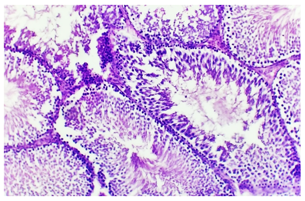 | Figure 1. Histomorphological and morphometric characteristics of the testes in 1-month-old rats following chronic CO exposure (H&E, ×20/×40) |
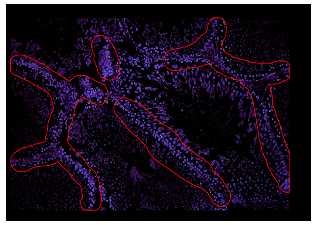 | Figure 2. Digital morphometric analysis showing polygonal segmentation of seminiferous tubules (red outline) and counting area (dark blue–black mask) |
4. Discussion
- The results demonstrate that chronic CO exposure induces progressive testicular damage that is both time- and age-dependent. Early hypoxic–dystrophic changes observed after 1 month suggest an initial adaptive response to reduced oxygen delivery. By 3 months, degenerative alterations indicate a transition from reversible damage to progressive structural impairment, while after 9 months, irreversible fibrosis and tubular involution reflect severe testicular atrophy.Mechanistically, these alterations are primarily due to CO binding to hemoglobin, forming carboxyhemoglobin and reducing oxygen availability, coupled with oxidative stress from reactive oxygen species. This hypoxic–oxidative environment leads to germ cell apoptosis, Sertoli cell dysfunction, and Leydig cell impairment, disrupting both spermatogenesis and endocrine function. [12]Morphometric data, including reductions in tubular diameter and epithelial thickness, along with interstitial expansion, confirm structural compromise of the testes. Decreased Leydig cell activity and interstitial fibrosis further impair the hormonal support required for normal spermatogenesis. These findings are consistent with previous studies on CO-induced reproductive toxicity in rodents, highlighting the vulnerability of the male reproductive system to chronic environmental and occupational exposure. [2]
5. Conclusions
- Chronic carbon monoxide exposure in male rats causes time-dependent and age-related histomorphological and morphometric alterations in the testes. Key effects include:1. Early hypoxic–dystrophic changes and delayed initiation of spermatogenesis (1 month).2. Progressive degeneration of germinal epithelium and impaired spermatogenesis (3 months).3. Severe tubular involution, interstitial fibrosis, and near-complete arrest of spermatogenesis (9 months).These findings indicate that chronic CO exposure disrupts both structural integrity and endocrine function of the testes, emphasizing its potential reproductive toxicity. The study provides important insights into the mechanisms underlying CO-induced male reproductive impairment and highlights the significance of monitoring environmental and occupational CO exposure.