-
Paper Information
- Next Paper
- Previous Paper
- Paper Submission
-
Journal Information
- About This Journal
- Editorial Board
- Current Issue
- Archive
- Author Guidelines
- Contact Us
American Journal of Medicine and Medical Sciences
p-ISSN: 2165-901X e-ISSN: 2165-9036
2026; 16(2): 421-425
doi:10.5923/j.ajmms.20261602.07
Received: Jan. 8, 2026; Accepted: Feb. 1, 2026; Published: Feb. 3, 2026

Hypoxia-Induced Esophageal Injury Following Chronic Carbon Monoxide Exposure: Morphological Changes and Phytocorrection with Silybum Marianum and Carthamus Tinctorius
 Abstract
 Reference
 Full-Text PDF
 Full-text HTML
Bakhronov B. B., Navruzov R. R.
Bukhara State Medical Institute, Bukhara, Uzbekistan
Correspondence to: Bakhronov B. B., Bukhara State Medical Institute, Bukhara, Uzbekistan.
| Email: |  |
Copyright © 2026 The Author(s). Published by Scientific & Academic Publishing.
This work is licensed under the Creative Commons Attribution International License (CC BY).
http://creativecommons.org/licenses/by/4.0/

Chronic carbon monoxide (CO) exposure remains an underrecognized cause of long-term hypoxic injury to the gastrointestinal tract, while esophageal morphological alterations and potential phytotherapeutic correction are insufficiently explored. To investigate structural and morphometric alterations of the esophagus induced by chronic CO exposure and to evaluate the corrective efficacy of Silybum marianum and Carthamus tinctorius extracts. An experimental study was conducted on 250 white outbred rats aged 3 and 9 months. Chronic CO exposure was modeled at 200–300 mg/m³. Animals were assigned to five groups: control, CO exposure, CO + Silybum marianum, CO + Carthamus tinctorius, and CO + combined phytocorrection. Histological, morphometric, and immunohistochemical analyses were performed, followed by statistical evaluation. Chronic CO exposure induced marked epithelial atrophy, interstitial edema, microcirculatory disturbances, and inflammatory infiltration of the esophageal wall, with more pronounced changes in older animals. Phytocorrection significantly attenuated inflammatory and dystrophic alterations and promoted tissue regeneration. Silybum marianum demonstrated superior anti-inflammatory and regenerative effects compared to Carthamus tinctorius, particularly in younger animals. Chronic CO exposure causes persistent hypoxia-driven esophageal injury. Phytocorrection, especially with Silybum marianum, effectively restores esophageal morphology and mitigates inflammation, highlighting its potential as a therapeutic strategy for hypoxia-induced esophageal damage.
Keywords: Carbon monoxide, Chronic hypoxia, Esophageal injury, Experimental toxicology, Phytocorrection, Silybum marianum, Carthamus tinctorius, Morphometric analysis, Inflammation, Tissue regeneration
Cite this paper: Bakhronov B. B., Navruzov R. R., Hypoxia-Induced Esophageal Injury Following Chronic Carbon Monoxide Exposure: Morphological Changes and Phytocorrection with Silybum Marianum and Carthamus Tinctorius, American Journal of Medicine and Medical Sciences, Vol. 16 No. 2, 2026, pp. 421-425. doi: 10.5923/j.ajmms.20261602.07.
1. Introduction
- Chronic exposure to carbon monoxide (CO) is traditionally regarded as a factor of systemic hypoxia exerting a pronounced impact on the central nervous and cardiovascular systems. According to experimental and clinical studies, the key mechanism of CO toxicity is the formation of carboxyhemoglobin, which reduces oxygen delivery to tissues and leads to the development of persistent tissue hypoxia [1,2]. In addition, CO induces mitochondrial dysfunction, activates free radical oxidation, and promotes endothelial dysfunction, thereby aggravating microcirculatory disturbances [3].In recent years, increasing attention has been directed toward the morphological consequences of chronic hypoxia in the organs of the gastrointestinal tract. Experimental models have demonstrated that prolonged hypoxic exposure is associated with impairment of the mucosal barrier function, activation of inflammatory responses, and an imbalance between cellular proliferation and apoptosis [4,5]. However, the majority of studies have focused on the stomach and intestines, whereas the esophagus remains a considerably less investigated target organ.Available evidence indicates that hypoxia and oxidative stress may induce epithelial atrophy of the esophagus, microcirculatory disorders, and fibrotic alterations in the submucosal layer [6]. Nevertheless, data on the morphometric characteristics of these changes, as well as their age-related dynamics, remain fragmented and insufficient to provide a comprehensive understanding of the pathogenesis of chronic hypoxic esophageal injury.In the context of searching for effective strategies to correct hypoxia-induced tissue damage, particular interest has been directed toward phytotherapeutic agents with pronounced antioxidant and anti-inflammatory properties. Extracts of Silybum marianum contain a complex of flavonolignans (silymarin) capable of stabilizing cellular membranes, inhibiting lipid peroxidation, and stimulating regenerative processes [7,8]. Extracts of Carthamus tinctorius, in turn, exhibit angioprotective and anti-inflammatory effects, contributing to the improvement of microcirculation and tissue oxygen exchange [9].Despite the proven efficacy of these phytobiostimulators in liver disorders, cardiovascular diseases, and experimental models of ischemic injury, data on their effects on the morphological state of the esophagus under conditions of chronic carbon monoxide intoxication remain extremely limited [10]. The available literature lacks comprehensive experimental studies combining histological, morphometric, and immunohistochemical approaches to evaluate the effectiveness of phytocorrection in chronic CO-induced esophageal injury.Thus, the existing gap in knowledge necessitates a systematic experimental investigation aimed at elucidating morphological and morphometric alterations of the esophagus following chronic carbon monoxide exposure and assessing the potential of phytocorrection using Silybum marianum and Carthamus tinctorius.
2. Materials and Methods of Research
- The experimental study was conducted on 250 white outbred rats of both sexes aged 3 and 9 months, weighing 200–250 g. Animals were obtained from a certified vivarium and housed under standard laboratory conditions (temperature 22–24 °C, relative humidity 50–60%, 12 h light/dark cycle) with free access to food and water. All experimental procedures were performed in accordance with international guidelines for the care and use of laboratory animals (ARRIVE guidelines) and were approved by the local institutional ethics committee.Experimental GroupsThe animals were randomly assigned into five experimental groups (n = 50 per group):1. Control group – intact animals not exposed to carbon monoxide.2. CO group – animals subjected to chronic carbon monoxide exposure without correction.3. CO + Silybum marianum – animals exposed to CO and treated with Silybum marianum extract.4. CO + Carthamus tinctorius – animals exposed to CO and treated with Carthamus tinctorius extract.5. CO + combined phytocorrection – animals exposed to CO and treated with a combination of Silybum marianum and Carthamus tinctorius extracts.Each group included animals of both age categories to assess age-related differences in morphological responses.Model of Chronic Carbon Monoxide ExposureChronic carbon monoxide intoxication was modeled by daily exposure of animals to CO in a sealed inhalation chamber at a concentration of 200–300 mg/m³ for a specified period, simulating prolonged low-dose environmental exposure. Gas concentration was continuously monitored using a calibrated gas analyzer to ensure stable exposure conditions.Phytocorrection ProtocolPhytocorrection was performed using standardized extracts of Silybum marianum and Carthamus tinctorius. The extracts were administered orally once daily at doses selected based on previous experimental studies demonstrating biological efficacy. In the combined phytocorrection group, both extracts were administered concurrently in equivalent therapeutic doses.Histological ExaminationAt the end of the experimental period, animals were euthanized under anesthesia, and esophageal tissue samples were harvested. Specimens were fixed in 10% neutral buffered formalin, processed using standard histological techniques, embedded in paraffin, and sectioned at a thickness of 4–5 μm. Sections were stained with hematoxylin and eosin for general morphological assessment.Morphometric AnalysisMorphometric evaluation was performed using digital image analysis software. Parameters assessed included epithelial thickness, degree of interstitial edema, inflammatory cell density, and structural integrity of the muscular and submucosal layers. Measurements were obtained from multiple randomly selected fields per specimen to ensure representativeness.Immunohistochemical AnalysisImmunohistochemical staining was conducted to assess proliferative and apoptotic activity in esophageal tissues. The expression of Ki-67 (proliferation marker) and Bcl-2 (anti-apoptotic marker) was evaluated using standard immunohistochemical protocols. The results were quantified as the percentage of positively stained cells.Statistical AnalysisStatistical analysis was performed using standard statistical software. Quantitative data were expressed as mean ± standard deviation (SD). Intergroup comparisons were conducted using appropriate parametric or non-parametric tests depending on data distribution. Differences were considered statistically significant at p < 0.05.
3. Results
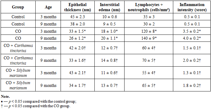
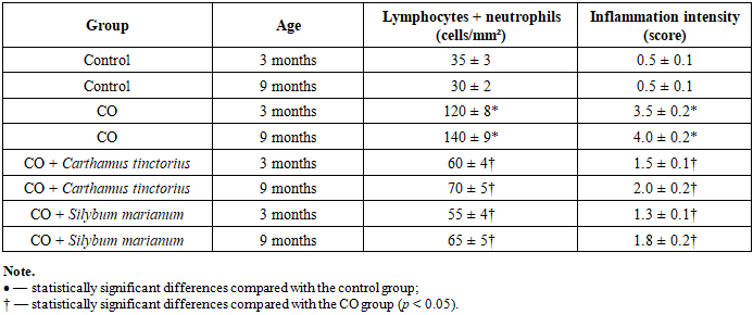
- Chronic exposure to carbon monoxide resulted in significant structural and morphometric alterations of the esophageal wall compared with the control groups (Table 1).
|
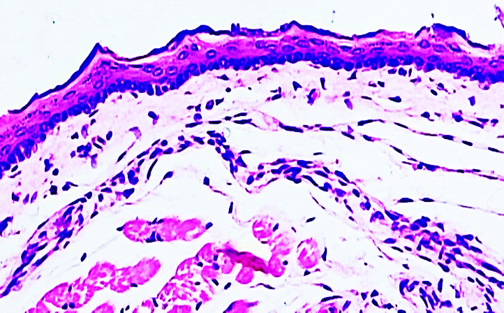 | Figure 1. Morphological and morphometric changes in the esophageal tissue of a 3-month-old white outbred rat from the control group. Hematoxylin–eosin staining. Magnification ×20, ×40 (objective) |
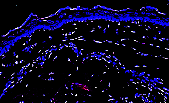 | Figure 2. Digital morphometric analysis of the selected area of esophageal tissue. The esophageal tissue was segmented using a polygonal method (red contour), and the measurement area is highlighted with a dark blue mask. |
 | Figure 3. Identification of morphological and morphometric changes observed in the esophageal tissues of 9-month-old white outbred rats subjected to chronic carbon monoxide exposure |
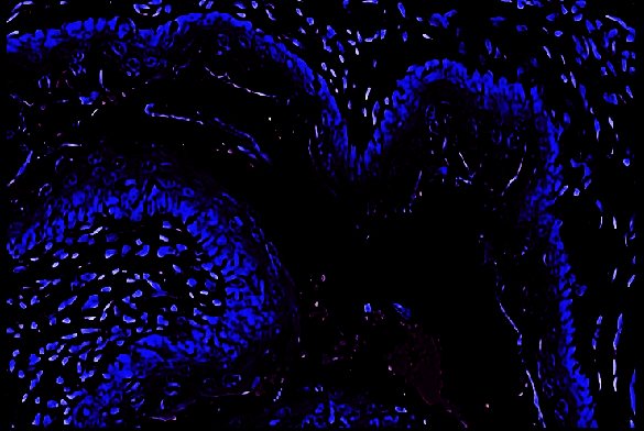 | Figure 4. Digital morphometric analysis of esophageal tissue in 9-month-old white outbred rats under chronic carbon monoxide exposure. Polygonal segmentation (red contour); measurement area marked in dark blue |
|
4. Conclusions
- Chronic exposure to carbon monoxide is associated with pronounced structural and morphometric alterations of the esophageal wall, reflecting the development of persistent hypoxia-induced tissue injury. The identified changes are characterized by epithelial atrophy, microcirculatory impairment, inflammatory infiltration, and disruption of tissue homeostasis. Notably, pathological alterations were more pronounced in older animals, indicating an age-dependent susceptibility of the esophagus to chronic hypoxic exposure.The application of phytocorrection using plant-derived biostimulators demonstrated a marked protective and restorative effect on esophageal morphology. Administration of Silybum marianum and Carthamus tinctorius extracts resulted in attenuation of inflammatory and dystrophic changes, improvement of microcirculatory parameters, and partial restoration of epithelial and submucosal architecture. The most pronounced regenerative and anti-inflammatory effects were observed with Silybum marianum, whereas combined phytocorrection provided additional normalization of structural integrity.Overall, the obtained results indicate that chronic carbon monoxide exposure leads to sustained hypoxia-induced esophageal injury, while phytocorrection represents a promising approach for reducing morphological damage and promoting regenerative processes. The present findings substantiate the rationale for further investigations aimed at dose optimization, elucidation of underlying mechanisms, and evaluation of the translational and clinical potential of phytobiostimulators.