-
Paper Information
- Next Paper
- Previous Paper
- Paper Submission
-
Journal Information
- About This Journal
- Editorial Board
- Current Issue
- Archive
- Author Guidelines
- Contact Us
American Journal of Medicine and Medical Sciences
p-ISSN: 2165-901X e-ISSN: 2165-9036
2026; 16(1): 204-207
doi:10.5923/j.ajmms.20261601.46
Received: Dec. 28, 2025; Accepted: Jan. 21, 2026; Published: Jan. 22, 2026

Clinical and Diagnostic Significance of κ-Free Light Chains and Oligoclonal Bands in Multiple Sclerosis in Individuals of Uzbek Ethnicity
 Abstract
 Reference
 Full-Text PDF
 Full-text HTML
Abdullazizova Umidakhon Saloxiddin qizi, Musayeva Yulduz Alpisovna
Department of Neurology, Tashkent State Medical University, Tashkent, Uzbekistan
Copyright © 2026 The Author(s). Published by Scientific & Academic Publishing.
This work is licensed under the Creative Commons Attribution International License (CC BY).
http://creativecommons.org/licenses/by/4.0/

Background: Multiple sclerosis (MS) is a chronic immune-mediated disease of the central nervous system, in which early and accurate diagnosis plays a crucial role in preventing irreversible neurological disability. Oligoclonal bands (OCB) in cerebrospinal fluid (CSF) are considered the gold standard laboratory marker of intrathecal immunoglobulin synthesis; however, their determination is invasive and not always widely available. In recent years, κ-free light chains (κFLC) have emerged as a promising quantitative biomarker of B-cell–mediated intrathecal immune activation. Data on their diagnostic value in different ethnic populations, including individuals of Uzbek ethnicity, remain limited. Objective: To evaluate the clinical and diagnostic significance of κ-free light chains and oligoclonal bands in patients with multiple sclerosis of Uzbek ethnicity. Methods: A single-center comparative study was conducted between 2023 and 2025 and included 60 patients with confirmed multiple sclerosis. Thirty patients underwent CSF and serum analysis, while 30 patients were examined using serum samples only. A control group consisted of 25 individuals without demyelinating diseases. κ-free light chains were measured using the Freelite® assay on an Optilite® analyzer, and the κFLC-index was calculated to assess intrathecal synthesis. Oligoclonal bands were detected by isoelectric focusing with immunoblotting. Statistical analysis included ROC curve evaluation, correlation analysis, and nonparametric testing. Results: All patients with multiple sclerosis demonstrated elevated κFLC-index values exceeding the diagnostic threshold (>5.9), whereas no pathological values were observed in the control group. Oligoclonal bands were detected in 87% of patients. The κFLC-index showed high diagnostic accuracy, with sensitivity ranging from 90% to 95% and specificity from 85% to 90%. Moderate correlations were identified between κFLC-index and chronic MRI markers of white matter damage, including T1 hypointense lesions. Quantitative assessment of κFLC demonstrated higher diagnostic informativeness compared with absolute CSF κFLC levels alone. Conclusion: κ-free light chains represent a highly informative quantitative biomarker of intrathecal immune activity in multiple sclerosis among individuals of Uzbek ethnicity. Combined assessment of κFLC and oligoclonal bands enhances diagnostic accuracy and is particularly valuable in the differential diagnosis of MS and other inflammatory disorders of the central nervous system, including CLIPPERS syndrome.
Keywords: Multiple sclerosis, κ-free light chains, Oligoclonal bands, Cerebrospinal fluid, Intrathecal immunity, Diagnostic biomarkers, Uzbek population, CLIPPERS syndrome
Cite this paper: Abdullazizova Umidakhon Saloxiddin qizi, Musayeva Yulduz Alpisovna, Clinical and Diagnostic Significance of κ-Free Light Chains and Oligoclonal Bands in Multiple Sclerosis in Individuals of Uzbek Ethnicity, American Journal of Medicine and Medical Sciences, Vol. 16 No. 1, 2026, pp. 204-207. doi: 10.5923/j.ajmms.20261601.46.
1. Introduction
- Multiple sclerosis (MS) is a chronic immune-mediated inflammatory disease of the central nervous system and remains one of the leading causes of non-traumatic disability in young and middle-aged individuals [1,2]. Early and accurate diagnosis of MS is of key importance, since timely initiation of disease-modifying therapy makes it possible to slow the progression of neurological deficit and improve long-term prognosis [1]. In this regard, sustained interest remains in the search for reliable laboratory markers reflecting the activity of the intrathecal immune process.At present, the detection of oligoclonal immunoglobulin bands (OCB) in cerebrospinal fluid (CSF) remains a generally accepted laboratory standard for confirming intrathecal immunoglobulin synthesis and is included in the McDonald diagnostic criteria [3,4]. At the same time, the method of OCB determination has a number of limitations, including the invasiveness of lumbar puncture, the labor-intensive nature of laboratory analysis, and limited availability in a number of clinical institutions, which stimulates the search for alternative or additional biomarkers [5,6].In recent years, special attention has been paid to kappa free light chains of immunoglobulins (κFLC), which are considered a quantitative marker of B-cell activation [7,8,9]. Increased levels of κFLC in CSF reflect an intrathecal immune response and demonstrate high diagnostic sensitivity in MS [7,9]. At the same time, the clinical significance of determining κFLC in serum as a less invasive and more accessible method is being discussed; however, data on its diagnostic informativeness remain limited and inconsistent [10,11,12]. It should be noted that most studies investigating κ-free light chains and oligoclonal bands have been conducted in European and North American populations, whereas data on other ethnic groups remain limited. As a result, the applicability of these findings to populations with different genetic and environmental backgrounds may be restricted. In addition, epidemiological and biomarker-related data on multiple sclerosis and other immune-mediated neurological disorders remain insufficient in several regions, including Central Asia [13]. In particular, individuals of Uzbek ethnicity are underrepresented in current research, which may limit the generalizability of existing diagnostic approaches. In this context, population-specific studies are important for improving the understanding of disease characteristics and for optimizing diagnostic strategies in routine clinical practice. In clinical practice, multiple sclerosis also requires careful differential diagnosis with other inflammatory disorders of the central nervous system, including CLIPPERS syndrome, in which diagnostic uncertainty may lead to fundamentally different therapeutic decisions [16,17]. Therefore, the assessment of immunological biomarkers acquires particular clinical relevance.
2. Materials and Methods
- Study design and sampleA single-center clinical and laboratory comparative study was conducted, the purpose of which was to assess the diagnostic significance of κ-free light chains of immunoglobulins and oligoclonal bands in multiple sclerosis. Patient recruitment and data analysis were carried out during 2023–2025. The study included 60 patients with an established diagnosis of multiple sclerosis. All examined individuals belonged to Uzbek ethnicity. The diagnosis was confirmed based on clinical presentation and magnetic resonance imaging data in accordance with current diagnostic criteria.Taking into account the scope of laboratory examination, patients were divided into groups. The main group included 30 patients who underwent examination of both cerebrospinal fluid and serum. The serum group consisted of 30 patients examined without lumbar puncture. A control group of 25 individuals without signs of demyelinating diseases was used to assess reference values of the κFLC-index. Patients with systemic autoimmune diseases, active infectious processes, pronounced renal dysfunction, as well as other conditions potentially affecting the concentration of immunoglobulin free light chains were excluded from the study.Laboratory methods Quantitative determination of κ-free light chains was performed in cerebrospinal fluid and serum using Freelite® kits on an Optilite® automated analyzer (The Binding Site, United Kingdom). The obtained values were expressed in milligrams per liter.To assess intrathecal synthesis of κ-free light chains, the κFLC-index was calculated based on the ratio of κFLC and albumin concentrations in CSF and serum. A threshold value of κFLC-index > 5.9 was used as diagnostically significant.Detection of oligoclonal immunoglobulin bands in cerebrospinal fluid was carried out using isoelectric focusing followed by immunoblot detection. Interpretation of the results was performed in accordance with the generally accepted international classification.Statistical analysisStatistical analysis was performed using IBM SPSS Statistics software (version 26.0). Measures of central tendency and dispersion were used to describe the data. Intergroup differences were assessed using nonparametric methods. Diagnostic informativeness of the studied markers was analyzed using ROC curve analysis with calculation of the area under the curve, sensitivity, and specificity. Differences were considered statistically significant at p < 0.05.
3. Results
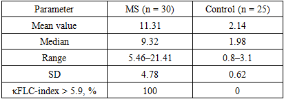
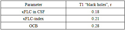
- Analysis of the κFLC-index revealed pronounced differences between patients with multiple sclerosis and the control group. In all patients with MS, κFLC-index values exceeded the diagnostic threshold, whereas no pathological values were recorded in the control group.
|
|
4. Discussion
- The obtained results confirm the high diagnostic significance of κ-free light chains in cerebrospinal fluid as a quantitative marker of intrathecal immune response in multiple sclerosis [7–9]. In contrast to qualitative detection of oligoclonal bands, quantitative assessment of κFLC allows a more objective characterization of the degree of immune activity. The results of the study acquire particular clinical significance in the context of differential diagnosis of multiple sclerosis with other inflammatory diseases of the central nervous system, in particular CLIPPERS syndrome [16,17]. Both conditions may have similar clinical and radiological manifestations but require fundamentally different therapeutic approaches. In clinical practice, we observed a patient with a clinical and radiological picture resembling CLIPPERS syndrome and multiple sclerosis, in whom comprehensive immunological diagnostics played a decisive role in the choice of treatment strategy [15].Thus, combined assessment of κ-free light chains and oligoclonal bands may contribute to clarification of the diagnosis and optimization of treatment tactics in demyelinating diseases of the central nervous system.
5. Conclusions
- Determination of κ-free light chains of immunoglobulins in cerebrospinal fluid has high clinical and diagnostic informativeness in multiple sclerosis in individuals of Uzbek ethnicity. Quantitative assessment of κ-free light chains reflects the intrathecal immune response and may be considered a reliable laboratory marker of the disease.The obtained results acquire particular clinical significance in the context of differential diagnosis of multiple sclerosis with other inflammatory diseases of the central nervous system, in particular CLIPPERS syndrome. Both conditions may be accompanied by activation of the B-cell immune response and demonstrate similar clinical and radiological manifestations but require fundamentally different therapeutic strategies [16,17]. In such clinical situations, comprehensive assessment of immunological biomarkers, including κ-free light chains and oligoclonal bands, may contribute to clarification of the diagnosis and selection of an optimal treatment strategy [15].