-
Paper Information
- Next Paper
- Previous Paper
- Paper Submission
-
Journal Information
- About This Journal
- Editorial Board
- Current Issue
- Archive
- Author Guidelines
- Contact Us
American Journal of Medicine and Medical Sciences
p-ISSN: 2165-901X e-ISSN: 2165-9036
2026; 16(1): 183-186
doi:10.5923/j.ajmms.20261601.41
Received: Dec. 28, 2025; Accepted: Jan. 17, 2026; Published: Jan. 20, 2026

Morphological and Functional Features of Fetal Internal Organs in Forensic Practice
 Abstract
 Reference
 Full-Text PDF
 Full-text HTML
Mansurova Dilafruz Odilovna1, Elieva Mehriniso Fakhridinnovna2
1Independent Researcher, Tashkent State Medical University, Tashkent, Uzbekistan
2Tashkent State Medical University, Tashkent, Uzbekistan
Correspondence to: Mansurova Dilafruz Odilovna, Independent Researcher, Tashkent State Medical University, Tashkent, Uzbekistan.
| Email: |  |
Copyright © 2026 The Author(s). Published by Scientific & Academic Publishing.
This work is licensed under the Creative Commons Attribution International License (CC BY).
http://creativecommons.org/licenses/by/4.0/

A forensic medical evaluation of the morphofunctional characteristics of fetal internal organs was conducted with the aim of improving the accuracy of gestational age determination based on a comprehensive analysis of forensic medical protocol data. The study material comprised 65 forensic medical reports (forensic examination protocols) of fetuses referred for expert evaluation due to intrauterine fetal death, stillbirth, and neonatal death of unknown etiology мicroscopic assessment included analysis of the degree of pulmonary alveolar differentiation, the intensity of hepatic hematopoiesis, the development of corticomedullary differentiation in the kidneys, the structural organization of the myocardium, and the degree of thymic maturity. Gestational age was determined by correlating the morphological characteristics of internal organs with generally accepted embryological and morphometric criteria of intrauterine development.
Keywords: Forensic medical examination, Gestational age, Fetus, Morphofunctional characteristics, Organogenesis, Histological diagnosis
Cite this paper: Mansurova Dilafruz Odilovna, Elieva Mehriniso Fakhridinnovna, Morphological and Functional Features of Fetal Internal Organs in Forensic Practice, American Journal of Medicine and Medical Sciences, Vol. 16 No. 1, 2026, pp. 183-186. doi: 10.5923/j.ajmms.20261601.41.
1. Introduction
- Determining the gestational age of the fetus is a key task in forensic medical examinations during the investigation of perinatal and intrauterine deaths, as well as in cases of criminal abortions, intrauterine death, and neonatal deaths of unknown etiology [1,2,3]. Accurate verification of the gestational age is essential for the correct legal classification of the case, establishing fetal viability, assessing the timeliness and adequacy of medical care, and for the differential diagnosis of antenatal and intrapartum deaths. Modern forensic practice demonstrates that traditional anthropometric indicators (weight, fetal length, head circumference) do not always reflect the actual gestational age, especially in cases of intrauterine growth restriction, chronic placental insufficiency, intrauterine hypoxia, and congenital pathology [4,5,6]. Under these circumstances, the role of morphofunctional criteria of internal organs, which more objectively reflect the degree of fetal maturity and the stages of organogenesis, increases. Morphological assessment of the degree of parenchymal organ differentiation, the maturity of the alveolar apparatus of the lungs, the corticomedullary differentiation of the kidneys, and the development of the thymus, adrenal glands, and liver allows for increased accuracy in gestational age determination, especially at the borderline stages of fetal viability [7,8]. A comprehensive analysis of macroscopic, histological, and, where necessary, immunohistochemical features forms a scientifically sound algorithm for forensic diagnostics. In the future, standardization of morphofunctional criteria for internal organs and their implementation in forensic practice will improve the objectivity of expert opinions, reduce diagnostic discrepancies, and ensure a unified interdisciplinary approach to assessing fetal gestational age, which has important medical, legal, and social implications.ObjectivesThe aim of this study was a forensic medical assessment of the morphofunctional characteristics of the internal organs of the fetus in order to improve the accuracy of determining gestational age based on a comprehensive analysis of forensic medical records.
2. Materials and Methods
- The study was based on the analysis of 65 forensic medical examination reports of fetuses submitted for expert evaluation due to intrauterine fetal death, stillbirth, or neonatal death of unknown etiology. All examinations were conducted in accordance with standard forensic medical protocols. In each case, a complete forensic autopsy was performed, including external examination and anthropometric assessment (body weight, crown–heel length, head circumference, and chest circumference). The morphofunctional condition of internal organs, particularly the lungs, liver, kidneys, heart, thymus, spleen, and adrenal glands, was evaluated.Tissue samples were fixed in 10% neutral buffered formalin and processed using routine histological techniques with hematoxylin and eosin (H&E) staining. Microscopic evaluation focused on the degree of pulmonary alveolar differentiation, hepatic hematopoietic activity, corticomedullary differentiation of the kidneys, myocardial organization, and maturity of lymphoid and endocrine organs. Gestational age was determined by correlating morphofunctional findings with established embryological and morphometric criteria. Cases were divided into two groups: second trimester (22–27 weeks) and third trimester (≥28 weeks). Statistical analysis was performed using Microsoft Excel, applying the Pearson χ² test and Mann–Whitney U test. Differences were considered significant at p < 0.05.
3. Results and Discussion
- Analysis of the examined cases revealed that morphofunctional characteristics of fetal internal organs provide reliable indicators of gestational maturity. Microscopic evaluation demonstrated consistent associations between the degree of organ differentiation and gestational age, irrespective of fetal anthropometric parameters. In the kidneys, marked variability in corticomedullary differentiation was observed. The predominance of a wide nephrogenic zone and the presence of immature glomeruli were characteristic of earlier gestational stages, even in fetuses with relatively higher body weight. These findings highlight the limited diagnostic value of anthropometric data alone and emphasize the importance of renal histological maturity as an indicator of gestational age. Pulmonary tissue exhibited gestational age–dependent differences in the development of the alveolar apparatus. In earlier stages, the lungs were characterized by an immature alveolar structure with poorly differentiated air spaces, whereas progressive gestational advancement was associated with increased alveolar complexity and the appearance of secondary alveoli. These changes reflect functional maturation of the respiratory system and are critical for assessing fetal viability in forensic practice.Hepatic morphology demonstrated a gradual decline in hematopoietic activity with increasing gestational age. Pronounced extramedullary hematopoiesis was a typical finding in less mature fetuses, while its reduction indicated advancing fetal development. Similar gestational trends were observed in the myocardium, where increasing structural organization and fiber alignment corresponded to later stages of intrauterine maturation.The thymus and adrenal glands showed distinct morphofunctional patterns related to gestational age. Insufficient corticomedullary differentiation of the thymus and predominance of the fetal zone in the adrenal glands were indicative of earlier developmental stages. In contrast, advanced gestation was associated with thymic structural maturation, reflecting functional development of the fetal immune system, and a relative reduction of the fetal adrenal zone.Comparative analysis between fetuses in the second and third trimesters revealed clear differences in the degree of morphofunctional maturity of internal organs. The second trimester was characterized by features of structural immaturity, including persistent nephrogenic zones in the kidneys, active hepatic hematopoiesis, and incomplete alveolar development. In the third trimester, organ morphology reflected functional readiness, with mature pulmonary architecture, reduced nephrogenesis, decreased hematopoietic activity in the liver, and advanced myocardial organization.Overall, the results demonstrate that a comprehensive morphofunctional assessment of fetal internal organs possesses high discriminatory capacity for distinguishing between gestational stages.
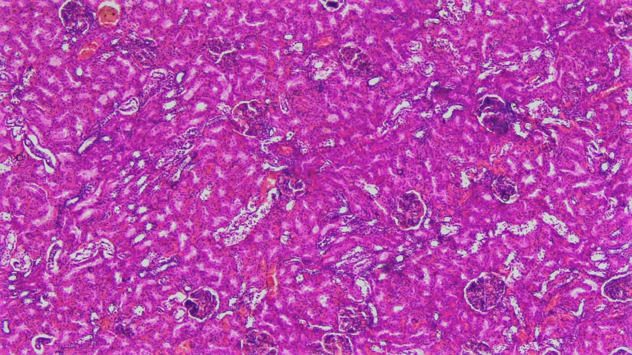
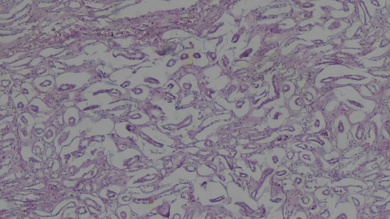
|
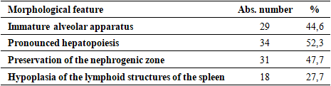
 | Figure 1. The criteria for immaturity of fetal kidney tissue are shown; there are embryonic glomeruli at 24 weeks, surrounded by heme-eosin, magnification: 10x10 |
 | Figure 2. Immunity of nephrons in a deceased fetus at 28 weeks of gestational age, surrounded by heme-eosin, magnification: 10x10 |
 | Figure 3. Lung. The alveolar structure is largely preserved in the lung tissue; however, the interalveolar septa are unevenly thickened due to severe edema, vascular congestion, and cellular infiltration. Heme-eosin staining. 10x10 |
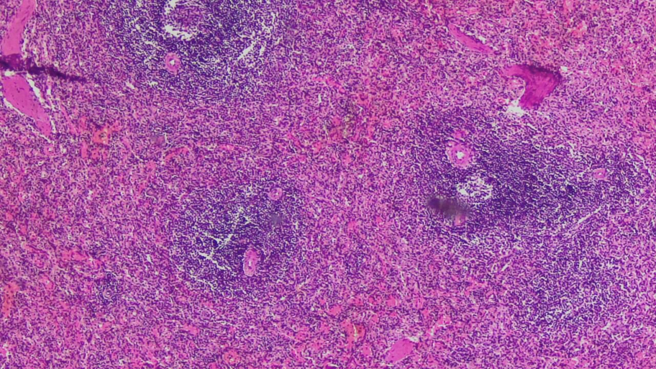 | Figure 4. The spleen contains secondary germinal centers of lymphoid follicles and vascular hyalinosis. Heme-eosin staining. 10x10 |
4. Conclusions
- The present study demonstrates that a comprehensive morphofunctional assessment of fetal internal organs is a highly informative and reliable approach for determining gestational age in forensic medical practice. The results confirm that morphological criteria of organ maturity provide more objective and accurate indicators of fetal development than isolated anthropometric parameters, particularly in cases complicated by intrauterine growth restriction, placental insufficiency, or hypoxic conditions. Distinct gestational age–dependent patterns were identified in the lungs, kidneys, liver, heart, thymus, spleen, and adrenal glands. Renal corticomedullary differentiation, pulmonary alveolar maturation, the degree of hepatic hematopoietic activity, myocardial structural organization, and the developmental status of lymphoid and endocrine organs demonstrated high discriminatory value in differentiating between the second and third trimesters of gestation. These findings emphasize the importance of internal organ morphology as a key determinant of fetal maturity and viability.The study highlights the limitations of relying solely on external anthropometric measurements for gestational age estimation and supports the integration of detailed histomorphological analysis into routine forensic examination algorithms. Standardization and systematic application of morphofunctional criteria will enhance the objectivity, reproducibility, and diagnostic accuracy of forensic expert conclusions, contributing to improved medico-legal decision-making in cases of perinatal and intrauterine death.Overall, the incorporation of expanded morphofunctional evaluation of fetal internal organs represents a scientifically grounded and practically valuable advancement in forensic diagnostics, with significant medical, legal, and social implications.