-
Paper Information
- Next Paper
- Paper Submission
-
Journal Information
- About This Journal
- Editorial Board
- Current Issue
- Archive
- Author Guidelines
- Contact Us
American Journal of Medicine and Medical Sciences
p-ISSN: 2165-901X e-ISSN: 2165-9036
2026; 16(1): 150-152
doi:10.5923/j.ajmms.20261601.33
Received: Sep. 23, 2025; Accepted: Oct. 28, 2025; Published: Jan. 20, 2026

Immunological Alterations in Children with Small Intestinal Diseases in the Aral Sea Region: A Pilot Study from Uzbekistan
 Abstract
 Reference
 Full-Text PDF
 Full-text HTML
Nargiza Abdurazzakovna Amanova, Nigora Rustamovna Aliyeva
Department of Hospital Pediatrics, Folk Medicine, Tashkent State Medical University, Uzbekistan
Copyright © 2026 The Author(s). Published by Scientific & Academic Publishing.
This work is licensed under the Creative Commons Attribution International License (CC BY).
http://creativecommons.org/licenses/by/4.0/

Background: Children living in the Aral Sea region are exposed to chronic environmental stressors that may contribute to gastrointestinal and immune disorders. Small intestinal diseases such as malabsorption syndromes, celiac disease, and chronic enteropathies remain understudied in this population [4]. Objective: To evaluate immunological profiles in children with small intestinal diseases residing in the Aral Sea region of Uzbekistan. Methods: We conducted a cross-sectional study involving 185 children aged 3–16 years. The main group consisted of 125 children with clinically confirmed small intestinal diseases; The comparison group (n=60) consisted of patients with diseases of the small intestine living in Tashkent; 40 healthy children from the same region served as controls. Clinical examination, complete blood count, biochemistry, and immunological assays were performed. Serum immunoglobulins (IgA, IgE) and cytokines (IL-1β, IL-4) were measured using ELISA. Statistical analysis included Mann–Whitney U test, Student’s t-test, and Spearman correlation. Results: Children with intestinal disorders demonstrated significant immunological alterations: decreased IgA while IgE was elevated (p<0.05). Pro-inflammatory cytokines IL-4 and IL-1β were elevated. The severity of immune disturbances correlated with clinical disease severity (r=0.62; p<0.01). Conclusion: Children with small intestinal diseases in the Aral Sea region exhibit marked immune dysregulation. These findings highlight the need for early immunological monitoring and targeted therapeutic strategies.
Keywords: Children, Small intestine, Immunity, Enteropathy, Aral Sea region, Uzbekistan
Cite this paper: Nargiza Abdurazzakovna Amanova, Nigora Rustamovna Aliyeva, Immunological Alterations in Children with Small Intestinal Diseases in the Aral Sea Region: A Pilot Study from Uzbekistan, American Journal of Medicine and Medical Sciences, Vol. 16 No. 1, 2026, pp. 150-152. doi: 10.5923/j.ajmms.20261601.33.
1. Introduction
- The Aral Sea ecological crisis has created one of the most environmentally affected regions worldwide. Chronic exposure to pollutants, malnutrition, and infectious agents has been linked to rising gastrointestinal morbidity among children [2,3]. However, the immunological mechanisms underlying small intestinal diseases in this population remain poorly understood. This study aims to fill this gap by evaluating immune alterations in children with enteropathies in the Aral Sea region. The problems of small intestine diseases in children living in ecologically unfavorable areas, such as the Aral Sea region, are becoming particularly relevant. These regions are characterized by a high level of anthropogenic stress, which affects the health of the younger generation, including the indicators of immune status. The study of the immunological status of children with pathologies of the small intestine in these conditions makes it possible to identify characteristic changes in the body's immune responses and assess their severity [1,5].
2. Materials and Methods
- Study design and participants: A cross-sectional study was carried out in 2023–2024 in Aral Sea region, Uzbekistan. Main group: 125 children aged 3–16 years with clinically and laboratory confirmed small intestinal diseases (allergic enterocolitis, celiac disease, chronic enteropathies). The comparison group (n=60) consisted of patients with diseases of the small intestine living in Tashkent. Control group: 25 age- and sex-matched healthy children.Immunological assays: ELISA: IgA, IgE; cytokines (IL-1β, IL-4).Statistics: Analysis was performed with SPSS 25.0. Differences between groups were assessed using t-test or Mann–Whitney U test. Correlations were evaluated with Spearman’s rank test. Significance was set at p<0.05.
3. Results
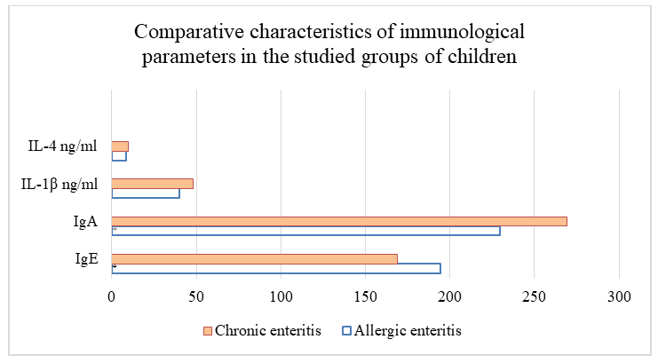
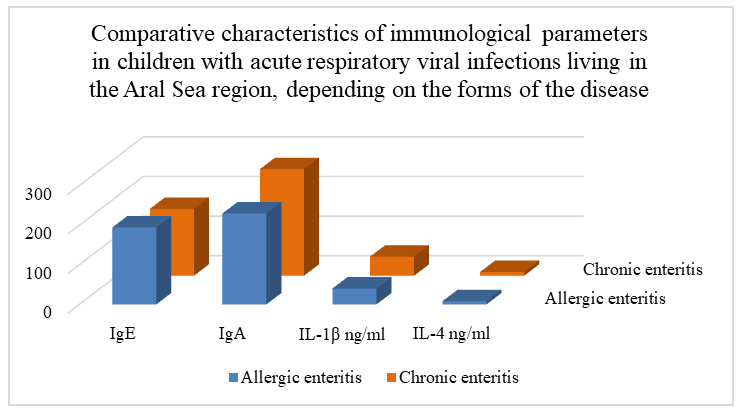
- Based on the objectives of this study, we conducted a comparative analysis of immunological parameters in children with small intestine diseases living in the Aral Sea region, the results of which are shown in the table 1.
 | Table 1 |
 | Table 2 |
4. Conclusions
- The cytokine status in children of the Aral Sea region with diseases manifested by maldigestion and malabsorption is characterized by an almost 3-fold increase in IL-1 β in relation to children with this pathology living in Tashkent against the background of a significant decrease in IL-4. These indicators reflected an increased inflammatory response in patients with intestinal diseases. A significantly significant increase in IgE and ID in peripheral blood in children of the Aral Sea region indicates an increased allergic background and activation of humoral immunity in children with intestinal diseases in this area in relation to children living in Tashkent. These markers may serve as predictors of disease severity and targets for immunomodulatory interventions.