-
Paper Information
- Next Paper
- Previous Paper
- Paper Submission
-
Journal Information
- About This Journal
- Editorial Board
- Current Issue
- Archive
- Author Guidelines
- Contact Us
American Journal of Medicine and Medical Sciences
p-ISSN: 2165-901X e-ISSN: 2165-9036
2026; 16(1): 22-25
doi:10.5923/j.ajmms.20261601.06
Received: Dec. 14, 2025; Accepted: Jan. 3, 2026; Published: Jan. 7, 2026

The Importance of Clinical and Laboratory Markers in Assessing the Development of Osteoporosis in Middle-Aged and Elderly Patients with Non-Alcoholic Fatty Liver Disease
 Abstract
 Reference
 Full-Text PDF
 Full-text HTML
Yuldasheva Dilnavoz Xasanovna1, Axmedov Shamshod Janshidovich2
1DSc, Dots. Head of the Department of Pharmacology of Bukhara State Medical Institute, Bukhara, Uzbekistan
2Assistеnt of the Department of Pharmacology of Bukhara State Medical Institute, Bukhara, Uzbekistan
Correspondence to: Yuldasheva Dilnavoz Xasanovna, DSc, Dots. Head of the Department of Pharmacology of Bukhara State Medical Institute, Bukhara, Uzbekistan.
| Email: |  |
Copyright © 2026 The Author(s). Published by Scientific & Academic Publishing.
This work is licensed under the Creative Commons Attribution International License (CC BY).
http://creativecommons.org/licenses/by/4.0/

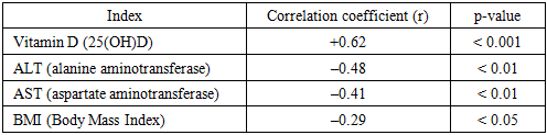
Nonalcoholic fatty liver disease is one of the most pressing problems in modern medicine, and its components, metabolic syndrome, insulin resistance, and chronic inflammation, negatively affect many systems, including bone metabolism. The study results revealed a reliable correlation between liver enzymes, vitamin D, parathyroid hormone, bone turnover biomarkers, and bone mineral density. Non-alcoholic fatty liver disease showed that individuals with nonalcoholic fatty liver disease are at high risk of developing osteoporosis. This study examined the prevalence of nonalcoholic fatty liver disease in middle-aged and elderly people. The risk of osteoporosis in patients with osteoporosis was comprehensively assessed using clinical, biochemical, and hormonal markers.
Keywords: Osteoporosis, Bone mineral density level, Osteoporosis, Vitamin D
Cite this paper: Yuldasheva Dilnavoz Xasanovna, Axmedov Shamshod Janshidovich, The Importance of Clinical and Laboratory Markers in Assessing the Development of Osteoporosis in Middle-Aged and Elderly Patients with Non-Alcoholic Fatty Liver Disease, American Journal of Medicine and Medical Sciences, Vol. 16 No. 1, 2026, pp. 22-25. doi: 10.5923/j.ajmms.20261601.06.
1. Introduction
- Nonalcoholic fatty liver disease (NAFLD) is a metabolic disease that occurs in approximately 25-30% of the world's population. Insulin resistance, lipid metabolism disorder, inflammatory process and oxidative stress play a key role in its pathogenesis. In recent years, NAFLD has become a topical issue not only in hepatology, but also in endocrinology, cardiology, and osteology. The results of scientific studies show that people with NAFLD have a high risk of developing low bone mineral density, osteopenia, and osteoporosis. [1,3,5,7,10]. Osteoporosis is a chronic systemic disease characterized by deterioration of the microarchitectural structure of bone tissue and a decrease in bone mineral density, which leads to increased bone fragility and fracture. This problem has a serious impact on the quality of life, especially in middle-aged and elderly people. Therefore, the use of clinical and laboratory markers for early detection and prognosis of bone changes associated with NAFLD is of great importance [2,4,6,8,9]. The aim of the study is to assess the risk of NAFLD in middle-aged and elderly patients with NAFLD. Comprehensive assessment of the risk of osteoporosis development in patients with osteoporosis using clinical, biochemical and hormonal markers.
2. Material and Methods
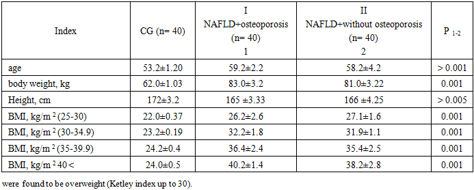
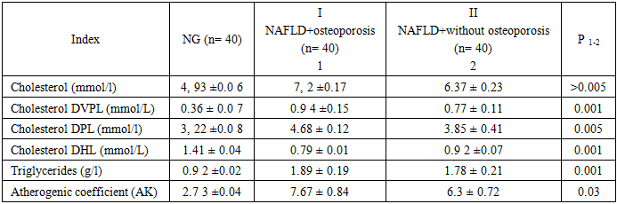
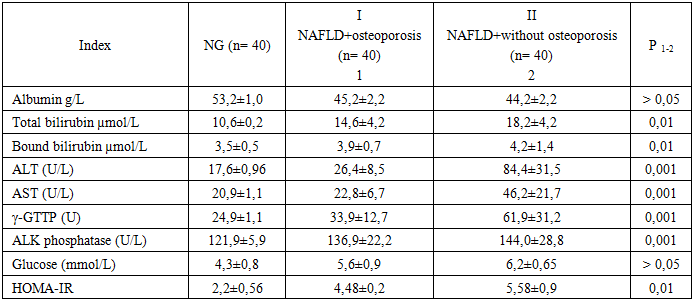
- 120 patients with nonalcoholic fatty liver disease were selected for the study, of which 46 (66 %) were women and 24 (34 %) were men, aged 45–75 years (mean age 59.2 ±4.2). All patients included in the study were divided into three groups. The first group of the study included patients with NAFLD + osteoporosis (40 patients); the second group included patients with group II – OA, but without osteoporosis (40 people); the third group of patients formed group III – healthy controls (40 people). The results of the study in all groups were evaluated by a clinical reference card (questionnaire). Consent was obtained from the members of the ethics committee established under the auspices of the Bukhara Medical Institute named after Abu Ali ibn Sina to carry out this research. In the study are as follows: with confirmed UTI; no alcohol consumption and no chronic hepatitis. Exclusion criteria: oncological diseases, steroid use, hormone therapy in menopause.The study was conducted in 2 stages. In the first stage, all patients in the main group were selected through a special questionnaire. Then, in the second stage of the study, patients in the main and control groups underwent laboratory and biochemical examinations. Checked parameters. Biochemical markers: In order to study the functional status of jigap, its lipid metabolism was investigated. The general cholesterol level (UXD) has been evaluated according to the classification of the European Atepoclepotic Society [11]: up to 5.2 mmol/l is the optimal level; 5.3-6.5 mmol/l — mild hypepxolectepinemia (GXC); 6.6-7.8 mmol/l — moderate, severe; Higher than 7.8 mmol/l — high. Expanded lipid lipoproteins were also studied: triglyceride (TG), cholecystokinin (XC) density pact lipopotein (DPL) and XC density high lipoprotein (DHL). Cholectepin very density pact lipopoteinlap (DVPL) invention was investigated. It is determined that TG is up to 1.7 mmol/l, cholectepin is higher than 2.6 mmol/l, cholectepin DHL is higher than 1.15 mmol/l. NAFLD To assess the functional state of the liver in patients, parameters of pigment metabolism, cytolysis and cholestasis were studied. C -reactive protein, Omega-3/6 ratio, markers of cholesterol metabolism: 25-hydroxyvitamin D (25(OH)D), parathyroid hormone (PTH), osteocalcin, CTX-1 (C-terminal telopeptide), P1NP (procollagen type 1 N-terminal propeptide) and instrumental testing methods: DEXA – lumbar spine and hip bone mineral density levels were assessed.
3. Results and Discussion
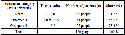
- Demographic and anthropometric indicators of patients were analyzed.
|
|
|
|
|