Ergashov Z. A., Nuralieva D. M.
Ministry of Health of the Republic of Uzbekistan, Military Medical Academy of the Armed Forces of the Republic of Uzbekistan
Copyright © 2025 The Author(s). Published by Scientific & Academic Publishing.
This work is licensed under the Creative Commons Attribution International License (CC BY).
http://creativecommons.org/licenses/by/4.0/

Abstract
The study presents the results of the investigation of morphometric characteristics of the thymus and biochemical indicators in patients with post-COVID dermatitis. Structural changes of the thymus gland were analyzed. It was established that patients who had COVID-19 and developed dermatological manifestations showed signs of thymic hypertrophy. The obtained data indicate the key role of immunometabolic alterations in the pathogenesis of post-COVID dermatitis and confirm the need for an integrated diagnostic approach including morphometric and biochemical assessments.
Keywords:
Thymus, Morphometry, Post-COVID dermatitis, Biochemical indicators, Immune system, Inflammation, COVID-19
Cite this paper: Ergashov Z. A., Nuralieva D. M., Morphometric of the Thymus and Characteristic Biochemical, Chronic Inflammation Coefficients Changes in Patients with Post-COVID Dermatitis, American Journal of Medicine and Medical Sciences, Vol. 15 No. 12, 2025, pp. 4476-4480. doi: 10.5923/j.ajmms.20251512.61.
1. Introduction
The COVID-19 pandemic, caused by the SARS-CoV-2 virus, has significantly affected not only the respiratory system but also the functioning of various organs and systems of the human body [3,7,10]. In recent years, increasing attention has been given to post-COVID complications, particularly dermatological manifestations, which are often associated with disturbances in immune and endocrine regulation. One of the key organs involved in immune response formation is the thymus gland [4,9,17]. The thymus plays a central role in the differentiation and maturation of T-lymphocytes, and its morphofunctional changes may reflect the state of systemic immunity [1,6]. After COVID-19 infection, signs of thymic dysfunction, atrophic processes, and remodeling of its morphometric parameters have been observed, possibly due to chronic inflammation and autoimmune reactions characteristic of post-COVID conditions [2,12].The study of thymic morphometry combined with biochemical analysis in post-COVID dermatitis patients is a relevant direction in modern medical science. It allows the establishment of links between immune and metabolic disorders, determines the thymus’s involvement in cutaneous reactions, and contributes to developing new diagnostic and therapeutic strategies for post-COVID syndromes [7,13,18].
2. Materials and Methods
The study was conducted in an outpatient setting at a military hospital between 2021 and 2024. It included 100% of patients with post-COVID dermatitis occurring 18–26 weeks after COVID-19 infection. Among them, 7.0% had lung lesions up to 80%, 26.5% up to 40%, and 34.0% up to 15%. In 18 patients (32.3%), no lung involvement was recorded. Diagnosis was confirmed by PCR, and no history of skin diseases was noted. Comprehensive testing included biochemical: vitamin D levels, liver enzymes (ALT, AST), bilirubin, urea, creatinine, and total protein. The control group consisted of 54 healthy individuals (78 men, mean age 39±3.4 years; 4 women, mean age 36±4.0 years). All patients were prescribed cholecalciferol + menaquinone-7 (125/50 µg per day) and magnesium arginine (644 mg daily) for 8 weeks. Analysis of clinical symptoms and quality of life in military patients with post-COVID dermatological syndrome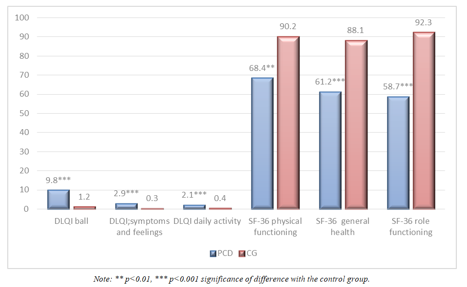 | Figure 1. Analysis of Quality of Life Indicators in Patients with Post-COVID Dermatitis |
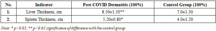
The total DLQI score was 9.8 compared to 1.2 in the control group (p<0.001). On the "Symptoms and Feelings" scale, patients scored 2.9 points, while the control group scored 0.3 (p<0.001). A similar trend was identified on the daily activity scale — 2.1 vs. 0.4 (p<0.001). According to SF-36 data, the differences were also statistically significant: physical functioning — 68.4 in patients vs. 90 in the control group (p<0.01); general health — 61.2 vs. 88.1 (p<0.001); role functioning related to emotional state — 58.7 vs. 92.3 (p<0.001).Post-COVID dermatitis leads to a significant decrease in the quality of life of patients across all studied scales. The obtained data indicate a pronounced negative impact of the disease on both the physical condition and the psycho-emotional well-being of patients, which emphasizes the need for a comprehensive approach to the treatment and rehabilitation of this category of patients.Characteristics of Peripheral Lymph Nodes in Patients with Post-COVID Dermatological SyndromeDuring the study, an increase in axillary lymph nodes was noted in 5.3% of patients, an increase in cervical lymph nodes in 3.5%, and an increase in supraclavicular lymph nodes in 4.7%.Table 1 presents data from an ultrasound examination of the liver and spleen sizes in military servicemen with post-COVID dermatological syndrome accompanied by post-COVID dermatitis, compared to the control group. The obtained results indicate significant differences between the study samples.Table 1. Indicators of Liver and Spleen Size in with Post-COVID Dermatitis According to Ultrasound Examination
 |
| |
|
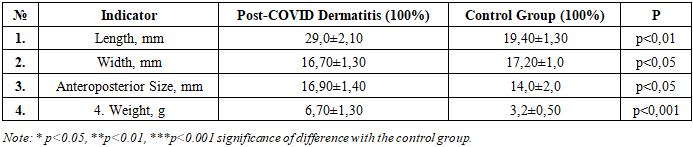
Thus, liver thickness in the main group of patients was 8.50±1.50 cm, which significantly exceeded the corresponding indicator in the control group (7.0±1.30 cm; p<0.01). A similar pattern was observed regarding the spleen: its thickness in patients with post-COVID changes reached 5.20±0.80 cm, while in the control group this indicator was 4.0±1.20 cm (p<0.05).Table 2 presents the results of an ultrasound examination of the thymus in military service patients with post-COVID dermatitis compared to the control group.Table 2. Morphometric Characteristics of the Thymus in Patients with Post-COVID Dermatitis
 |
| |
|
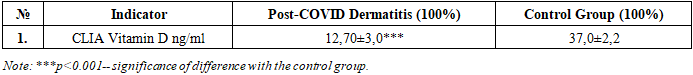
The obtained data indicate a significant increase in the size and mass of the thymus in patients of the main group. Thus, the length of the organ in patients with post-COVID dermatitis was 29.0±2.10 mm, which is significantly higher than the control values (19.40±1.30 mm; p<0.01). A similar trend was revealed when assessing the anteroposterior size of the thymus (16.70±1.40 mm vs. 17.2±1.0 mm; p<0.05). Slightly smaller but statistically significant differences were noted when comparing the width of the thymus (16.90±1.40 mm in the main group vs. 14.20±2.0 mm in the control; p<0.05). This fact indicates that hyperplastic changes in post-COVID pathology are predominantly longitudinal-axial, affecting the length and depth of the organ. The most pronounced differences were revealed in the analysis of thymus mass. In patients with post-COVID dermatitis, this indicator reached 6.70±1.30 g, which is almost twice the corresponding values in the control group (3.2±0.50 g; p<0.001). During the study, a comparative analysis of vitamin D levels was conducted in military servicemen who had COVID-19 and have post-COVID dermatitis, compared to a control group of healthy individuals (Table 3). According to the obtained data, the concentration of vitamin D in the blood serum of patients with skin manifestations of the post-COVID period was significantly reduced and averaged 12.7±3.0 ng/ml. At the same time, in the control group, this indicator was more than two and a half times higher — 37.0±2.2 ng/ml. The established differences between the study groups have high statistical significance (p<0.001), which confirms the reliability of the obtained results. The identified vitamin D deficiency in patients with post-COVID dermatitis can be considered as one of the unfavorable factors contributing to the formation and maintenance of pathological skin changes.Table 3. Characteristics of Vitamin D Levels in Patients with Post-COVID Dermatitis
 |
| |
|
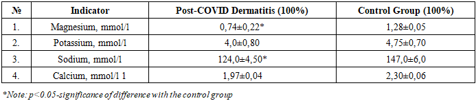
During the study, a comparative assessment of the levels of electrolytes (magnesium, potassium, sodium, and calcium) was conducted in military servicemen suffering from post-COVID dermatitis and in the control group. It was found that the concentration of magnesium in the blood serum of patients with post-COVID dermatitis was 0.74 ±0.22 mmol/l, which is significantly lower than the values in the control group (1.28 ±0.05 mmol/l; p<0.05). The decrease in magnesium levels may be associated with impaired metabolic processes and increased body demand for trace elements against the background of inflammatory reactions. Potassium levels in the main group (4.0±0.80 mmol/l) were also somewhat lower than in the control group (4.75±0.70 mmol/l), but no statistically significant difference was found. More pronounced changes were observed in sodium content: in patients with post-COVID dermatitis, its level was significantly reduced (124.0±4.50 mmol/l) compared to control values (147.0±6.0 mmol/l; p<0.05). This fact indicates possible disturbances in the water-salt balance in this category of patients. Calcium levels were also reduced in patients with post-COVID dermatitis (1.97±0.04 mmol/l), while in the control group it was 2.30 ± 0.06 mmol/l. Although the differences did not reach high statistical significance, the identified trend indicates the potential involvement of calcium metabolism in the pathogenesis of the disease.Table 4. Characteristics of Electrolyte Levels in Patients with Post-COVID Dermatitis
 |
| |
|
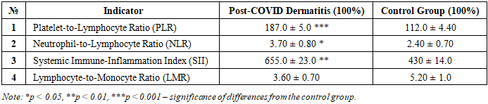
During the comparative analysis of chronic inflammation indicators in servicemen with post-COVID dermatitis and individuals from the control group, significant differences were identified across several indices (Table 5). The platelet-to-lymphocyte ratio (PLR) in patients with post-COVID dermatitis was statistically significantly higher than in the control group (187.0 ± 5.0 vs. 112.0 ± 4.40; p < 0.001). This finding indicates an enhancement of the platelet component of inflammation and the formation of a systemic inflammatory response. The neutrophil-to-lymphocyte ratio (NLR) was also significantly increased in the main group (3.70 ± 0.80 vs. 2.40 ± 0.70; p < 0.01), reflecting a tendency toward activation of the neutrophil component of innate immunity and a reduction in the immunoregulatory role of lymphocytes. The systemic immune-inflammation index (SII) in patients with post-COVID dermatitis reached 655.0 ± 23.0, which considerably exceeded the value in the control group (430 ± 14.0; p < 0.01). This result indicates pronounced chronic inflammatory activity and immune maladaptation. At the same time, the lymphocyte-to-monocyte ratio (LMR) was significantly lower in servicemen with post-COVID dermatitis compared to the control group (3.60 ± 0.70 vs. 5.20 ± 1.00; p < 0.05). The decrease in this index confirms the presence of monocytic predominance and the formation of chronic inflammation. In summary, the combination of obtained data indicates pronounced disturbances in immune homeostasis in servicemen with post-COVID dermatitis, manifested by activation of the systemic inflammatory response and a decrease in the lymphocytic component of immune regulation.Table 5. Characteristics of Chronic Inflammation Coefficients in patients with Post-COVID Dermatitis
 |
| |
|
Thus, the increase in mass and size of the thymus in patients with post-COVID dermatitis can be considered as a morphological reflection of disturbances in immune homeostasis. The thymus, being the central organ of T-lymphopoiesis, actively reacts to systemic inflammation and immune dysregulation, which is characteristic of the post-COVID period. On the one hand, hyperplasia of the gland may play a compensatory role, promoting increased production of T-cells and restoration of the immune response. On the other hand, excessive activation of the thymus can maintain the pathological hyperreactivity of the immune system underlying post-COVID dermatitis. Of particular importance is the fact that the most pronounced differences were noted precisely in the mass of the organ. This indicator integrally reflects not only morphological but also functional changes in the thymus. Consequently, the mass of the gland can be considered as a key criterion for assessing the degree of involvement of the organ in the pathological process in post-COVID complications. The obtained data are consistent with previously identified immune response disorders and complement the understanding of the pathogenesis of post-COVID dermatological manifestations. The results of the study indicate a possible pathogenetic role of vitamin D deficiency in the development of skin complications in military servicemen in the post-COVID period. The obtained data should be taken into account when developing comprehensive therapeutic and preventive measures, including correction of the vitamin status of this category of patients. Indicate significant shifts in the electrolyte composition of the blood in military servicemen with post-COVID dermatitis. The greatest changes concern the levels of magnesium and sodium, which may play an important role in the clinical course of the disease and require consideration when developing therapeutic measures.
References
| [1] | Iwasaki M, Saito J, Zhao H, Sakamoto A, Hirota K, Ma D. Inflammation Triggered by SARS-CoV-2 and ACE2 Augment Drives Multiple Organ Failure of Severe COVID-19: Molecular Mechanisms and Implications. Inflammation. 2021; 44(1): 13-34. https://doi.org/10.1007/s10753-020-01337-3. |
| [2] | World Health Organization. Clinical management of COVID-19: interim guidance. Geneva: WHO [1]; 2021. |
| [3] | Nalbandian A, Sehgal K, Gupta A, et al. Post-acute COVID-19 syndrome. Nat Med. 2021; 27(4): 601–615. |
| [4] | Yong SJ. Long COVID or post-COVID-19 syndrome: putative pathophysiology, risk factors, and treatments. Infect Dis. 2021; 53(10): 737–754. |
| [5] | Freeman EE, McMahon DE, Lipoff JB, et al. The spectrum of COVID-19–associated dermatologic manifestations: An international registry. J Am Acad Dermatol. 2020; 83(4): 1118–1129. |
| [6] | Suchonwanit P, Leerunyakul K, Kositkuljorn C. Cutaneous manifestations in COVID-19: lessons learned from current evidence. J Am Acad Dermatol. 2020; 83(1): e57–e60. |
| [7] | Genovese G, Moltrasio C, Berti E, Marzano AV. Skin manifestations associated with COVID-19: current knowledge and future perspectives. Dermatology. 2021; 237(1): 1–12. |
| [8] | Tang K, Wang Y, Zhang H, et al. Cutaneous manifestations of the coronavirus disease 2019 (COVID-19): a brief review. Dermatol Ther. 2020; 33(4): e13528. |
| [9] | McMahon DE, Amerson E, Rosenbach M, et al. Cutaneous manifestations of COVID-19: a systematic review. JAAD Int. 2021; 2: 119–132. |
| [10] | Herman A, Peeters C, Verroken A, et al. Evaluation of chilblains as a manifestation of the COVID-19 pandemic. JAMA Dermatol. 2020; 156(9): 998–1003. |
| [11] | Gupta A, Madhavan MV, Sehgal K, et al. Extrapulmonary manifestations of COVID-19. Nat Med. 2020; 26(7): 1017–1032. |
| [12] | Mazurov V.I., Petrova O.V. Immunopathogenesis and clinical manifestations of COVID-19. Therapeutic archive. 2021; 93(3): 4–11. |
| [13] | Fedorova N.Yu., Bazhenov D.V. Dermatological aspects of the new coronavirus infection. Russian Journal of Skin and Venereal Diseases. 2021; 24(3): 155–161. |
| [14] | Ahmed H, Patel K, Greenwood DC, et al. Long-term clinical outcomes in survivors of severe acute respiratory syndrome and Middle East respiratory syndrome coronavirus outbreaks after hospitalization: a systematic review and meta-analysis. J Rehabil Med. 2020; 52(5): jrm00063. |
| [15] | Kim J, Kim S, Kim HJ, et al. Long COVID in South Korea: national cohort study on risk of autoimmune skin diseases. JAMA Dermatol. 2023; 159(5): 490–498. |
| [16] | Lallas A, Kyrgidis A, Papageorgiou C, et al. COVID-19 and the skin: pathophysiology and clinical management. Clin Dermatol. 2021; 39(1): 82–89. |
| [17] | Russian Society of Dermatovenerologists. Guidelines for the management of patients with skin manifestations of COVID-19. Moscow; 2022. |
| [18] | Lee SW, Yang JM, Moon SY, et al. Association between COVID-19 infection and new-onset autoimmune diseases: a nationwide cohort study. Lancet Rheumatol. 2023; 5(2): e109–e118. |

 Abstract
 Reference
 Full-Text PDF
 Full-text HTML