American Journal of Medicine and Medical Sciences

p-ISSN: 2165-901X    e-ISSN: 2165-9036

2025;  15(12): 4422-4430

doi:10.5923/j.ajmms.20251512.50

Received: Nov. 12, 2025; Accepted: Dec. 2, 2025; Published: Dec. 12, 2025


Clinical and Genetic Parallels of Uterine Fibroids

Dinara Irnazarova1, Dilchekhra Yuldasheva1, Akmal Irnazarov2

1Department of Obstetrics and Gynecology, Tashkent State Medical University, Tashkent, Uzbekistan

2Department of Faculty and Hospital Surgery №1, Tashkent State Medical University, Tashkent, Uzbekistan

Correspondence to: Dinara Irnazarova, Department of Obstetrics and Gynecology, Tashkent State Medical University, Tashkent, Uzbekistan.

Email:

Copyright © 2025 The Author(s). Published by Scientific & Academic Publishing.

This work is licensed under the Creative Commons Attribution International License (CC BY).
http://creativecommons.org/licenses/by/4.0/

Abstract

Uterine fibroids are the most common neoplasm in women in gynecological practice, causing significant morbidity and impacting negatively on a woman's quality of life. Despite its widespread prevalence, uterine fibroids remain a relatively understudied disease. At the Multidisciplinary Clinic of the Tashkent Medical Academy, we studied 200 women of reproductive and premenopausal age in 2021-2023. All women underwent a general clinical and molecular genetic examination. The polymorphic marker of the estrogen and progesterone gene in the women studied is associated with the development of uterine fibroids.

Keywords: Uterine fibroids (UF), Genetic polymorphism, Estrogen receptor genes (ER), Progesterone receptor genes (PR)

Cite this paper: Dinara Irnazarova, Dilchekhra Yuldasheva, Akmal Irnazarov, Clinical and Genetic Parallels of Uterine Fibroids, American Journal of Medicine and Medical Sciences, Vol. 15 No. 12, 2025, pp. 4422-4430. doi: 10.5923/j.ajmms.20251512.50.

1. Introduction

Uterine fibroids (UF), also called uterine leiomyomas, are benign smooth muscle tumors of the uterus affecting reproductive and perimenopausal women. [1]. UFs are the most common solid and symptomatic tumors in women and are the main indication for hysterectomy, the definitive and effective radical surgical treatment for leiomyomas. However, many women have future reproductive goals and wish to preserve the uterus [2]. The prevalence of UF ranges from 12-25 to 70-80% of all gynecological diseases, reaching a maximum in late reproductive and premenopausal age [3].
Despite the numerous studies conducted in this field, a consensus among scientists regarding the pathogenesis of UF remains elusive. The etiology of UF remains a subject of considerable debate, with a multitude of factors contributing to its development. These factors are believed to interact in a complex manner, influenced by both genetic and epigenetic elements. Despite their benign nature, uterine leiomyomas are symptomatic and cause various symptoms such as abdominal pain, heavy menstrual bleeding, pelvic pain and infertility [4,5]. The growth of these tumors is positively regulated by the hormones estrogen and progesterone, so the tumors usually decrease in size after the onset of menopause [5]. Myomas are comprised of disordered bundles of smooth muscle cells, accompanied by an abundance of various ECM proteins, including collagens, fibronectins and laminins [6]. The etiology of leiomyomas is considered to be monoclonal tumors resulting from the transformation of a solitary myometrial progenitor cell, influenced by various genetic and epigenetic alterations such as sex steroid hormones, cytokines, and growth factors [4,7]. Despite the advancement of research in this field, the existing knowledge concerning the pathophysiology of leiomyoma remains limited and our understanding of the underlying causes of the disease remains deficient. A recent meta-analysis of epidemiological and sequencing data has indicated that mutations in mediator complex subunit 12 (MED12) may be more prevalent in leiomyomas affecting black women. A more profound comprehension of the protein dysregulation associated with these leiomyoma subtypes may facilitate the identification of non-surgical targeted therapeutic strategies in future studies.
The function of progesterone signaling in the development and growth of UFs is a pivotal area of investigation. The role of progesterone in promoting UF cell proliferation is well-documented, with the activation of several signaling pathways at both the genetic and epigenetic level having been demonstrated, including the Akt/MEK/ERK pathway. A further observation of particular relevance in the present study is that there is significantly higher progesterone receptor expression in UF tissue samples than in normal myometrium tissue samples [9]. The underlying causes of fibroids are not yet fully established, however, a mounting body of evidence from epidemiological, clinical, and experimental sources support a pivotal role for ovarian steroid hormones in the growth and pathogenesis of UF [10].
It has been determined that certain factors have the capacity to influence the molecular and genetic processes of proliferation, apoptosis, hypertrophy and hyperplasia of UF. Among these factors, the most significant are estrogens, progesterone, and their respective receptors [11,12]. The molecular mechanism through which estrogen stimulates myoma growth involves the estrogen receptor genes (ER), which is induced by estrogen through the expression of progesterone receptor genes (PR), thereby allowing leiomyomas to respond to progesterone [12]. Conversely, progesterone has been observed to induce leiomyomas cell growth by means of the expression of a specific set of genes that regulate the processes of apoptosis and proliferation [13]. The role that estrogens play is multifaceted, and involves progesterone, growth factors, genetic and epigenetic factors [14].
The combined action of these two sex hormones is a prerequisite for UF growth, i.e. the stimulatory effects of estrogen and progesterone are complementary. The action of estrogen is known to form the conditions for progesterone-mediated growth in target tissues [15,16]. Upon internalization, the ER, facilitating its entry into the nucleus and subsequent gene expression, activates the hormone estrogen (estradiol) within the cytoplasm of the cell. In the nucleus, the receptor complex then stimulates the expression of estrogen-dependent genes. Increased expression of ERα and ERβ is realized through overproduction of estrogens and prostaglandins, which entails disruption of apoptosis mechanisms leading to accumulation of DNA errors. It is vital to note that cells evade apoptosis, and estrogen-dependent proliferation of transformed cells results in tumor formation [17]. Aberrations in various chromosomes have been observed in the ER and signaling pathways, implicating them in the pathobiology of fibroid [14,18].
The presence of estrogen, in conjunction with ERα, has been demonstrated to render UF susceptible to the effects of progesterone through the induction of PR expression (Picture I). As illustrated in Picture I, estrogen, in conjunction with ERα, induces PR expression, thereby rendering UF susceptible to the effects of progesterone. PR subsequently binds to numerous DNA sites within leiomyoma smooth muscle cells, regulating a multitude of genes and promoting proliferation, survival, and the aberrant production of the extracellular matrix [19].
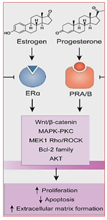
Figure 1. Нormonal action on myometrial tissue as a cause of UF [19]
Some reports suggest that estrogen and progesterone expression is higher in the fibroid than in the intact myometrium [19,20], whereas serum estrogen and progesterone levels do not differ between women with and without fibroids [16]. Progesterone is actively involved in peripheral myoma growth under the influence of estrogens, as the latter are able to increase PR expression both in the myometrium and in the myoma [15,21,22]. In addition, steroid hormones, by binding to the ER, PR located in the nucleus, differentially activate growth factors (transforming growth factor (TGF), fibroblast growth factor (FGF), platelet-derived growth factor (PDGF), epidermal growth factor (EGF), insulin-like growth factor-1 (IGF-1), tumor necrosis factor (TNF-α), vascular endothelial growth factor (VEGF), glycoprotein CD24 [16,20].
At the current stage of development of medical science, the etiopathogenesis of UF can be considered from the position of the multifactorial nature of the disease, which requires further studies.
Thus, the literature review showed that despite the high prevalence of UF, the pathogenesis, development and risk factors of this disease are not fully understood. The study of genetic associations related to epigenetic mechanisms will help to clarify the pathogenic aspects of UF development and course. Further search for genetic markers associated with the risk of developing the disease seems promising. All this points to the relevance of the problem and served as the basis for the present study.

2. Aim of the Work

The purpose of the research was to study the clinical course of uterine fibroids and the association of genetic polymorphism of ESR1 (rs2228480/594) and PGR (rs1042838) receptor genes with this pathology.

3. Materials and Methods

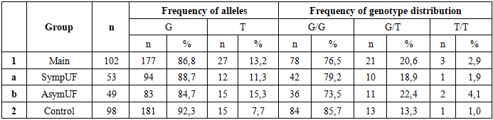
In order to accomplish this objective, an open prospective cohort study was conducted from 2021 to 2023, which was based on clinical and laboratory examinations of 200 women of reproductive and premenopausal age who were admitted to the Women's Health Centre and Gynecology Department of the Multidisciplinary Clinic of the Tashkent Medical Academy. The participants were stratified into three distinct groups: the first cohort consisted of patients diagnosed with myomas (n=102), which were further categorized as symptomatic (n=53) or asymptomatic (n=49). The second cohort comprised healthy subjects (n=98) who served as the control group. All the women who were the subject of this study underwent a comprehensive examination including clinical and anamnestic, laboratory, instrumental (ultrasound duplex scanning), histological, molecular genetic (PCR) methods of research. Following database formation, a statistical method of data processing was employed.
In the course of the study, the ethical principles stipulated by the World Medical Association Declaration of Helsinki "Ethical Principles for Scientific and Medical Research Involving Human Subjects" (revised in 2024) [23] and diagnostic measures were observed in accordance with of the National Clinic Protocol for Diagnostic Investigations of the Ministry of Health of the Republic of Uzbekistan [24].

4. Results

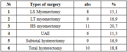
The investigation of the age demographics of the subjects revealed that the mean age was between 18 and 54 years. Specifically, the mean age was determined to be 42.6±1 years (n=49) in women within the primary cohort who were characterized as having asymptomatic fibroids, and 43.5±0.2 years (n=53) in those who were symptomatic. Notably, the mean age in the control group (n=98) was found to be 38.7±0.9 years (p<0.001).
Furthermore, a risk analysis of symptomatic fibroids development was performed in the study population. The analysis revealed that 1.9% of women (n=53) exhibited high risk (31 points), 84.9% exhibited medium risk, and 11.3% exhibited low risk (p<0.001). In contrast, the analysis of asymptomatic fibroids development revealed that the proportion of women with high risk (p<0.001) was non-zero; more than half of the women (52.2%, n=49) exhibited medium risk, and 47.8% exhibited low risk. Risk factors predisposing to the development of the disease are hereditary predisposition, aggravated gynecological history, obesity and lifestyle of the examined persons [25].
The present study set out to analyses the outcomes of surgical intervention for women with symptomatic UF (n=53). The selection of surgical methodology for the treatment of symptomatic fibroids was determined by various factors, including but not limited to: age, the presence of reproductive goals, and the severity of clinical symptoms exhibited by the study participants. The presence of anemia in woman with abnormal uterine bleeding (AUB), symptoms of rapid growth and pelvic pain were also considered (Table 1).
Table 1. Types of Surgical Interventions in Women with Symptomatic UF, n=53, (abs, %)
     
When analyzing the performance of radical operations, organ-preserving operations such as uterine artery embolization (UAE), hysteroscopy and conservative myomectomy were performed in 34 (64.15%) women of reproductive age (18 to 42 years), who did not fulfil their reproductive function. On the other hand, organ-removing procedures as subtotal and total hysterectomy were performed in 19 (35.85%) of women of late reproductive and perimenopausal age who achieved reproductive function, and there was no effect of medical treatment.
As the main method for primary screening and diagnosis of UF, as well as for monitoring the disease, we performed pelvic ultrasound, which revealed the number and location of myomatous nodes. We analyzed the association between the number and location of myomatous nodes and the presence and absence of myoma clinic. In women with symptomatic UF (n=53), 1/3 (32.1%) had multiple form (more than 2 myomatous nodules) and 2/3 (67.9%) had a solid tumor, whereas in asymptomatic UF patients, multinodular nodes was found 2 times less often compared to symptomatic UF (14.3% and 85.7%, respectively). The number of myomatous nodes in the women studied in the main multinodular group varied from 2 to 6 nodes. According to the location of myomatous nodes in the thickness of the uterus, intramural nodes were predominant in both groups of the main group (71.7% and 63.2%, respectively), submucosal nodes (7.15% and 6.12%) were equally frequent, and subserosal nodes (7.15% and 24.5%) were 3 times more frequent in women with asymptomatic UF. Mixed myomatous nodules were 2 times more frequent in women with symptomatic UF than in women with asymptomatic (13.2% and 6.12%, respectively). The median uterine volume at ultrasound examination (Brunn, 1981) in the group with symptomatic UF was 237.54 cm3, in asymptomatic - 103.45 cm3, and in the control group - 52.1 cm3 (p<0.01).
In order to optimize the diagnosis of women with UF during their reproductive and perimenopausal years, it is necessary to search for new non-invasive methods of diagnosis and prognosis of the clinical course and complications of the disease in order to prevent both the development and progression of the disease. UF is a hormone-dependent tumor, in the pathogenesis of which the imbalance between steroid hormones plays an important role. Taking into account the comorbid conditions in the studied women - the presence of risk factors for fibroid development and complications of the disease, leading to radical management tactics, led us to conduct molecular genetic studies and study the associative role of genetic polymorphism of the ESR1 (rs2228480/594) and PGR (rs1042838) receptor genes with this pathology. The distribution of genotypes in the control and main groups for the genes studied is consistent with the Hardy-Weinberg equilibrium (HWE).
This study investigates the distribution of allelic and genotype frequencies of ESR1 receptor gene polymorphism (rs2228480/594) in patients diagnosed with UF. The association of polymorphic loci of genes involved in the pathogenesis of myoma, such as ESR1, is being actively studied worldwide. The present study aims to contribute to this global research effort by determining the significance of the ESR1 receptor gene polymorphism (rs2228480/594) in the development of UF. The distribution of genotypes in the control and main groups corresponded to XWE. The subsequent analysis of the allele and genotype frequency distribution of the ESR1 gene polymorphism (rs2228480/594) revealed that the G allele frequency was 90.8% and 84.8% in the control and main groups, respectively. Conversely, the unfavorable A allele exhibited a 1.6-fold decrease (9.2%), in contrast to its prevalence of 15.2% within the main group. The predominant genotype in the study was the favorable G/G genotype in the control group (83.7%) and in the main group (72.5%). The frequency of the unfavorable heterozygous G/A genotype was found to be 1.5 times more prevalent in the main group (22.5%) than in the control group (14.3%). Conversely, the homozygous mutant A/A genotype was observed to be 4.1 times more prevalent in the asymptomatic UF group (8.2% vs. 2.0%) and 4.3 times more prevalent in the symptomatic UF group (8.2% vs. 1.9%) when compared to the control group. Consequently, the homozygous AA genotype accounted for 4.9% of subjects in the main group (Table 2).
Table 2. Frequency of Distribution of Alleles and Genotypes of rs2228480/594 Polymorphism of ESR1 gene in the Main Group with UF and Controls (abs, %)
     
The association between the rs2228480 polymorphism of the ESR1 gene and the risk of UF development was studied based on the allele and genotype frequency distribution (Table 3). The results obtained revealed an effect of rs2228480 of ESR1 gene: the presence of G/G genotype leads to a statistically significant decrease in the probability of disease (χ2<3.6; P=0.06; RR=0.9; 95%CI 0.7478-1.005; OR=0.5; 95%CI 0.2587-1.028). As illustrated in Table 3, there is a discrepancy in the frequency of alleles and genotypes of the rs2228480/594 polymorphism of the ESR1 gene in the group of patients with UF and the control group.
Table 3. Differences in the Frequency of Alleles and Genotypes of the rs2228480/594 Polymorphism of the ESR1 Gene in the Group of Patients with UF and Controls
     
Concurrently, the G/G genotype exhibited a protective effect in the development of UF. However, upon comparison of heterozygous unfavorable G/A genotypes, a 1.7-fold increase was observed in women diagnosed with UF (OR=1.7; 95% CI 0.8402-3.632).
When analyzing the difference in allele and genotype frequencies of the rs2228480/594 polymorphism of the ESR1 gene in the group of women with symptomatic UF and the control sample, it was found that the risk of developing fibroids with the heterogeneous unfavorable G/A genotype was 2.1 times higher than in the control group (χ2=3.3; P=0.07; RR=1.8; 95%CI 0.954-3.581; OR=2.1; 95% CI 0.9360-4.95). In addition, the G/G genotype played a protective role in the development of UF (Figure 2).
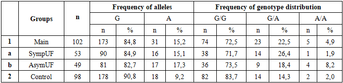
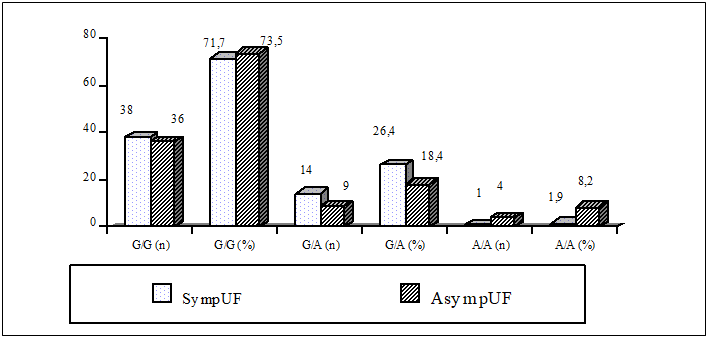
Figure 2. The Differences in the Frequency of Alleles and Genotypes of the rs2228480/594 Polymorphism of the ESR1 Gene in Symptomatic and Asymptomatic UF Groups
The observed variations in the prevalence of the wild-type genotype among women diagnosed with UF can be elucidated by the authors' findings, which indicate that the ESR1 polymorphism exerts an indirect influence on myoma development. This suggests that in women with UF, there is a tendency to increase the frequency of carrying 'mutant' alleles and genotypes for the genes under study compared to women from the population sample. The manifestation of symptomatic UF is associated with the presence of multiple epigenetic factors, while the absence of symptoms in women with asymptomatic UF and low-risk factors does not necessarily imply clinical inactivity.
Consequently, the polymorphic marker rs2228480/594 of the ESR1 gene in the studied women is associated with the development of fibroids, though not always accompanied by clinical symptoms, in women with abnormal mutant homozygous genotypes.
Study of allele and genotype distribution frequency of PGR receptor gene polymorphism (rs1042838) in UF. Currently, the presence of the cellular receptors ER1 and PR play an important role in the development of the disease. To date, one of the major genetic factors in the development of UF is the polymorphism of the PGR receptor gene (rs1042838). Progesterone, through PR-A and PR-B receptors, plays a key role in the initiation of the cascade of biological disorders and, together with estradiol, is a regulator of this process. Progesterone inhibits the expression of the ER and acts directly through the PR. It is actively involved in peripheral myoma growth with the help of estrogens (Table 4).
Table 4. Frequency of Distribution of the Alleles and Genotypes of the rs1042838 Polymorphism of the PGR Gene in Groups of Patients with UF and Groups of Controls
     
A thorough examination of allelic and genotypic variants of the G/T polymorphism (s1042838) of the PR gene revealed no statistically significant variation in the prevalence of the G allele in the primary sample (86.8%) and control (92.3%) groups (χ2=3.3; P=0.07; RR=1.6; 95%CI 0.949-3.51; OR=1.8; 95%CI 0.943-3.577), and the T allele in 13.2 and 7.7%, respectively (Tables 4-5).
Further investigation into the effect of G/T gene polymorphism (rs1042838) of the PR gene revealed that the probability of detecting the wild-type G allele in the primary cohort was statistically significantly higher in comparison to the control group (χ²=3.3; P=0.07; RR=1.7; 95% CI 0.949-3.151; OR=1.8; 95% CI 0.9473-3.577). Concurrently, the unfavorable T allele was observed to be more prevalent (OR=1.72) in comparison to the control group. However, a significant discrepancy was identified in the frequency of detection of the heterozygous unfavorable G/T genotype (1.7 times more frequent (OR=1.7; 95%CI 0.796-3.609) and (2.9 times more frequent) homozygous mutant T/T genotype in UF patients compared to the control group (OR=2.9; 95%CI 0.371-28.75) (Table 5). This finding suggests a potential lack of response to conventional progesterone therapy in symptomatic fibroids. As the subjects who exhibited symptoms of the disease were predominantly those with the T/T genotype, and these subjects had a high number of risk factors, this finding is of particular significance.
Table 5. Differences in the Frequency of Alleles and Genotypes of the PGR gene rs1042838 Polymorphism in a Group of UF Patients and a Control Sample
     
The predominant genotype in the study was the favorable G/G genotype in the control group (85.7%) and in the main group (76.5%). The population sample group exhibited the highest number of individuals with homozygous GG genotype. The prevalence of the GG genotype in the group of conditionally healthy donors confirms the protective function of this genotype in comparison with the group of patients (OR=0.5).
The allele frequency of the rs1042838 polymorphism of the PGR gene was analyzed in the primary cohort. The mutant genotype was identified at a significantly higher frequency in the symptomatic T/T UF group (n=10) than in the asymptomatic group (n=15) (OR=2.2; 95%CI 0.194-25.2).
The distribution analysis of the PGR gene rs1042838 polymorphism demonstrated that the homozygous G/G genotype exhibited a protective effect in relation to the disease, as evidenced by the statistically significant decrease in the frequency of detection of this genotype in the patient sample compared to the control group (OR=0.5; 95%CI 0.261-1.121). Conversely, G/T (OR=1.7; 95%CI 0.796-3.609) and T/T genotypes (OR=2.9; 95%CI 0.371-28.75) of PGR exhibited a promoter effect in relation to UF, as the probability of detecting this genotype was statistically significantly higher in the patient population compared to the control group, thereby increasing the risk of developing the disease from 1.7 to 2.9 times.
Consequently, population analysis of the studied polymorphisms of key UF regulator genes indicates that polymorphisms of VDR (rs10735810), ESR1 (rs2228480/594) and PGR (rs1042838) receptor genes exhibit an associative role of genetic polymorphism in the development of the disease. Given the multifactorial etiopathogenesis of the disease, it is not sufficient to assess the influence of individual polymorphic loci in predicting the genetic risk of fibroids in women; gene-gene interaction analysis is therefore necessary.
The interaction of favorable and unfavorable genotypes of the ESR1 and PGR genes is a subject of current research. In order to predict the genetic risk of myoma development, the current priority in the molecular genetic direction is to study the associations of polymorphic loci of genes involved in the pathogenesis of the disease. Consequently, unfavorable genotypes of polymorphism polymorphisms of various genes encoding different links of this pathological process can be detected in individual patients with UF with symptomatic and asymptomatic course. This may explain the fact that some genotypes, including homozygous genes, do not reach the level of reliable differences. A significant component of our study involves the identification of interactions between ESR1 rs2228480/594 and PGR rs1042838 genes, with the objective of determining combinations of unfavorable genotypes (haplotypes) that would have the greatest pathogenetic significance in the development of UF. It was observed that there was a significant tendency for an increase in the proportion of carriers of the combination of functional favorable homozygous genotypes A/A+G/G+G/G/G in the group of conditionally healthy women. This phenomenon can be attributed to the protective effect exerted by these carriers, who exhibited enhanced resistance to the development of UF.
The most prevalent unfavorable haplotype was identified as homozygote+heterozygote in both the main and control groups, accounting for 47.2% and 37.7% of the subjects, respectively. Concurrent carriage of homozygote (ESR1 rs2228480/594) and heterozygote (PGR rs1042838) haplotypes was observed in the symptomatic UF group. Furthermore, co-occurrence analysis of unfavorable genotypes of the studied genes among fibroids patients and control sample in genetic association with the risk of developing this pathology showed a significant role of association of these polymorphisms (OR=4.4; 95%CI 1.829-10.81). The area under the curve (AUC) was found to be up to 0.7, indicating a noteworthy prognostic performance of the investigated markers for patients with UF.
In order to study the prognostic significance of the investigated receptor gene polymorphisms for UF risk prediction, Se, Sp and prognostic efficiency of the marker - AUC were determined (Table 6).
Table 6. Indicators of Prognostic Significance in the Studied Groups
     
The population analysis of the studied polymorphisms of key genes that regulate UF demonstrated the associative roles of genetic polymorphisms of ESR1 (rs2228480/594) and PGR (rs1042838) receptor genes in the development of this pathology.
The study's findings on the primary characteristics of the polymorphic variants of the aforementioned genes in women with fibroids and in the control sample demonstrated a statistically significant increase in the risk of developing this disease when carrying the heterozygous unfavorable A/A rs2228480 genotype of the ESR1 gene (OR=4.3; 95% CI 0.753-24.16), G/T (OR=1.7; 95%CI 0.796-3.609) and T/T (OR=2.9; 95%CI 0.371-28.75) of rs1042838 of the PGR gene had a promoter effect in relation to UF, as. It is evident that patients in the primary cohort demonstrate a heightened probability of detecting these specific haplotypes. This heightened probability is statistically significant, with a calculated increase in the risk of disease development from 1.7 to 4.7 times.

5. Discussion

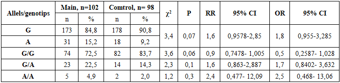
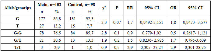
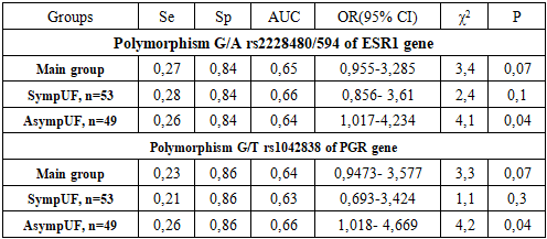
Uterine fibroid is more frequent in late reproductive and perimenopausal age, which is consistent with the literature. Women with symptomatic fibroids and a high index of risk factors for the disease have a significantly impaired quality of life. As a result, these women develop clinical symptoms such as abnormal uterine bleeding leading to severe anemia, rapid growth of the myoma, pelvic pain and infertility. On ultrasound in women with a mixed form of myomatous nodes, the median uterine volume is greater than 2 times that of asymptomatic UF. This, in turn, leads to the use of radical, organ-removing surgical methods of treatment of this pathology.
The current understanding of the role of genetic abnormalities in UF development is insufficient. The extent to which chromosomal abnormalities in tumor cells contribute to the manifestation of clinical symptoms as a result of molecular genetic processes at the level of genes and intragenic interactions remains to be fully elucidated. In order to ascertain the role of polymorphic variants of predictor genes in the development of fibroids, molecular-genetic studies of ESR1 and PGR receptor genes were carried out, and their gene-gene interaction in the development and clinical course of UF was studied.
The investigation of allelic variants and genotype frequency of ESR1 receptor gene polymorphism (rs2228480/594) in women diagnosed with UF revealed that individuals carrying a heterogeneous unfavorable G/A genotype exhibited a 2.1-fold higher risk of developing myoma when compared to the control group (OR=2.1; 95% CI 0.936-4.95), whereas individuals possessing the G/G genotype appeared to demonstrate a protective effect against fibroids development (OR=0.5). The involvement of polymorphic loci of ESR1 genes in the pathogenesis of myoma has been demonstrated in a number of studies, which corroborates the findings of the present study. The authors' data further demonstrates that the presence of mutant genotype in women diagnosed with UF can be explained by the influence of ESR1 Polymorphism on myoma development, a process that occurs indirectly.
In the study of allele and genotype frequency distribution of the PGR receptor gene polymorphism (rs1042838) in UF, it was established that the homozygous G/G genotype exhibited a protective effect concerning myoma (OR=0.5; 95% CI 0.261-1.121), while the G/T (OR=1.7; 95% CI 0.796-3.609) and T/T (OR=2.9; 95% CI 0.371-28.75) PGR genotypes demonstrated a promoter effect in relation to UF. The probability of identifying this genotype was found to be statistically significant within the patient population of the primary cohort, resulting in an elevated risk of developing the disease from an initial value of 1.7 to a value of 2.9.
This finding suggests the potential inefficacy of gestagen-based preparations in women with symptomatic UF, highlighting the necessity of further investigation. Progesterone, through PR-A and PR-B receptors, plays a pivotal role in instigating the sequence of biological disorders, and, along with estradiol, functions as a controller of this process. It has been demonstrated that progesterone exerts its inhibitory effect on ER expression and functions directly through PR. The active involvement of progesterone in the peripheral growth of myomas is well-documented, with estrogens being instrumental in augmenting the expression of the PR in both the myometrium and myoma [15,22].
Consequently, population analyses suggest that polymorphisms in ESR1 and PGR genes are associated with susceptibility to the disease, as evidenced by genetic polymorphisms. In light of the multifaceted etiopathogenesis of UF, a gene-gene interaction analysis was conducted with the aim of predicting the genetic risk of UF development in women. In this study, the interaction between the favorable and unfavorable genotypes of the ESR1 rs2228480/594 and PGR rs1042838 genes was analyzed in patients with UF and in control groups, to identify the haplotype combinations. This approach enabled us to identify unfavorable genotypes of polymorphism polymorphisms of diverse genes implicated in various aspects of this pathological process, in patients exhibiting both symptomatic and asymptomatic symptoms. It was observed that the presence of certain genotypes, including homozygous genes, did not reach a level of statistical significance. An analysis of the combination of unfavorable genotypes of the studied genes among patients with UF and a control sample demonstrated that the risk for myoma development in women with symptomatic (OR=4.7) and asymptomatic fibroids (OR=4.2) was significantly associated with having two or more unfavorable gene combinations, fourfold higher than in the control group. The analysis also revealed a high prognostic significance of unfavorable haplotypes in the development of UF, with an area under the curve (AUC) value of 0.7.
Conducted molecular genetic studies have confirmed that evaluating polymorphic loci in isolation is insufficient for predicting the risk of UF development. The gene-gene interaction of ESR1 and PGR has been demonstrated to have a pronounced independent effect on the development of symptomatic myoma, in addition to contributing to the phenotypic development of the disease complication. The observed stability in the connection between polymorphisms in receptor genes in symptomatic and asymptomatic subjects indicates the value of genetic testing, particularly in instances of a family history of UF, in predicting the likelihood of UF development and progression among women in the reproductive and perimenopausal ages.
Prediction of UF patients will allow for the anticipation of the manifestation of disease symptoms and its complication, the implementation of timely conservative or organ-preserving surgical procedures, and enhancement of the quality of life for women afflicted by UF. The investigation of epigenetic and genetic factors associated with the development of myomas, which can lead to a heightened risk of disease, constitutes a crucial step in enhancing our comprehension of cellular transformation processes and the identification of diagnostic biomarkers, facilitating early diagnosis of UF. This, in turn, represents a significant advancement in the realm of personalized predictive medicine.

6. Conclusions

The risk of developing uterine fibroids depends on the presence of cumulative risk factors, which are more pronounced in women with symptomatic fibroids. The associative role of genetic polymorphism of genes: A/A ESR1 (rs2228480/594) (OR=2.1) and G/T and T/T PGR (rs1042838) (OR=1.9) with uterine fibroid development was demonstrated. Two intragenic combinations of unfavorable genotypes of G/A ESR1 rs2228480/594 + G/T PGR rs1042838 predictor genes in patients increased the risk of myoma development from 3.6 to 4.4 times (OR=3.6-4.4; 95% CI 1.829-10.81). At this stage of the work, it would be interesting to further investigate the association of epigenetic risk factors with genetic determinants of fibroids.

ACKNOWLEDGEMENTS

We would like to express our deep gratitude to all the patients who gave informed consent and participated in this study, and to the staff of the Department of Obstetrics and Gynecology of Family Medicine, Tashkent Medical Academy, and the Department of Molecular Genetics, Institute of Hematology, Uzbekistan.

References

[1]  S. Cramer, A. Patel. “The frequency of uterine leiomyomas”. American Journal Clinic Pathology, vol 94, pp. 435–8, 1990.
[2]  Practice Bulletin. “Management of Symptomatic uterine leyomyomas”, ACOG, № 228, pp. 3-11, 2021. doi: 10.1097/AOG.0000000000004401.
[3]  Clinical guidelines. “Uterine myoma”. RUOG. Mosco, pp. 5-7, 2021.
[4]  C. Walker, E. Stewart. “Uterine fibroids: the elephant in the room”. Science, 308: pp. 1589–92, 2005. https://doi.org/10.1126/science.1112063.
[5]  W. Parker. “Etiology, symptomatology, and diagnosis of uterine myomas”. Fertility and Sterility, Vol. 87, pp. 725-36, 2007.
[6]  A. Commandeur, A. Styer, J. Teixeira. “Epidemiological and genetic clues for molecular mechanisms involved in uterine leiomyoma development and Growth”. Human Reproduction Update, Vol. 21, Issue 5, pp. 593–615, 2015. https://doi.org/10.1093/humupd/dmv030.
[7]  T. Takao, M. Ono, Y. Yoshimasa, H. Masuda, T. Maruyama, “A mediator complex subunit 12 gain-of-function mutation induces partial leiomyoma cell properties in human uterine smooth muscle cells”. Fertility and Sterility, Vol. 3 №3, pp. 288-298, 2022. https://doi.org/10.1016/j.xfss.2022.04.002.
[8]  N. Bateman, T. Abulez, C. Tarney, et al. “Multiomic analysis of uterine leiomyomas in selfdescribed Black and White women: molecular insights into health disparities”. Am J Obstet Gynecol, pp. 231: 321. e1-1, 2024.
[9]  M. Ali, M. Ciebiera, S. Vafaei, et al. “Progesterone Signaling and Uterine Fibroid Pathogenesis; Molecular Mechanisms and Potential Therapeutics”. Cells 12, 1117. 2023, https://doi.org/10.3390/cells12081117.
[10]  M. Moravek, S. Bulun. “Endocrinology of uterine fibroids: Steroid hormones, stem cells, and genetic contribution”. Curr. Opin. Obstet. Gynecol., vol. 27, pp. 276–283, 2015.
[11]  L. Saisai, Ch. Beilei, Sh. Bo, W. Jing, Zh. Xueqiong. “The associations between serum vitamin D, calcium and uterine fibroids in Chinese women: a case-controlled study”. Journal of International Medical Research. Vol. №48 (5), pp. 1–11, 2020. DOI: 10.1177/0300060520923492.
[12]  A. Tinelli, M. Vinciguerra, A. Malvasi, M. Andji´c, I. Babovic, R. Sparic. “Uterine Fibroids and Diet”. Int. J. Environ. Res. Public Health. №18, Vol. 1066, pp. 1-7, 2021. https://doi.org/10.3390/ijerph18031066.
[13]  M. Moravek, P. Yin, M. Ono et al. “Ovarian steroids, stem cells and uterine leiomyoma: therapeutic implications”. Hum Reprod Update, №21, pp. 1-12, 2015.
[14]  M. Borahay, M. Asoglu, A. Mas, S. Adam, G. Kilic, A. Al-Hendy. “Estrogen Receptors and Signalingin Fibroids: Role in Pathobiologyand Therapeutic Implications”. Reproductive Sciences, Vol. 24, № 9, pp. 1235-1244, 2017. https://doi.org/10.1177/1933719116678686.
[15]  S. Bulun. “Uterine fibroids”. N. Engl. J. Med., № 369(14), pp. 1344–1355, 2013. https://doi.org/10.1056/NEJMra1209993. PMID: 24088094.
[16]  M. McWilliams, V. Chennathukuzhi. “Recent advances in uterine fibroid etiology”. Semin. Reprod. Med., №35, pp. 181–189, 2017.
[17]  A. Khashukoeva, S. Khlynova, I. Ilyina, S. Kerchelaeva. “Estrogen-dependent conditions of the female reproductive system: the possibilities of non-hormonal therapy with the use of indole-3-carbinol system: the possibilities of non-hormonal therapy with the use of indole-3-carbinol”. Obstetrics and Gynaecology. №5, pp. 65-69, 2020.
[18]  P. Gordon, B. Alicia, S. Deloris, F. Norris et al. “The Life Cycle of the Uterine Fibroid Myocyte”. Current Obstetrics and Gynecology Reports. № 7, pp. 97-105, 2018. https://doi.org/10.1007/s13669-018-0241-7.
[19]  A. Machado-Lopez, C. Simón; A. Mas. “Molecular and Cellular Insights into the Development of Uterine Fibroids”. Int. J. Mol. Sci. 2021, 22, 8483. https://doi.org/10.3390/ijms22168483.
[20]  M. Ciebiera, M. Włodarczyk, S. Zgliczy´nski, T. Łozi´nski, et al. “The Role of miRNA and Related Pathways in Pathophysiology of Uterine Fibroids. From Bench to Bedside”. Int. J. Mol. Sci. № 21, pp. 3014-3019, 2020. doi: 10.3390/ijms21083016 www.mdpi.com/journal/ijms.
[21]  E. Shtokh, V. Tskhai. “Uterine myoma. Current understanding of pathogenesis and risk factors”. Siberian Medical Review, pp. 22-28, 2015.
[22]  H. Chill, M. Safrai, A. Reuveni Salzman, A. Shushan. “The rising phoenix-progesterone as the main target of the medical therapy for leiomyoma”. Biomed. Res. Int., 4705164, pp. 1-8, 2017.
[23]  WMA, Declaration of Helsinki Medical Research Involving Human Participants. 2024 https://www.wma.net/what-we-do/medical-ethics/declaration-of-helsinki/.
[24]  “Standards of diagnostics and treatment of gynaecological diseases in medical institutions of the health care system of RUz”. MHRUz, pp. 23, 79-80, 2017.
[25]  D. Irnazarova, D. Yuldasheva, D. Najmutdinova et al. “Effect of Excess Body Weight and Deficiency of Vitamin D on the Clinical Course of Uterine Myoma”. Europ. J. Mol. Clin. Med., Vol. 7, №3, pp. 3317-3329, 2020. https://ejmcm.com/article_4197.html.