-
Paper Information
- Next Paper
- Paper Submission
-
Journal Information
- About This Journal
- Editorial Board
- Current Issue
- Archive
- Author Guidelines
- Contact Us
American Journal of Medicine and Medical Sciences
p-ISSN: 2165-901X e-ISSN: 2165-9036
2025; 15(12): 4404-4409
doi:10.5923/j.ajmms.20251512.46
Received: Nov. 19, 2025; Accepted: Dec. 10, 2025; Published: Dec. 12, 2025

Features of Quantitative Indicators of UBM in the Study of Structures of the Anterior Segment of the Eye in Primary Zug with Pupillary Block
 Abstract
 Reference
 Full-Text PDF
 Full-text HTML
Shakhnoza Iskandar kizi Rustambekova1, Azizbek Fazilovich Ikramov2, Otabek Azizbekovich Ikramov3
1Assistant, Department of Ophthalmology, Andijan State Medical Institute, Andijan, Uzbekistan
2DSc., Professor Head of Department of Ophthalmology, Andijan State Medical Institute, Andijan, Uzbekistan
3PhD., Associate professor, Department of Ophthalmology, Andijan State Medical Institute, Andijan, Uzbekistan
Correspondence to: Azizbek Fazilovich Ikramov, DSc., Professor Head of Department of Ophthalmology, Andijan State Medical Institute, Andijan, Uzbekistan.
| Email: |  |
Copyright © 2025 The Author(s). Published by Scientific & Academic Publishing.
This work is licensed under the Creative Commons Attribution International License (CC BY).
http://creativecommons.org/licenses/by/4.0/

The article discusses modern research methods in ophthalmology, such as ultrasound biomicroscopy (UBM). UBM allows you to accurately assess the state of the anatomical structures of the anterior eye. The use of these modern research methods makes it possible to carry out differential diagnosis in complex clinical cases, as well as to predict the outcomes and course of ophthalmopathologies. In recent years, technologies for diagnostics and visualization of eye structures have been intensively developed. All this has significantly expanded the understanding of pathogenesis, clinical course options of various ophthalmopathologies, and contributes to the development of more effective methods of their treatment.This review will present the diagnostic capabilities of the most commonly used diagnostic research methods - ultrasound biomicroscopy (UBM) of the anterior segment of the eye.
Keywords: Ultrasound biomicroscopy, Ophthalmopathology
Cite this paper: Shakhnoza Iskandar kizi Rustambekova, Azizbek Fazilovich Ikramov, Otabek Azizbekovich Ikramov, Features of Quantitative Indicators of UBM in the Study of Structures of the Anterior Segment of the Eye in Primary Zug with Pupillary Block, American Journal of Medicine and Medical Sciences, Vol. 15 No. 12, 2025, pp. 4404-4409. doi: 10.5923/j.ajmms.20251512.46.
1. Introduction
- Ultrasound examination of the eyes as a diagnostic method has been used in ophthalmology since the 50s of the last century. The need for this method is determined by its advantage over other methods of ophthalmological diagnostics, including the usual examination of the structures of the anterior segment of the eye on a slit lamp. UBM is much better at visualizing opaque fabrics because it uses high-energy sound waves [1,2,3]. That is, its advantage lies in the possibility of lifetime visualization of all anatomical structures of the anterior segment (conjunctiva, cornea, anterior chamber, sclera, iris, lens, ligamentous apparatus, ciliary body, anterior vitreous), including in conditions of reduced transparency of optical media [5,6,7]. However, due to the limited penetration depth of UBM into tissue structures (5 mm penetration depth), it is mainly used in ophthalmology to visualize anterior structures such as the angle and ciliary body. The UBM method significantly expands the possibilities of studying the structures of the anterior segment of the eye [9,10,11].ObjectiveTo study theprevalence of quantitative indicators of UBM in the study of the structures of the anterior segment of the eye in primary ZUG with pupillary block.
2. Research Methods
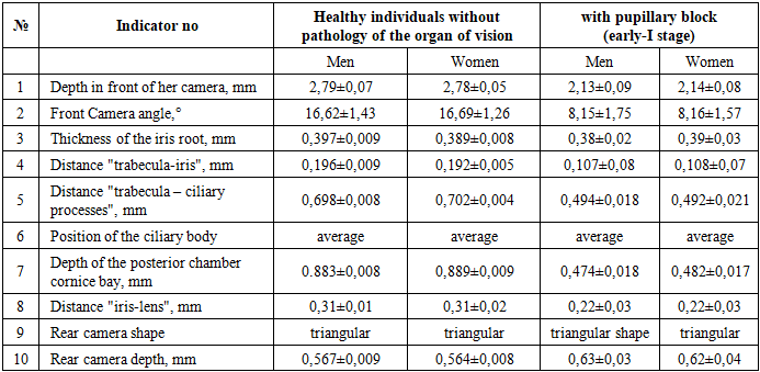
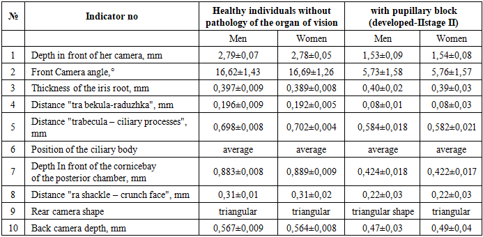
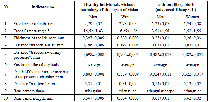
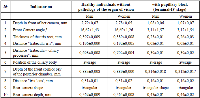
- The study uses the methods of epidemiological, general clinical, biochemical, instrumental, special and statistical studies. The age-and gender-specific UBM indicators of primary ZUG were studied by the mechanism of blocking the anterior chamber angle, which determines not only the increase in IOP, but also the subsequent course and outcome of the disease. On the basis of which it is necessary to choose the treatment of primary ZUG [12,13,14]. In patients with primary angle-closure glaucoma, several pathogenetic variants of the disease were identified that differ in the mechanism of formation of anterior chamber angle block: pupillary block, flat iris (iris plateau type), vitreous-crystalline block corresponding to the malignant form of glaucoma, and a shortened anterior chamber angle known as" creeping " glaucoma [15,16,17].Traditional methods of ophthalmic examination — biomicroscopy and gonioscopy-did not allow to fully differentiate the mechanism of blockage of the anterior chamber angle due to limited visualization of deep structures. However, the use of ultrasound biomicroscopy (UBM) in combination with morphometric analysis of linear and angular parameters made it possible to study in detail the spatial relationships of anatomical formations of the anterior segment of the eye. This approach significantly increased the accuracy of topographic diagnostics and allowed a reasonable approach to the choice of individual treatment tactics for different types of angle-closure glaucoma [18,19,20].The gonioscopy data obtained by us in PSOG shows that the number of patients with pupillary block is about 75% and the second advanced stage prevails. Functional block most often develops in eyes with relative pupillary block, when the angle of the anterior chamber is closed by the protruding anterior root of the iris. In addition, in eyes with a narrow angle of the anterior chamber, its sharp apex, or the posterior position of the Schlemm canal, the trabecular zone may close when the pupil dilates with the basal fold of the iris (Fuchs fold). At the same time, the iris is not protruding, flat, and the anterior chamber is of medium depth [21].Usually in clinical practice, it is observed that the course of this form of glaucoma is undulating, attacks are replaced by calm, asymptomatic intervals. As a result of each attack, spikes in the drainage system remain, which later lead to chronically high ophthalmotonus and changes in the visual fields characteristic of glaucoma. UBM study of patients with angle-closure glaucoma was performed mainly in the early-I and advanced-II stages of the disease. The first group included 103 patients (183 eyes) who showed characteristic anatomical and topographic changes in the anterior segment of the eye, including a shallow anterior chamber, a convex contour of the iris, its thinning in the basal zone, as well as a narrow or completely closed angle of the anterior chamber in combination with an increase in the depth of the posterior chamber. In ultrasound biomicroscopy, the posterior chamber had a triangular shape, and the combination of the above morphological features corresponded to the ultrasound picture of the pupillary block — one of the key pathogenetic variants of angle-closure glaucoma. UBM study in the early – 1 stage in PSG with pupillary block is characterized with the following characteristic features: In patients of the first group with pupillary block, according to ultrasound biomicroscopy (UBM), characteristic morphometric features of the structures of the anterior segment of the eye were noted. The anterior chamber of the eye was characterized by a shallow depth and a narrow or closed angle. The averagevalues of anatomical parameters were: anterior chamber depth-2.13±0.09 mm in men and 2.14±0.08 mm in women; anterior chamber angle-8.15±1.75° and 8.16±1.57°, respectively. The thickness of the iris root was 0.38±0.02 mm in men and 0.39±0.03 mm in women. The distance "trabecula-iris" was sharply reduced — 0.107±0.08 mm and 0.108±0.07 mm, respectively, which indicates a pronounced narrowing of the angle. The distance "trabecula–ciliary processes" was 0.494±0.018 mm in men and 0.492±0.021 mm in women. The depth of the anterior cornice bay of the posterior chamber reached 0.474±0.018 mm and 0.482±0.017 mm, respectively, and the iris-lens distance was 0.31±0.02 mm for men and 0.32±0.03 mm for women. The rear camera had a typical triangular shape.
|
|
|
3. Conclusions
- 1. Based on the most informative parameters of the UBM study, we have developed a simplified scheme for UBM diagnostics: According to the depth of the anterior chamber; according to the width of the CCP; according to the position of the ciliary body: medium; posterior and anterior; according to the shape and depth of the posterior chamber: triangular deep; triangular medium; triangular small and arched very small. (characteristic of "creeping" glaucoma) and by the degree of structural damage to the anterior segment (atrophy of the iris) and iridociliary zone by the thickness of the iris of 3 degrees.2. According to ultrasound biomicroscopy data, all patients with primary angle-closure glaucoma revealed closure of the anterior chamber angle with the basal zone of the iris adjacent to the trabecula and corneal endothelium by an average of 1.33±0.02 mm. Reducing the distance "trabecula-iris" serves as an objective indicator of the degree of convergence of the iris with the trabecular apparatus. The complete absence of this distance indicates the contact of the basal part of the iris with the trabecular zone and, consequently, the complete closure of the angle of the anterior chamber. This feature is of great diagnostic importance, as it allows not only to confirm the presence of a block, but also to accurately determine its anatomical localization and extent within the angle of the anterior chamber.3. Indicators of UBM in the study of age and gender in primary ZUG, shows that the mechanism of blocking the angle of the anterior chamber determines not only the increase in IOP, but also the subsequent course and outcome of the disease. Our new simplified scheme of UBM in primary glaucoma may be useful in the early diagnosis of OCD and determination of treatment tactics. Of particular importance is the choice of the surgical method of anti-glaucomatous surgery.
References
| [1] | Gundorova P. A., Kodzov M. B., Dzhanelidze I. V. The significance of ultrasound studies in the intraoperative diagnosis of foreign bodies. 2014, No. 3, pp. 112-113. |
| [2] | Dorofeev D., et al. Efficiency and safety of preservative-free antihypertensive drug in long-term therapy // Bulletin of Ophthalmology-2022. - 138 (5) pp. 178-184. |
| [3] | Dzhalalova D., et al. Modern aspects of diagnosis and treatment of glaucoma optical neuropathy (review) // inLibrary (Ophthalmology) – 2025. – 2 (1) pp. 78-85. |
| [4] | Egorova E. V., Ioshin N. E. Clinical and functional results of implantation of collagen lenses in the absence of a posterior capsule // Actual problems of ophthalmology: Mat. conf. on ophthalmology, Ufa, 2016, p. 23. |
| [5] | Ermakova N. A. General ideas about the pathogenesis of uveitis. 2013, no. 4, pp. 141-143. |
| [6] | Ermakova N. A. Klassifikatsiya i klinicheskaya otsenka uveitov [Classification and clinical assessment of uveitis]. 2013, no. 4, pp. 146-149. |
| [7] | Sarukhanyan A. A. Anatomical and topographic features of the anterior segment of the eye in the progression of cataracts combined with glaucoma and pseudoexfoliative syndrome, according to ultrasound biomicroscopy: Abstract of the dissertation of the Candidate of Medical Sciences, Moscow, 2017, 28 p. |
| [8] | Senchenko N. Ya. Pathogenetic substantiation of principles of surgical correction of post-traumatic and postoperative aphakia in children: Dis. ... kand. med. nauk. Irkutsk, 2015, 157 p. (in Russian) |
| [9] | Strakhov V. V., Buzykin M. A., Mineeva L. A. Involutional changes in the accommodation apparatus of the human eye according to ultrasound biometrics and biomicroscopy. ophthalmol. - 2017. - No. 4. - p. 32-35. Tamarova P. M. Optical devices for eye research, Moscow: Meditsina Publ., 2012, 176 p., ill. |
| [10] | Takhchidi Kh. P., Khodzhaev N. S., Egorova E. V., Uzunyan D. G., Ovchinnikova A.V. Ultrasound biomicroscopy in the assessment of stabilization of surgically formed drainage pathway and formation of additional outflow mechanisms after non-penetrating deep sclerectomy. 2016, No. 4, pp. 16-24. |
| [11] | Uzunyan D. G. Ultrasound biomicroscopy in evaluating the effectiveness of non-penetrating deep sclerectomy: Dis. ... kand. med. nauk, Moscow, 2017. 150 p. |
| [12] | Fedorov S. N., Egorova E. V. Errors and complications in the implantation of an artificial lens. - Moscow, 2011. - 244 p., ill. |
| [13] | Kharlap S. I. Biometric relations and hemodynamic characteristics of the vascular system of the eye and orbit in normal and pathological conditions based on the results of modern methods of ultrasound clinical spatial analysis: Dis. ... d-ra med. nauk, Moscow, 2013, 294 p. |
| [14] | Shirshikov Yu. K., Kharlap S. I. Acoustic B-scanning with a gray scale. Vestn. ophthalmol. 2017, No. 2, pp. 40-42. |
| [15] | Shkrebets G. V., Dolzhich G. I. Clinical and immunological prognostic criteria for the development of glaucoma in peripheral uveitis. - 2017. - No. 2. - pp. 28-31. |
| [16] | Kraus W., Narl-Elias A., Schramm P. Diagnostic progress in malignant melanomas by high-resolution real-time sonography // Hautarzt. - 2015. - Vol. 36. - P. 386-392. |
| [17] | Kremmer S., Schiefer U., Wilhelm H. Mobilization of intraocular foreign bodies by magnetic resonasnce tomography // Klin. Mbl. AugenheiIk. - 2016. - Vol. 31. - P. 201-202. |
| [18] | Laganowski H.C., Kerr Muir M.G., Hitchings R.A.Glaucoma and the iridocorneal endothelial syndrome // Arch Ophthalmol. - 2012. - Vol. 110. - P. 346-350. |
| [19] | Langner S., Martin H., Terwee T., Koopmans S.A., Kriiger P.C., Hosten N., Schmitz K.P., Guthoff R.F., Stachs O. 7.1 T MRI to Assess the Anterior Segment of the Eye // Invest. Ophthalmol. Vis. Sci. - 2010. - Vol. 51. - P. 65756581. |
| [20] | Lee H.S., Lew H., Yun Y.S. Ultrasonographic measurement of upper eyelid thickness in kiorean children with epicanthus // Korean J Ophthalmol. - 2016. - Vol 20. - P. 79-81. |
| [21] | Li S., Wang H., Mu ., Fu J., Wang X., Wang J., Wang N. Prospective evaluation of changes in anterior segment morphology after laser iridotomy in Chinese eyes by rotating Scheimpflug camera imaging // Clin Experiment Ophthalmol. - 2010. - Vol. 38. - P. 10-14. |