-
Paper Information
- Next Paper
- Previous Paper
- Paper Submission
-
Journal Information
- About This Journal
- Editorial Board
- Current Issue
- Archive
- Author Guidelines
- Contact Us
American Journal of Medicine and Medical Sciences
p-ISSN: 2165-901X e-ISSN: 2165-9036
2025; 15(12): 4719-4724
doi:10.5923/j.ajmms.20251512.115
Received: Aug. 25, 2025; Accepted: Sep. 17, 2025; Published: Dec. 31, 2025

Effectiveness of Pomegranate Seed Oil in the Correction of Morphological Changes in Thymic Tissue During Experimental Chemotherapy
 Abstract
 Reference
 Full-Text PDF
 Full-text HTML
Rasulova N. R., Temirova N. R.
Bukhara State Medical Institute, Uzbekistan
Correspondence to: Rasulova N. R., Bukhara State Medical Institute, Uzbekistan.
| Email: |  |
Copyright © 2025 The Author(s). Published by Scientific & Academic Publishing.
This work is licensed under the Creative Commons Attribution International License (CC BY).
http://creativecommons.org/licenses/by/4.0/

During chemotherapy, cytostatic drugs exert a significant toxic effect on the thymic tissue, which is one of the main organs of the immune system. As a result, a decrease in lymphoid elements, cortical zone atrophy, dystrophic changes in the medullary areas, as well as a relative increase in stromal components, are observed. These alterations lead to a decline in immunological defense mechanisms, increasing the susceptibility of the organism to various infectious and inflammatory processes. In recent years, the use of natural antioxidants and immunomodulatory agents has been considered one of the important approaches to reducing adverse effect of chemotherapy. In this study, morphological and morphometric changes in thymic tissue caused by chemotherapy in experimental animals were investigated, and the possibilities of their correction using pomegranate seed oil were evaluated. The results demonstrated that in the groups treated with pomegranate seed oil, the thymic cortical layer was relatively better preserved, the activity of the germinal centers of the follicles was restored, and the compensatory remodeling of stromal components approached physiological norms.
Keywords: Chemotherapy, Thymus, Pomegranate seed oil, Immunocorrection, Morphometry, Antioxidant, Immunomodulator
Cite this paper: Rasulova N. R., Temirova N. R., Effectiveness of Pomegranate Seed Oil in the Correction of Morphological Changes in Thymic Tissue During Experimental Chemotherapy, American Journal of Medicine and Medical Sciences, Vol. 15 No. 12, 2025, pp. 4719-4724. doi: 10.5923/j.ajmms.20251512.115.
1. Introduction
- Today, oncological diseases are among the most pressing health problems worldwide, causing high morbidity and mortality rates. One of the main methods used in the treatment of malignant tumors is chemotherapy. Cytostatic drugs inhibit the division of tumor cells and lead to their destruction. [1] However, these drugs also negatively affect normal tissues, especially organs with rapidly dividing cells, such as bone marrow, intestinal epithelium, and thymus. The thymus, as the central organ of the immune system, ensures T-lymphocyte differentiation and immune surveillance. During chemotherapy, degenerative changes in lymphoid cells, significant narrowing of the cortical layer, decreased activity of germinal centers in follicles, and relative proliferation of stromal elements occur in thymic tissue. These morphological changes weaken the patient’s immunobiological stability and create conditions for the development of secondary infections, autoimmune processes, and relapses. Therefore, supporting the immune system during and after chemotherapy is both a relevant scientific and practical issue. [2]In recent years, scientific research has focused on methods of protecting and restoring the immune system using biologically active substances of natural origin. Pomegranate seed oil (Punica granatum L.) contains a high amount of polyphenols, flavonoids, tocopherols, punicic acid, and other unsaturated fatty acids. Their strong antioxidant, anti-inflammatory, and cytoprotective properties have been scientifically proven. For this reason, pomegranate seed oil may reduce oxidative stress, slow down the process of apoptosis in immune cells, and stimulate the regeneration of lymphoid tissues in the thymus.Thus, the issue of correcting chemotherapy-induced morphological changes in thymic tissue with the help of pomegranate seed oil is relevant not only for fundamental morphology but also for practical medicine. The results of this study provide a theoretical basis for the use of natural antioxidants in alleviating the adverse effects of chemotherapy and may, in the future, allow their broad application in clinical practice as an adjunct therapy to support the immune system.Purpose of the study. To identify morphological and morphometric changes occurring in thymic tissue under experimental chemotherapy conditions and to evaluate the effectiveness of their correction using pomegranate seed oil.
2. Materials and Methods
- This research was conducted at the Scientific Research Laboratory of Bukhara State Medical Institute named after Abu Ali ibn Sina. The experimental stages were organized in accordance with a pre-developed plan based on the requirements of research methodology. Particular attention was paid to ensuring a stepwise approach, adherence to bioethical standards, and achieving sufficient statistical sample size to obtain reliable results.Experimental DesignThe experimental work was based on the principles of empirical medicine. At the initial stage, laboratory animals (white outbred rats) were bred and randomly divided into groups. The groups were formed as control and experimental groups, which helped ensure the representativeness and reliability of observations. During the preparation phase of the experiment, feeding, testing conditions, and necessary factors were standardized.Experimental GroupsThree main groups were formed within the framework of the study:1. Control group (n=10): Healthy white outbred rats used for comparison with the experimental groups.2. Experimental group 1 (n=10): Rats in which mammary gland carcinoma was induced using 7,12-dimethylbenz[a]anthracene (DMBA). The obtained results were confirmed by the CA-15-3 (Cancer Antigen 15-3) tumor marker.3. Experimental group 2 (n=10): Rats with mammary gland carcinoma that were administered paclitaxel at a dose of 0.4 mg/kg intravenously and a dose of 0.2 mg/kg and 0.7 ml of pomegranate seed oil intragastrically via a stomach metal probe for 21 days. Collection of BiomaterialAt the end of the experimental period, the animals were sacrificed by decapitation. The abdominal cavity was opened, and the thymus glands were excised. From each thymus, tissue fragments measuring approximately 5 × 3 × 3 mm were cut and prepared for histological examination. The samples were fixed in 10% neutral formalin, ensuring that the volume of fixative exceeded the tissue volume by at least 20–25 times.Histological ProcessingAfter fixation, the material was dehydrated in a graded series of ethanol (from 40° to 96°). To ensure the quality of dehydration, the method described by Yanin V.L. et al. (2015) was used. Following dehydration, the material was embedded in paraffin blocks. For optimal sectioning, 5 g of wax was added to 100 g of paraffin.Sections with a thickness of 5–7 nm were prepared and stained with histological dyes. Ehrlich’s hematoxylin was used as the primary stain, and eosin as the counterstain. Additionally, the Van Gieson staining method was applied to visualize stromal and connective tissue elements. The stained preparations were mounted with Canada balsam. Prepared micropreparations were examined under an NLCD-307B light microscope, and photomicrographs were taken. Morphometric parameters (thickness of the thymic capsule, ratio of cortical and medullary zones, follicle diameter, etc.) were calculated using specialized software.All stages of the study were pre-approved by the Ethics Committee, and all procedures were carried out in accordance with biosafety regulations. The results were recorded under the supervision of a scientific advisor and subjected to statistical analysis at the final stage. [3]
3. Results and Discussions
- When studying the morphological changes in thymic tissue under the influence of chemotherapeutic drugs using hematoxylin–eosin staining, the following alterations were most prominent. Under the effect of chemotherapeutic agents, a reduction in thymic size was observed, along with fatty changes in the surrounding tissue and slight thickening of the capsule. Examination of histological preparations under higher magnification revealed that the main structural components of the thymic lobules—the cortex and medulla—were stained uniformly, which was the first noticeable feature. This phenomenon indicated that T-lymphocytes in the cortical region had been released into the peripheral blood and dependent regions, resulting in a reduced number of mature T-lymphocytes. At the same time, thickening of the cytoreticulum, i.e., the blood-thymus barrier, reflected impaired blood supply to the tissue. In turn, this condition, combined with the toxic effects of chemotherapeutic agents, led to the early toxic apoptosis of lymphoblasts. [7]
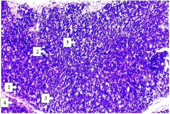 | Figure 1. Microscopic view of the thymus of a 3-month-old white outbred rat after experimental chemotherapy. Hematoxylin and eosin staining. Magnification 10x20. 1 – Decreased thymocytes around reticuloepithelial cells; 2 – Uniform staining of the cortical and medullary regions of the lobules, indicating a reduction in lymphocytes in both zones; 3 – Reduction of T lymphocytes in the cortical region of the lobules as a result of increased apoptosis; 4 – Shrinkage of lobules; 5 – Increased number of macrophages |
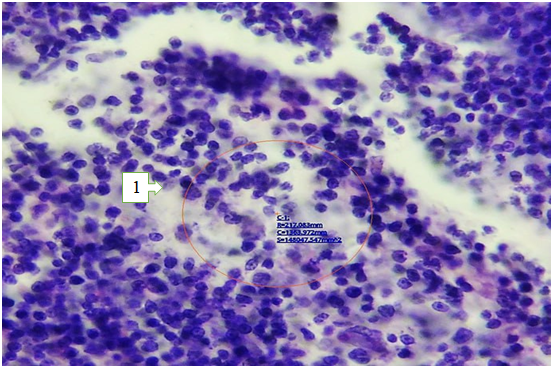 | Figure 2. Morphometry of thymocytes in the cortical region of the thymus of a 3-month-old white outbred rat after experimental chemotherapy. Hematoxylin and eosin staining. Magnification 20x20. 1 – Morphometric measurements of T lymphocytes (thymocytes) in the cortical region of the thymus |
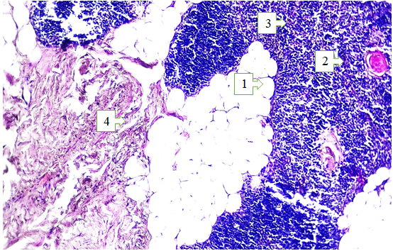 | Figure 3. Microscopic structure of the thymus in 3-month-old white outbred rats after experimental chemotherapy combined with pomegranate seed oil. Hematoxylin and eosin staining. Magnification 10x20. 1 – Dense accumulation of T lymphocytes in the cortical region; 2 – Enlarged and expanded Hassall’s corpuscles; 3 – Enlarged reticuloepithelial cells; 4 – Reduced adipose tissue, replaced by connective tissue |
 | Figure 4. Morphometry of thymocytes in the cortical region of the thymus of 3-month-old white outbred rats after experimental chemotherapy combined with pomegranate seed oil. Hematoxylin and eosin staining. Magnification 20x20. 1 – Morphometric measurements of cortical T-lymphocytes (thymocytes) |
4. Conclusions
- Experimental studies demonstrated that administration of cytostatic drugs such as paclitaxel causes profound morphological and morphometric changes in the thymic tissue. In treated animals, thymus size and weight were reduced, with early signs of organ involution observed. The thymic capsule and trabeculae thickened significantly, blood vessels dilated, and in certain areas hemodynamic disturbances and stasis developed. In addition, the cortical region showed a sharp decline in T-lymphocyte numbers, with increased apoptotic forms and intensified phagocytosis. These changes disrupted the thymus’s primary immunogenetic function—the differentiation and maturation of thymocytes. [12]Morphometric parameters in the paclitaxel group also shifted negatively: capsule and trabecular thickness increased markedly beyond physiological limits, vascular diameters widened, and thymocyte density decreased significantly. These alterations form the morphological basis for immunosuppression and increased susceptibility to infectious and inflammatory processes during chemotherapy. [13]In the second phase of the experiment, when paclitaxel was administered together with pomegranate seed oil, the severity of thymic pathomorphological changes was notably reduced. Thymic lobules were better preserved, cortical and medullary zones were clearly demarcated, T-lymphocytes were densely arranged, and their proliferative activity was relatively maintained. Hassall’s corpuscles were enlarged, showing signs of functional activity. The integrity of the blood–thymus barrier was comparatively well preserved, and hemodynamic disturbances were minimal. [14]Morphometric analysis confirmed these positive changes: thymic capsule and trabecular thickness approached physiological values, vascular diameters were significantly reduced, and thymocyte density was much higher compared to the paclitaxel-only group. These findings indicate that pomegranate seed oil, owing to its antioxidant, anti-inflammatory, and immunomodulatory properties, serves as an effective protective factor for thymic structure and function. [15]Thus, pomegranate seed oil alleviates chemotherapy-induced morphological and morphometric changes in the thymus, supports the differentiation and maturation of thymic lymphoid cells, and contributes to restoring the body’s immunological stability. These results provide an important scientific basis for the development of strategies to incorporate adjuvant therapeutic agents into clinical oncology practice.