-
Paper Information
- Next Paper
- Paper Submission
-
Journal Information
- About This Journal
- Editorial Board
- Current Issue
- Archive
- Author Guidelines
- Contact Us
American Journal of Medicine and Medical Sciences
p-ISSN: 2165-901X e-ISSN: 2165-9036
2025; 15(12): 4674-4679
doi:10.5923/j.ajmms.20251512.106
Received: Nov. 13, 2025; Accepted: Dec. 6, 2025; Published: Dec. 26, 2025

The Prevalence and Epidemiological Control of Taeniasis (Teniarhynchosis) in Uzbekistan
 Abstract
 Reference
 Full-Text PDF
 Full-text HTML
Yusupov Shavkat Shuxratovich1, Madreimov Amet2, Rasulov Shomurod Maxmudovich3
1Urgench Ranch Technological University, Assistant Department of Medical and Biological Sciences, Urgench, Uzbekistan
2Karakalpakstan Medical Institute, Professor Department of Hygiene, Environment and Epidemiology, Republic of Karakalpakstan, Uzbekistan
3Head of Department, Department of Microbiology, Public Health, Hygiene and Management, Termez Branch of Tashkent State Medical University, Termez, Uzbekistan
Correspondence to: Yusupov Shavkat Shuxratovich, Urgench Ranch Technological University, Assistant Department of Medical and Biological Sciences, Urgench, Uzbekistan.
| Email: |  |
Copyright © 2025 The Author(s). Published by Scientific & Academic Publishing.
This work is licensed under the Creative Commons Attribution International License (CC BY).
http://creativecommons.org/licenses/by/4.0/

The article presents a retrospective epidemiological analysis of the distribution of teniarinchiasis in the Republic of Uzbekistan and its administrative territories, including laboratory examinations, the incidence rate of the disease among children and adults, as well as among urban and rural populations, preventive measures, and the implementation of epidemiological surveillance.
Keywords: Teniarynchosis, Epidemiology, Epidemic process, Retrospective analysis, Morbidity, Prevention, Epidemiological control
Cite this paper: Yusupov Shavkat Shuxratovich, Madreimov Amet, Rasulov Shomurod Maxmudovich, The Prevalence and Epidemiological Control of Taeniasis (Teniarhynchosis) in Uzbekistan, American Journal of Medicine and Medical Sciences, Vol. 15 No. 12, 2025, pp. 4674-4679. doi: 10.5923/j.ajmms.20251512.106.
Article Outline
1. Introduction
- Teniarhynchiasis is a parasitic disease caused by the biogelmint Taeniarhynchus saginatus, a tapeworm belonging to the family Taeniidae. Currently, this parasite is known as Taenia saginata. The parasite uses cattle as intermediate hosts, while humans serve as the definitive hosts [2,5].The disease is more prevalent in Africa, parts of Eastern Europe, Asia, and the Philippines than in Latin America. The parasite is found anywhere beef is consumed, including in countries with strict sanitation policies, such as the United States. The disease is most prevalent in sub-Saharan Africa and the Middle East [19,21,25].According to the results of studies conducted in our country, teniasis accounts for 0.2% of all helminthiasis. Almost 1/5 of all teniasis cases detected in the republic are in the Khorezm region. Khorezm, Navoi, Bukhara and Samarkand regions account for almost 55% of all teniasis cases [1,8,14,15,23].As for livestock products, in accordance with the Decree of the President of the Republic of Uzbekistan No. PF-5696 dated March 28, 2019 "On measures to radically improve the system of state management in the field of veterinary medicine and livestock breeding", preventive work is being carried out to ensure epizootic welfare, as well as timely detection, diagnosis and prevention of the spread of infectious diseases of animals.Thus, as a result of studying the literature on the prevalence of teniarynchosis and cysticercosis, it can be concluded that the prevalence of this parasite differs between countries. Also, incidence rates may vary in different regions of the same country. There are contradictions regarding the prevalence of the disease in different age and gender groups.The cattle tapeworm is one of the longest tapeworms, reaching a length of 4-12 m, sometimes reaching 25 m. It is 12-14 mm wide. The head is square-oval, 1.5-2 mm in diameter. There are 4 suckers on the head, the pronotum is rudimentary or absent, and there are no hooks. The anterior segments are short and wide, and the posterior segments are long and thin. The length of the mature segments is 16-20 mm, and the width is 5-7 mm. The eggs are oval, yellowish-brown in color, measuring 46-50 x 39-41 μm [4]. The cattle tapeworm, when mature in the intestines of humans, can excrete up to 5-15 pieces per day through feces, and they contain up to 150-175 thousand eggs. In one year, about 440 million eggs can be released. Its eggs persist in the external environment for a long time. Eggs released through feces can remain viable for several weeks or months in wastewater or pastures. Eggs remain viable at temperatures from -10°C to 17°C. These data indicate that one person infected with the parasite can infect many cattle with cystocercosis.According to other scientific literature, the optimal environmental conditions for T. saginata eggs in the soil are a temperature of 25–30°C and a humidity level of 80%.Some researchers believe that cysticercosis can occur in humans if the parasite eggs are accidentally ingested through the mouth, but this is extremely rare. However, other groups of researchers believe that T. saginata does not cause cysticercosis in humans. In 2022, a patient with suspected T. saginata cysticercosis was identified in China. However, due to the limited availability of molecular genetic testing, a definitive diagnosis has not been made. The ability of T. saginata to cause cysticercosis in humans remains incompletely understood and controversial [18,20,24,25].Cattle become infected by consuming feed or water contaminated with human feces containing T. saginata eggs. Eggs that enter the intestine through the mouth penetrate the intestinal wall, enter the mesenteric venous capillaries or lymphatic vessels, and move throughout the body for 8-10 weeks, settling in the intermuscular connective tissue and transverse muscle (mainly the tongue, masticatory muscles, heart, but also the muscles of the body, arms and legs, diaphragm), connective tissue of internal organs, subcutaneous tissue, eyes, and brain, where they form cysts (cysticerci). Cysticerci remain viable for 8-9 months [11,13,19,21].However, one study conducted in recent years found that the proportion of infected cattle increases with age. It was also found that male cattle were more likely to be infected, although no statistically significant difference was found. The authors believe that this may be due to the fact that bulls move around more in pastures. Researchers have also observed that the prevalence of cysticercosis varies among different breeds of cattle [21].Disease distribution and economic damage: Numerous helminthological investigations have shown that cattle cysticercosis is found in farms of all regions. 2.6 - 8.9% of cattle are affected in ordinary farms where this disease is spread, while in livestock complexes it occurs in 1.3 - 10.3%.Cattle diseases also contribute to the development of infestation, as people with taeniarhinosis shed adult parasites and eggs for a long time, and these can survive in the environment for up to 12 months, in livestock buildings for up to 18 months, in manure and urine for up to 8 months, in water for up to 6 months, and in grass and hay for up to 4 months.In Uzbekistan, an average of 24 people out of every 10,000 people suffer from taeniarhinosis per year. In Uzbekistan, cysticercosis, which is most common in cattle, is found in about 10% of slaughtered cattle. Veterinary specialists consider 1% of the meat of slaughtered cattle infected with this disease to be unfit for consumption. In addition, young cattle in captivity with cysticercosis lose an average weight of 40 kg due to obesity.Thus, since taeniarhiniasis differs from other helminthiasis in that it proceeds without clinical symptoms, there is no accurate information about the level of incidence of this disease. Therefore, early diagnosis, conservative treatment, and improvement of preventive measures for taeniarhiniasis are among the urgent problems facing professionals in the field today.
2. Purpose of the Research
- The aim of the study was to analyze the prevalence and epidemiological characteristics of the development of the causative agent of teniarinchiasis in the Republic of Uzbekistan and its administrative territories, and to improve preventive measures against the disease.
3. Materials and Methods
- The study utilized official reports on teniarinchiasis from 2010 to 2023 provided by the Sanitary and Epidemiological Welfare and Public Health Committee of the Republic of Uzbekistan and its regional departments; epidemiological investigation maps of infectious disease foci; and patient case records. Epidemiological and statistical methods were employed in the research. The epidemiological analysis was carried out dynamically, taking into account various indicators such as natural, social, occupational, age, and gender factors.
4. Results and Discussion
- In particular, the analysis of the long-term dynamics of the disease allows us to study the trend, periodicity and irregular (epizootic) fluctuations of the epidemic process. In order to study the long-term dynamics of the incidence of teniarrhythmia in the Republic of Uzbekistan, official data of the Committee of the Republic of Uzbekistan for Sanitary and Epidemiological Wellbeing and Public Health for 2010-2023 were retrospectively analyzed.In the Republic of Uzbekistan, a total of 6,607 patients with taeniarhiniasis were registered during 2010-2023, and the intensity index of the disease per 100,000 population was found to be between 2.7 and 0.6 in different years (Figure 1).
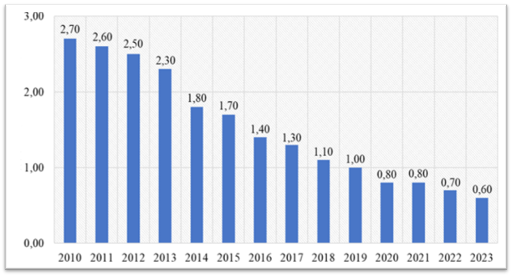 | Figure 1. Long-term dynamics of teniarinchiasis in the Republic of Uzbekistan (incidence rate per 100,000 population, 2010–2023) |
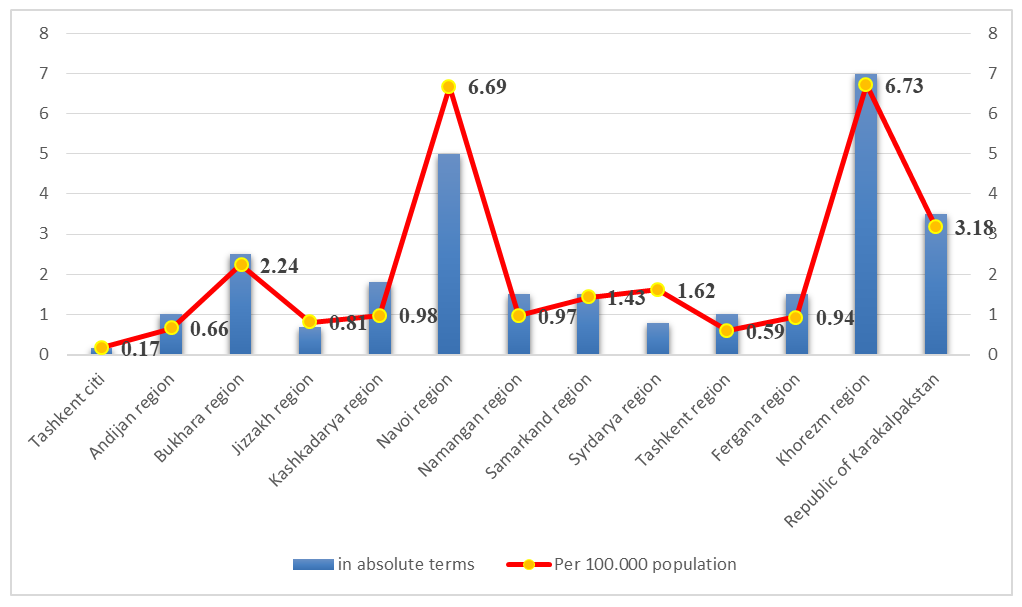 | Figure 2. Incidence rates of teniarinchiasis by administrative territories of the Republic of Uzbekistan (2010–2023) |
|
|
5. Conclusions
- Thus, an analysis of the literature on measures aimed at preventing taeniarhiniasis shows a number of problems and shortcomings. The problem is mainly related to identifying the source of the disease, since infected people may not show obvious clinical signs, which means that they can spread parasite eggs for a long time. In addition, the effectiveness of laboratory diagnosis of patients cannot be called high. Even when conducting a veterinary examination of large-horned cattle for cysticercosis, fins are not fully detected. In rural areas, most households have livestock, and they always consume animals slaughtered for their own needs without undergoing veterinary examination. The nutritional and medical culture of the population is also of great importance in the fight against this parasite.Epidemiological control of teniarhynchosis. To prevent people from contracting taeniarhiniasis, measures against this disease should be taken in two directions. First, it is necessary to identify the source of the disease early, make an accurate diagnosis, and treat it.Secondly, it is necessary to take into account that butchers have a great epidemiological importance in the fight against teniarhinosis in livestock areas. Therefore, they should know the ways of transmission of teniarhinosis, factors and measures to combat its spread. In addition to butchers, shepherds and all agricultural workers should also know this. Teniarhinosis is most often contracted by rural residents or those who prepare dishes and salads from meat products. All waste generated during the slaughter and dressing of cattle infected with teniarhinosis should be disinfected.To avoid infection, it is necessary to follow personal preventive measures. These include taking care of pets, as they may have pinworm eggs on their fur. After each pet care, hands should be washed thoroughly with soap.It should be remembered that children constantly put their hands and various objects in their mouths, which may be contaminated with tapeworm eggs. This shows how important it is to maintain good hygiene in children. Since people are often infected through their hands, it is advisable not to shake hands.It is difficult to give specific advice for every situation in life, of course, but from what has been said, it is clear that personal hygiene is of great importance in the fight against tapeworm disease.Mass medical examination of the population in endemic foci will allow for early detection of the disease. Data on animal infestation, data from medical and preventive institutions and the level of organization of medical care in the region will be developed, as well as effective measures will be taken and the organization of medical care will be improved.Epidemiological analysis is carried out at a certain time interval (month, half-year, year) in the outbreak and a retrospective epidemiological analysis should be conducted. For this, it is necessary to use all elements of epidemiological analysis: where (place, region), when (month, seasonality), in what form (in an epizootic outbreak, during an epidemic, epidemic) and who (patients, gender, age) became ill or are at risk of contracting taeniarhinosis.Epidemiological surveillance of taeniarhinosis should be carried out taking into account the pathogenetic characteristics of this invasion, the interrelationship of epizootic and epidemic situations, the type of circulating pathogen, as well as the social and environmental conditions that ensure a certain level of disease transmission among animals and humans. The results of regular epidemiological surveillance form the basis for planning rational, targeted measures against taeniarhinosis. In addition, they allow us to identify changes in the trend of epidemic and epizootic situations.In conclusion, the complete eradication of this disease from all livestock farms through preventive measures is an urgent issue of national economic importance, which, along with strengthening the livestock economy, is also a struggle to protect human health.