-
Paper Information
- Next Paper
- Previous Paper
- Paper Submission
-
Journal Information
- About This Journal
- Editorial Board
- Current Issue
- Archive
- Author Guidelines
- Contact Us
American Journal of Medicine and Medical Sciences
p-ISSN: 2165-901X e-ISSN: 2165-9036
2025; 15(11): 4142-4146
doi:10.5923/j.ajmms.20251511.86
Received: Sep. 4, 2025; Accepted: Oct. 2, 2025; Published: Nov. 25, 2025

Management of Pregnant Women with Varicose Veins in Samarkand
 Abstract
 Reference
 Full-Text PDF
 Full-text HTML
Khudoyarova Dildora Rakhimovna1, Yusupov Orzumurod Shomurodovich2
1DcS, Professor, Academician of the Turon Academy of Sciences, Head of the Department of Obstetrics and Gynecology No. 1, Samarkand State Medical University, Uzbekistan
2Free Applicants of the Department of Obstetrics and Gynecology No. 1, Samarkand State Medical University, Obstetrician-Gynecologist of the Samarkand Branch of the Republican Specialized Scientific and Practical Medical Center for the Protection of Maternal and Child Health, Uzbekistan
Copyright © 2025 The Author(s). Published by Scientific & Academic Publishing.
This work is licensed under the Creative Commons Attribution International License (CC BY).
http://creativecommons.org/licenses/by/4.0/

The aim of the study was to consider the features of management of pregnant women with varicose veins in the conditions of clinics of the city of Samarkand, to determine modern approaches to diagnostics, therapy and prevention of complications, and to evaluate the outcomes for the mother and fetus. The study involved 132 pregnant women who were divided into the main and control groups. Pregnant women with varicose veins have an increased load on the systemic venous return, which affects the development of edema, pain and deterioration in the quality of life. The main principles of management include: early diagnostics and dynamic monitoring of the venous system, ensuring medium or high compression, individualized planned physical activity and rest regimen, correction of risk factors (overweight, prolonged standing, sitting with crossed legs). If necessary, safe treatment methods for the perinatal period are used to minimize the risk to the fetus: sclerotherapy of limited areas, treatment of varicose veins of the lower extremities in phases and, if possible, delayed surgical intervention until the postpartum period. Prevention of thrombosis and symptom control play an important role in order to reduce the incidence of complications, including varicose-ulcerative lesions and trophic changes. Conclusions: the proposed algorithm was successfully used in Samarkand, effective management of pregnant women with varicose veins requires screening at early stages, prevention of complications, individualized choice of treatment methods and timely planning of delivery. The use of proven strategies in Samarkand reduces the risk of feto-material complications and improves outcomes for both mother and child.
Keywords: Pregnancy, Varicose veins, Prothrombin G2021OA, Antithrombin III, Factor V Leiden mutation, Thrombodynamics, Management
Cite this paper: Khudoyarova Dildora Rakhimovna, Yusupov Orzumurod Shomurodovich, Management of Pregnant Women with Varicose Veins in Samarkand, American Journal of Medicine and Medical Sciences, Vol. 15 No. 11, 2025, pp. 4142-4146. doi: 10.5923/j.ajmms.20251511.86.
Article Outline
1. Relevance
- Varicose veins are a significant problem in obstetrics and gynecology, especially among pregnant women. With the increasing frequency of this disease, it is necessary not only to study its pathogenesis and clinical manifestations, but also to develop optimal approaches to its prevention and treatment.There is a lot of information about the increasing incidence of varicose veins in the world community. According to phlebologists, in recent decades, cases of varicose veins (VV) of the internal organs and pelvic organs have been increasing [6]. As is known, VV of the genitals is an expansion of the veins of the external and internal genital organs (labia majora and minora, vagina, appendages of the uterus and uterus), which, according to some authors, is classified as an atypical form of venous disease [5,15]. According to the international epidemiological study of the Vein Consult program, which included 99,359 patients from different countries, the prevalence of varicose veins of the legs reaches 51.9-70.18%, depending on the region [3,12]. According to a study by Vuylsteke M.E., age and female gender are correlated with more severe clinical manifestations of the disease (p<0.001). Patients who regularly exercise and do not have a family history of this pathology are included in the lower CEAP classification for chronic venous diseases, group C. This study also provides information on the relationship of the degree of varicose veins with a high body mass index, lack of regular physical activity, the number of pregnancies, hereditary predisposition and the age of the patients [12]. According to the studies of Novikov B.N. (2011), 70-90% of women associate the appearance of varicose veins with pregnancy. This scientist's research is confirmed by both international and domestic scientists, but pregnancy cannot be a direct cause of varicose changes [10]. The development of advanced technologies in modern medicine has introduced many methods for diagnosing varicose veins. With the help of modern technologies, innovative methods for diagnosing varicose veins of the legs and pelvis have been introduced, and there are methods that provide high accuracy and safety for patients, including pregnant women. However, they are aimed at diagnosing the disease after it has developed.A 2021 study (Trombos Research), a 2023 study found that thrombodynamics is 30% more accurate than D-dimer in predicting thrombosis in pregnant women [11]. In the work of Smith J.R. et al. (2022), the method was successfully used to monitor anticoagulant therapy in patients with varicose veins [14]. In this regard, finding adequate diagnostic methods that are able to identify the risk of developing varicose veins and maintain the stability of hemodynamic parameters is a very important task.The purpose of the study management of pregnant women with various manifestations of varicose veins in the city of Samarkand.
2. Research Materials and Methods
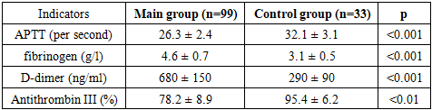
- This research was conducted on the basis of the Samarkand branch of the Republican Specialized Scientific and Practical Center for Maternal and Child Health and the maternity complex of the Multidisciplinary Clinic of the Samarkand State Medical University. 132 pregnant women were selected to participate in the study. The main group was formed by pregnant women with varicose veins (n=99), who were divided into three subgroups depending on the manifestation of varicose veins: group 1 included 33 pregnant women with varicose veins of the legs, group 2 included 33 pregnant women with varicose veins of the pelvis, and group 3 included 33 pregnant women with varicose veins of both the legs and the pelvis. The clinical and laboratory control group included 33 practically healthy pregnant women.Examination and treatment were carried out in accordance with the standards of medical care, as new methods for diagnosis, thrombodynamic blood analysis and methods for determining the prothrombin G2021OA, antithrombin III, Leiden factor V mutation were used.The working principle of the thrombodynamic analysis is as follows: a coagulation activator (usually tissue factor) is added to a blood plasma sample. The blood is placed in a special chamber with a two-dimensional cavity modeled (imitation of blood vessels). The process of fibrin clot formation is recorded in real time using a fluorescence microscope or optical sensors. We study this analysis based on such indicators as the thrombus growth rate, the delay in its development, the initial growth rate, the steady-state growth rate of the thrombus, the thrombus volume after 30 minutes, the thrombus density, and the time of spontaneous thrombus formation.From instrumental methods, an ECG was performed according to the standard, and Dopplerography of the pelvic organs and leg vessels was performed using UTT, as well as the state of the fetus and feto-placental system was studied. According to the results of a comprehensive examination, a decision was made on the need for VC therapy and prevention of thrombosis and thromboembolism.Variational and statistical processing of the study results was carried out using the Statistica 6.0 program with the determination of the main variability indicators: mean values (M), errors of the mean value (m), standard deviation (p). The reliability of the results was determined using the Student t-test. The difference between two mean values is considered significant if the p-parameter is less than 0.05. The level of confidence was at least 95%. The correlation between the indicators was calculated using the Excel 2010 spreadsheet.
3. Research Results and Discussion
- All pregnant women studied were of active reproductive age, i.e. from 19 to 44 years. The average age was 27±0.7 in the main group and 26.2±0.5 in the control group.Analysis of the social status of women in the main group showed that in group 1, 45.45% of women were employees, of which 27.27% were workers requiring heavy labor, 24.24% were unemployed (housewives), 30.3% were students, and in group 2, 35.35% were employees, workers - 18.18%, housewives - 42.42% and students - 4.05%. It is seen that the prevalence of varicose veins in the legs is more pronounced in workers with heavy labor and students. In the third group of women, workers accounted for 54.54%, housewives for 21.21%, and students for 24.24%. Women in the control group did not differ significantly from the previous two groups in terms of social status (p>0.05).The main and control groups were compared according to the main parameters, including social status, menstrual and reproductive history, obstetric and gynecological condition, somatic diseases, contraceptive methods used, etc.The average number of pregnancies was 2.8±0.2 in the main group of women, and the number of births was 2.4±0.2. The average gestational age in the main group was 30.3 ± 1.0 weeks. According to statistical data, the average BMI varied from 17 to 35, but averaged 28.4 ± 0.4. High BMI values were more common in women of group 3, who had mixed forms of varicose veins.Analysis of complaints showed that the most common symptom in women of the main group was the presence of a feeling of heaviness and pain in the legs, which was noted by 83.83% of women in group 2 and 100% in groups 1 and 3. Subjective symptoms in the form of heaviness in the legs, dilation and pain in the area of varicose veins were noted in 100% of women in all subgroups of the main group.The CEAP classification of varicose veins in pregnant women was carried out. 30 women in the control group were assigned to category C0, and 3 women in the control group to category C1. Pregnant women in the main group were divided into categories from C1 to C5. In group 1, telangiectasias and venous plexuses (C1) were detected in 12.12%, in group 2 - in 15.15%, and in group 3 - in 9.09%. Category C5, i.e., trophic ulcers, occurred in 15.15% and 21.21% of representatives of groups 1 and 3, respectively.
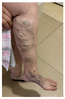 | Figure 1. Group 1 patient in category C-5 (E.F. case history No. 116/533) |
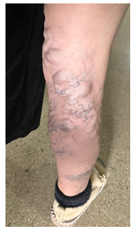 | Figure 2. Group 3 patient of category C-5 (D.K. case history No. 180) |
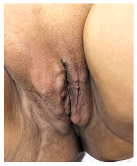 | Figure 3 |
|
4. Conclusions
- An algorithm for diagnosing and preventing varicose veins in women during pregnancy is proposed, including the tactics of differential treatment and management of pregnant women with varicose veins, taking into account the form and degree of the disease, the presence of complications, if necessary, the identification of polymorphisms of their genes and the appointment of additional therapy.The use of the algorithm for diagnosing varicose veins in women during pregnancy increases the effectiveness of therapy and treatment outcomes in women with varicose veins and reduces the number of complications of the disease, especially thrombotic complications that can lead to maternal death. The medical effectiveness of the algorithm for diagnosing and preventing varicose veins in women during pregnancy is characterized by a 50% reduction in varicose vein complications. The use of the proposed algorithm has high social effectiveness in terms of preventing the transition of the disease to a complicated form, maintaining reproductive health and improving the quality of life of women, and improving diagnostic and preventive work using primary medical and sanitary services.