-
Paper Information
- Previous Paper
- Paper Submission
-
Journal Information
- About This Journal
- Editorial Board
- Current Issue
- Archive
- Author Guidelines
- Contact Us
American Journal of Medicine and Medical Sciences
p-ISSN: 2165-901X e-ISSN: 2165-9036
2025; 15(11): 3998-3999
doi:10.5923/j.ajmms.20251511.55
Received: Oct. 25, 2025; Accepted: Nov. 12, 2025; Published: Nov. 14, 2025

Changes in the Structural and Functional Parameters of the Adrenal Glands on the 7th Day After Experimental Modeling of Traumatic Brain Injury
 Abstract
 Reference
 Full-Text PDF
 Full-text HTML
Kadirova Laylo Valijanovna
Assistant at the Department of Pathological Physiology, Bukhara State Medical Institute, Uzbekistan
Correspondence to: Kadirova Laylo Valijanovna, Assistant at the Department of Pathological Physiology, Bukhara State Medical Institute, Uzbekistan.
| Email: |  |
Copyright © 2025 The Author(s). Published by Scientific & Academic Publishing.
This work is licensed under the Creative Commons Attribution International License (CC BY).
http://creativecommons.org/licenses/by/4.0/

According to the literature, there is growing interest in studying the pathogenesis of brain injury that leads to structural remodeling of the organs of the endocrine system, caused by various etiological factors. The hypothalamic–pituitary–adrenal (HPA) axis is the primary system responsible for the stress response, and it undergoes structural and functional changes that result in impaired hormone production. Traumatic brain disease is the body's systemic response to CNS injury, characterized by disrupted homeostasis and the development of a specific reaction that mobilizes the hypothalamic–pituitary–adrenal axis, accompanied by endogenous release of catecholamines and adrenocorticotropic hormone (ACTH), which reflect adrenal gland function. The main part of the adrenal cortex consists of cuboidal and polygonal cells forming the fascicular zone, which produces glucocorticoid hormones.
Keywords: Traumatic brain injury, Hypothalamic–pituitary–adrenal axis, Adrenal cortex, Fascicular zone, Adrenocorticocytes
Cite this paper: Kadirova Laylo Valijanovna, Changes in the Structural and Functional Parameters of the Adrenal Glands on the 7th Day After Experimental Modeling of Traumatic Brain Injury, American Journal of Medicine and Medical Sciences, Vol. 15 No. 11, 2025, pp. 3998-3999. doi: 10.5923/j.ajmms.20251511.55.
1. Introduction
- Studies on the pathogenesis of traumatic brain injury (TBI) have shown that the initial local signs of traumatic damage to the brain appear within the first day and reach their peak expression between the 4th and 8th days after the injury [7]. This article presents information on the morphostructural changes occurring in the layers of the adrenal cortex, adrenocorticocytes [1], and blood vessels of the adrenal glands in white outbred rats on the 7th day after experimental modeling of traumatic brain injury. [2]
2. Materials and Methods
- The research was conducted at the Bukhara State Medical Institute named after Abu Ali ibn Sina. For this experimental scientific study, white outbred sexually mature rats of both sexes were selected as subjects, weighing between 130 and 150 grams and aged 90 days. Traumatic brain injury (TBI) was experimentally modeled using a device designed to simulate a road traffic accident, thereby inducing TBI in the laboratory rats. On the 7th day after the experiment, the animals were dissected, and their adrenal glands were extracted for further examination of the morphostructural changes in the layers of the adrenal cortex. The morphological appearance of the adrenal glands in white outbred rats on the 7th day after experimental TBI was as follows: Upon macroscopic examination, the adrenal glands were found to be reduced [4] in size compared to earlier time points. The usual rounded shape was absent, particularly in comparison with day 3 after TBI. In the subcapsular region of the cortex, thickened layers were identified in the intercellular spaces, and a slight, localized edema of the stromal tissue was observed. The adrenal capillaries were dilated and hyperemic, mainly located in the fascicular zone. [3] The number of hyperemic capillaries per 0.01 mm², evenly distributed in both the fascicular and reticular zones, was 2.7. Tissue edema and hypertrophy of adrenocorticocytes contributed to a slight thickening of both the fascicular and reticular zones. [6] In the inner part of the fascicular zone, the typical columnar structure was disrupted. Lipid accumulation in cortical cells was observed in all experimental animals. [5]
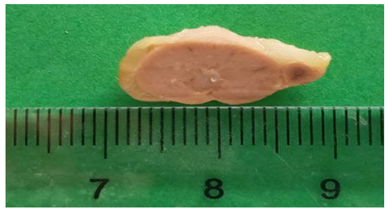 | Figure 1. Macroscopic specimen. Adrenal gland on the 7th day after experimental modeling of traumatic brain injury (TBI) |
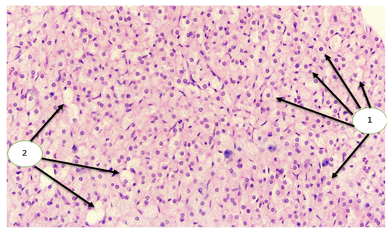 | Figure 2. Microscopic specimen. Adrenal cortex on the 7th day after TBI. Hypertrophy of adrenocorticocytes (1), stromal edema, and thickening of the fascicular and reticular zones (2) are observed. Stained with H&E. Magnification: objective 40×, ocular 20× |
3. Conclusions
- In the pathogenesis of traumatic brain injury (TBI), the most pronounced changes in brain tissue occur due to the direct impact of the traumatic agent and as a result of secondary changes characterized by neuroinflammation at the injury site. These lead to disturbances in the hypothalamic–pituitary–adrenal (HPA) axis. Macroscopic examination of the adrenal glands of rats on the 7th day after TBI shows a reduction in adrenal size compared to the 1st and 3rd days after injury. The adrenal capillaries are dilated and congested, with hyperemia especially pronounced in the fascicular zone. Amid circulatory disturbances, a slight and limited edema is observed. Changes in the HPA axis during the acute phase after TBI manifest as dysfunction of anterior pituitary hormones, which is reflected in the morphostructural appearance of the adrenal glands studied on the 7th day after TBI. These changes are associated with pituitary dysfunction and suppression, which describe the pathogenesis of pituitary alterations characteristic of the acute post-traumatic period.