-
Paper Information
- Next Paper
- Previous Paper
- Paper Submission
-
Journal Information
- About This Journal
- Editorial Board
- Current Issue
- Archive
- Author Guidelines
- Contact Us
American Journal of Medicine and Medical Sciences
p-ISSN: 2165-901X e-ISSN: 2165-9036
2025; 15(11): 3958-3963
doi:10.5923/j.ajmms.20251511.47
Received: Oct. 9, 2025; Accepted: Nov. 6, 2025; Published: Nov. 14, 2025

Immunohistochemical Characteristics of Vessels in Pulmonary Artery Thromboembolic Developed After Surgery
 Abstract
 Reference
 Full-Text PDF
 Full-text HTML
Kurbanov Jalalkhan Bakhromovich1, Mamajonov Bokhadirjon Solijanovich2
1Fergana Public Health Medical Institute, Fergana, Uzbekistan
2Andijan State Medical Institute, Fergana, Uzbekistan
Copyright © 2025 The Author(s). Published by Scientific & Academic Publishing.
This work is licensed under the Creative Commons Attribution International License (CC BY).
http://creativecommons.org/licenses/by/4.0/

Thromboembolic complications after surgery are important in determining the molecular substrates underlying damage to the pulmonary arteries in thromboembolism. In particular, detection of blood clotting factors in vascular endothelium using SD 31 and SD 63 markers is an important link in the assessment of gene expression.
Keywords: Thromboembolism, Immunohistochemical examination, Pathomorphology, Thrombosis
Cite this paper: Kurbanov Jalalkhan Bakhromovich, Mamajonov Bokhadirjon Solijanovich, Immunohistochemical Characteristics of Vessels in Pulmonary Artery Thromboembolic Developed After Surgery, American Journal of Medicine and Medical Sciences, Vol. 15 No. 11, 2025, pp. 3958-3963. doi: 10.5923/j.ajmms.20251511.47.
1. Introduction
- Pulmonary embolism (PE) is the final stage of venous thromboembolism (VTE) and is the third leading cause of death among circulatory diseases worldwide, after myocardial infarction and stroke, with an estimated 0.1% of the world's population, or 8.5 million people, dying from this disease each year [1,2]. In the United States, approximately 600,000 patients are hospitalized with PE each year, 33% of whom die in critical condition [3,4]. PE remains one of the leading causes of death in surgical hospitals. It has been reported that it occurs in 4.1% of patients in vascular surgery, 14.3% in abdominal surgery, 33.3% in surgical surgery, 30.7% in neurosurgery, and 40% in proctology. Thromboembolic complications remain one of the leading causes of sudden death, particularly in the elderly. Despite significant progress in understanding coagulation and vascular-thrombotic mechanisms, the pathogenetic features of age-related changes in the vascular wall and endothelial response during thromboembolism remain insufficiently studied. One of the key links in the pathogenesis of thrombogenesis is the interaction between endothelial cells, platelets, and their activation markers, among which CD63 - a tetraspanin protein localized in the granules of platelets and endothelial cells, including Weibe-Palade bodies [5,6,7] - plays a major role. It is involved in the processes of degranulation, adhesion, and secretion of biologically active substances, including von Willebrand factor and P-selectin, which regulate the adhesive and aggregative properties of the vascular wall. However, in clinical and morphological practice, the dynamics of CD63 expression in blood vessels and thrombi depending on patient age and stage of the thromboembolic process remain poorly understood. In particular, there is a lack of data on: the morphofunctional characteristics of endothelial cells and their secretory activity in thromboembolic events among different age groups; changes in CD63 expression as a marker of platelet and endothelial cell degranulation depending on the severity and localization of thrombosis; the relationship between age-related vascular involution and decreased expression of adhesion and secretion markers such as CD63, von Willebrand factor, and P-selectin; pathomorphological signs of “aging endothelium” in the context of increased thromboembolic susceptibility. With advancing age, a decline in the metabolic and secretory activity of the endothelium is observed, along with a reduction in the number of Weibel–Palade bodies, degeneration of platelet granular structures, and disruption of intercellular interactions, which collectively increase the risk of unregulated thrombus formation [8,9,10,11]. These processes form the basis of age-dependent vascular involution, yet their morphological and immunohistochemical characteristics in thromboembolic complications have been only fragmentarily described [12,13,14]. Thus, the relevance of the present study is determined by: the need for an in-depth investigation of the molecular and morphological mechanisms of vascular wall aging and their role in the development of thromboembolic complications; the absence of systematized data on CD63 expression and other endothelial markers across various age groups in acute and chronic thromboembolism; the limited diagnostic value of conventional histological techniques for identifying age-related endothelial changes and the need for immunohistochemical markers (CD63, vWF, P-selectin) to assess the degree of vascular aging; the practical importance of defining age-related expression patterns of CD63, which may contribute to clarifying the morphogenesis of thromboembolism and predicting thrombotic risk in both clinical and forensic pathology. Taken together, these factors emphasize the necessity of a comprehensive morphological and immunohistochemical analysis of vascular–platelet interactions in thromboembolic complications, taking into account age-related vascular alterations.Aim of the StudyThe aim of this study is to investigate the age-related immunohistochemical features of CD63 expression in vascular endothelium and thrombi in cases of thromboembolic complications, in order to identify the molecular and morphological mechanisms of endothelial aging and its role in the pathogenesis of thrombus formation and vascular damage.
2. Materials and Methods
- The study used morphological, morphometric, immunohistochemical, and statistical methods to improve the assessment of age-related pathomorphological changes in pulmonary arteries and lung tissue in patients with pulmonary thromboembolism that developed after surgery. The material was pulmonary arteries and lung tissue from autopsies of 78 women who died of pulmonary embolism.
3. Results and Discussion
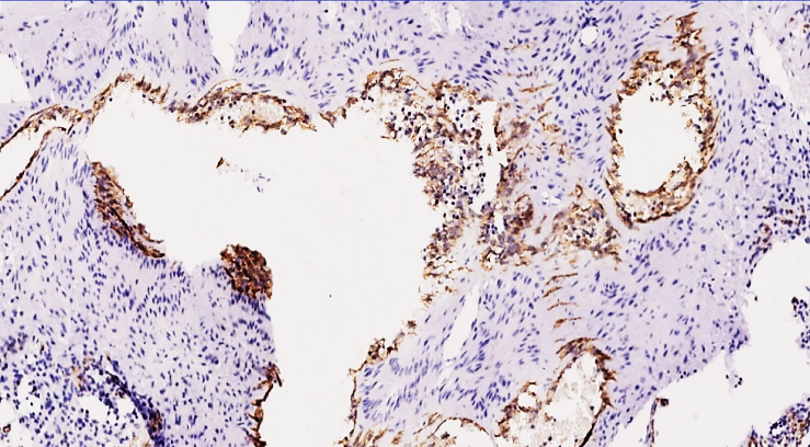
- Paraffin-embedded biopsy materials were subjected to immunohistochemical examination using monoclonal antibodies using standard methods. The CD31 PECAM marker is a marker indicating the level of platelet-1, which adheres to the endothelium. The glycoprotein protein is mainly expressed on vascular endothelial cells, platelets, granulocytes, monocytes, and some T lymphocyte coreceptors, indicating the development of new vessels, and if it is present on the inner surface of the vessel, then thromboplastin is formed and the damage process on the inner surface of the vessel is acute.This glycoprotein is a transmembrane protein, and normally it does not react with the CD 31 PECAM marker and is not masked. Since the free location of these glycoproteins indicates the absence of intercellular contact, in our study, the complication of thromboembolism indicates the degree of vascular damage.This also means that this glycoprotein in the perivascular areas accelerates the process of angiogenesis by connecting other types of mesenchymal cells, and if the yellow-golden expression is detected on the inner surface of the vessels, it proves that the process of injury and thrombogenesis is taking place in these vessels.If, upon examination, it is detected in a scattered and irregular appearance in the stroma of the prevascular and intramural tissue, this means that the process of transformation of endothelial cells from mesenchymal cells and neoangiogenesis is taking place.In our study, there were 3 groups, and the lung tissue and small-caliber vessels of 18-44, 45-59, 60-74 years old were stained with the CD 31 PECAM marker. The following immunohistochemical analysis showed a high positive expression of the CD 31 PECAM marker in 6 out of 26 patients aged 18-44 years, which clinically and morphologically indicates a predominance of vascular damage at this age, and this indicates an acceleration of the process of thrombogenesis due to damage to the inner surface of small-caliber blood vessels of the lungs. The systemic appearance of leukotrienes and cytokines released from damaged tissues in the period after any type of surgical intervention indicates a predominance of the ability of the body to cause a systemic vascular response, mainly in 18-44 years, which also indicates a predominance of the body's age-related hyperergic response and the fact that in the period after thromboembolic complications, small-caliber vessels of the lungs are damaged mainly from small-caliber vessels.
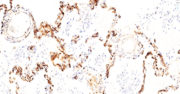 | Figure 1. A 41-year-old patient died of pulmonary embolism on the 4th day after surgery. Moderately positive expression of the CD 31 PECAM marker is detected in the intima of the alveolar walls and small-caliber vessels of the lung tissue. Staining is Dab chromogen. Size 20x10 |
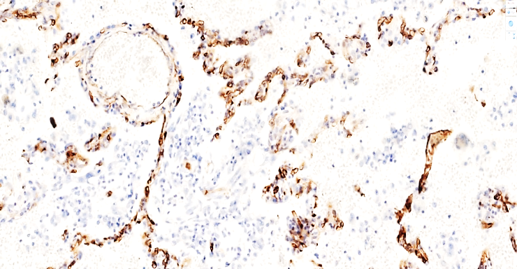 | Figure 2. A 40-year-old patient died of pulmonary embolism on the 3rd day after surgery. Moderately positive expression of the CD 31 PECAM marker is detected in the intima of the alveolar walls and small vessels of the lung tissue. Staining is Dab chromogenic. Size 20x10 |
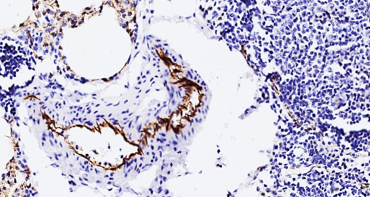
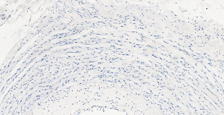 | Figure 3. A 58-year-old patient died of pulmonary embolism on the 3rd day after surgery. Lung tissue shows a negative reaction of the CD 31 PECAM marker in the pulmonary trunk and the vessel wall. Staining is Dab chromogenic. Size 20x10 |
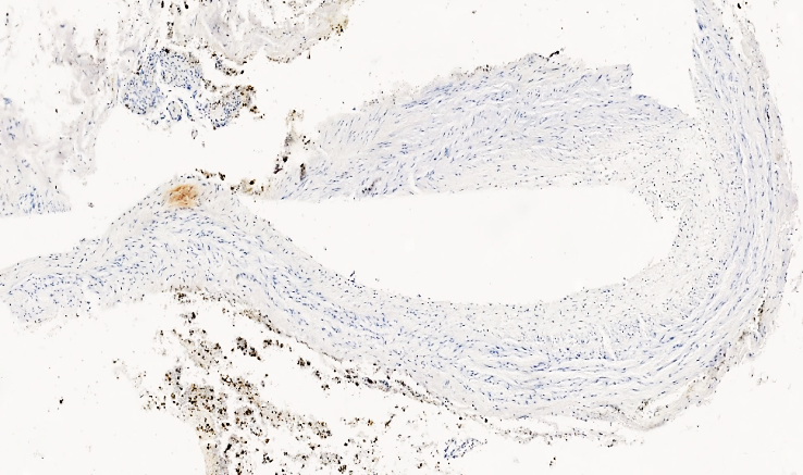 | Figure 4. A 72-year-old patient died of pulmonary embolism on the 4th day after surgery. Lung tissue shows a negative reaction of the CD 31 PECAM marker in the pulmonary trunk and the vessel wall. Staining is Dab chromogenic. Size 20x10 |
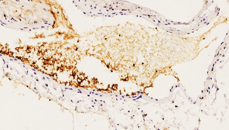
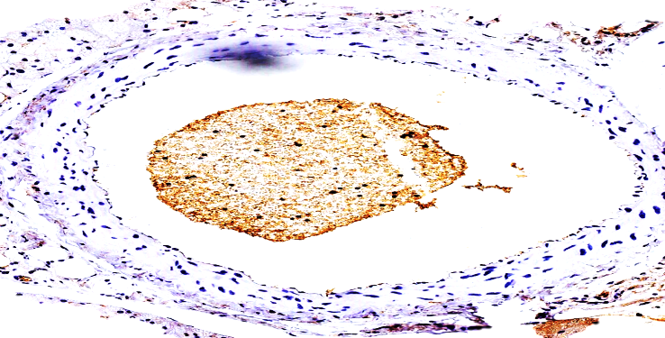
 | Figure 5. 41-year-old 4 days after surgery. High positive expression of the SD 63 marker is determined mainly on the surface of the inner surface of the vessel endothelium and thromboblastin substrate. This also means that the vessel is damaged and the process of thrombogenesis is advanced. Paint Dab chromogenic. Size 4x10 |
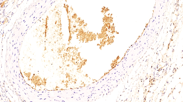
 | Figure 6. 44-year-old 5 days after surgery. High positive expression of SD 63 marker. On the surface of the vascular endothelium, platelet adhesion aggregation and sediments in the granular form of different sizes are determined. Aggregation of platelets preserved in thrombi formed in the vessel cavity is determined in granular form. Dab chromogen. The size is 20x10 |
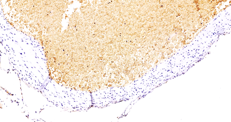
 | Figure 7. 42-year-old woman. 5 days after surgery. High positive expression of the CD63 marker. Adhesive aggregation of platelets in the form of plaques on the surface of the vascular endothelium and Weibel-Palade bodies in the cytoplasm of endothelial cells are detected. Staining is Dab chromogen. Size 20x10 |
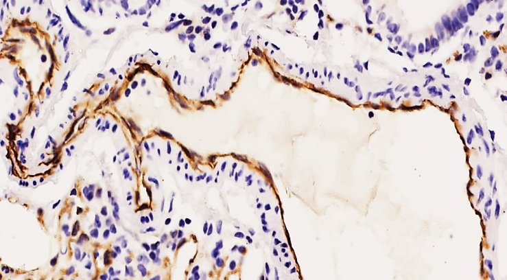 | Figure 8. 37-year-old woman. 4 days after surgery. Moderately high positive expression of the CD63 marker. Adhesive aggregation of platelets in the form of flat sheets on the surface of the vascular endothelium and Weibel-Palade bodies in the cytoplasm of endothelial cells are detected. Staining is Dab chromogenic. Size 40x10 |
 | Figure 9. Group 2. 51-year-old woman. Positive expression of thromboplastins in the form of adhesive granular thromboplastins in the vascular lumen. No changes in the vascular endothelium are detected. Staining is Dab chromogenic. Size 20x10 |
 | Figure 10. Group 2. 49-year-old woman. Moderate positive expression of the CD 63 marker is detected in the thrombus in the vascular lumen and in granular platelets adhered to the vascular wall. Focal changes are detected in the vascular endothelium. Staining Dab chromogenic. Size 20x10 |
 | Figure 11. Group 3. 74-year-old woman. Massive obturating pulmonary artery anterior wall thrombus, massively adhered granular platelets in the thrombus, high positive expression of the CD 63 marker. Staining is Dab chromogenic. Size 20x10 |
4. Conclusions
- In pulmonary artery thromboembolism, it was found that the immunohistochemical CD 31 PECAM marker of the pulmonary arteries showed low positive expression in 55-59-year-olds and medium positive expression in 18-44-year-olds, indicating a high degree of damage to the vascular endothelium, which was explained by the production of biologically active substances by endothelial cells, and manifested as a vascular response. Based on the immunohistochemical analysis of CD63 marker expression in different age groups, distinct age-related differences in vascular and thromboembolic processes were identified. In group 2 (45–59 years), moderate positive expression of CD63 was detected both in the thrombus and in platelets adhered to the vascular wall, with focal endothelial changes. The frequency of positive endothelial reactions was 1.5 times lower than in younger individuals (group 1), indicating the beginning of endothelial involutional changes. The formation of thromboembolic complications in this group was primarily associated with vascular damage caused by migrated thrombi, while local endothelial secretory activity and reparative capacity were partially preserved. In group 3 (60–74 years), CD63 expression decreased sharply. Moderate positivity was observed only in 31.13% of cases, mainly within thrombi and granular platelets, whereas endothelial cells on the vascular surface exhibited negative or minimal reactions. The content of Weibel–Palade bodies and von Willebrand factor was significantly reduced, confirming endothelial functional exhaustion. The diminished endothelial secretory response and increased vascular rigidity reflect age-dependent biological involution of the vascular wall. Overall, with aging, there is a progressive reduction in endothelial reactivity, suppression of CD63 expression, and depletion of secretory granules containing adhesion molecules such as von Willebrand factor and P-selectin. These changes lead to reduced vascular resilience, increased susceptibility to thromboembolic complications, and impaired thrombus resolution. Thus, the obtained data morphologically and immunohistochemically substantiate that vascular aging is accompanied by a decline in CD63-mediated platelet-endothelium interactions, resulting in impaired regulation of thrombus formation and contributing to the increased incidence and severity of thromboembolic events in elderly individuals.