-
Paper Information
- Next Paper
- Previous Paper
- Paper Submission
-
Journal Information
- About This Journal
- Editorial Board
- Current Issue
- Archive
- Author Guidelines
- Contact Us
American Journal of Medicine and Medical Sciences
p-ISSN: 2165-901X e-ISSN: 2165-9036
2025; 15(10): 3724-3727
doi:10.5923/j.ajmms.20251510.96
Received: Oct. 6, 2025; Accepted: Oct. 22, 2025; Published: Oct. 31, 2025

Forensic Medical Diagnosis of Injuries to the Nose, Ear, and Larynx: Modern Methods and Algorithms
 Abstract
 Reference
 Full-Text PDF
 Full-text HTML
Abdumalikov I. M.
Fergana Medical Institute of Public Health, Fergana, Uzbekistan
Correspondence to: Abdumalikov I. M., Fergana Medical Institute of Public Health, Fergana, Uzbekistan.
| Email: |  |
Copyright © 2025 The Author(s). Published by Scientific & Academic Publishing.
This work is licensed under the Creative Commons Attribution International License (CC BY).
http://creativecommons.org/licenses/by/4.0/

Background: Injuries to the nose, ear, and larynx are among the most common reasons for forensic medical examination. Accurate diagnosis of such injuries requires a combination of clinical, instrumental, and laboratory approaches. The purpose of this study was to improve the accuracy and objectivity of forensic evaluation of ENT injuries by integrating advanced diagnostic algorithms. Methods: The research analyzed 100 forensic medical cases involving ENT trauma using clinical observation, computed tomography (CT), ultrasound, anterior active rhinomanometry, and morphological as well as immunohistochemical analyses. Tissue samples were stained for fibronectin, collagens, and cytokines (IL-1β, IL-6, TNF-α) to determine the injury’s age and healing stage. Results: Nasal injuries accounted for 70% of cases, predominantly caused by blunt force trauma. CT and ultrasound demonstrated over 90% diagnostic accuracy in identifying fractures. Rhinomanometry effectively quantified nasal breathing resistance, while immunohistochemical testing provided precise timing of injury formation. The study also confirmed the value of systematic photographic documentation in expert conclusions. Conclusions: Combining instrumental imaging, functional assessment, and morphological techniques significantly enhances the precision of forensic medical evaluations. The proposed diagnostic model aligns with modern international standards and improves the reliability of expert judgments.
Keywords: Forensic medical examination, ENT injuries, Nose, Ear, Larynx, Computed tomography, Rhinomanometry, Morphological analysis, Immunohistochemistry, Photographic documentation
Cite this paper: Abdumalikov I. M., Forensic Medical Diagnosis of Injuries to the Nose, Ear, and Larynx: Modern Methods and Algorithms, American Journal of Medicine and Medical Sciences, Vol. 15 No. 10, 2025, pp. 3724-3727. doi: 10.5923/j.ajmms.20251510.96.
1. Introduction
- Injuries to ENT organs (ears, nose, larynx, etc.) are frequently the subject of forensic medical examinations. According to the literature, nasal injuries account for up to 91% of all ENT injuries [1]. Ear injuries are less common - about 5-6% of all injuries in otorhinolaryngology, primarily occurring in young men and in domestic settings [2]. Blunt and stab wounds of the larynx and pharynx are relatively rare but potentially dangerous due to the risk of airway obstruction. Despite the frequency of such injuries, the criteria for their forensic medical assessment - severity, time since injury, and mechanism of formation - remain topics of discussion [1,2,3].To objectively evaluate nasal breathing disorders during examinations, instrumental methods are used, including anterior active rhinomanometry and acoustic rhinometry [4]. Computed tomography and ultrasound demonstrate high sensitivity in detecting nasal bone fractures [3,5]. Morphological and immunohistochemical methods, including the detection of inflammatory marker expression, have gained significant importance in determining the time elapsed since injury [6].
2. Materials and Methods
- The study incorporated materials from forensic medical examinations of ENT injuries in recent years, as well as data from literature. For illustration, generalized clinical and expert data from 100 cases are presented. During the examination of victims, a physical assessment was conducted, documenting the clinical picture, presence of hematomas, edema, wounds, and deformities. Instrumental methods employed included: facial skeleton radiography, computed tomography (CT) of the nasal and temporal bones when fractures were suspected, endoscopic rhinoscopy and laryngoscopy, and audiometry and tympanometry to assess hearing.To objectively evaluate nasal breathing, anterior active rhinomanometry (AARM) was used - a dynamic method for assessing the resistance of nasal passages to airflow [7]. Photographic documentation of injuries was mandatory at all stages of the examination [8]. In cases where CT was contraindicated, ultrasound of the nasal bones was used to clarify the diagnosis.Morphological examination included histology of wound zone tissues with immunohistochemical staining (fibronectin, collagens, pro-inflammatory cytokines, etc.) to assess the age of injuries [9].Examination methodology: following the physical assessment and history taking, macroscopic examination, photography, and a series of instrumental diagnostic procedures were performed. If a fracture was suspected, CT of the paranasal sinuses was conducted. In cases of breathing difficulties, AARM was employed, while for ear injuries, audiometry and tympanometry were used. Morphological analysis included histological and immunohistochemical examination of tissues.
3. Results
- In the examined sample, nasal injuries predominated (70%), mainly in young men (average age 25-32 years), which aligns with the data from Mohammadi and Ghasemi-Rad's study. The primary mechanism of injuries was blunt domestic impacts and falls (65-80% of cases), while stab and cut wounds were rare. Among nasal injuries, 75% were fractures of the nasal bones involving the nasal septum, with contusions and injuries to the external nose observed less frequently. Table 1 presents the summary clinical and expert indicators.
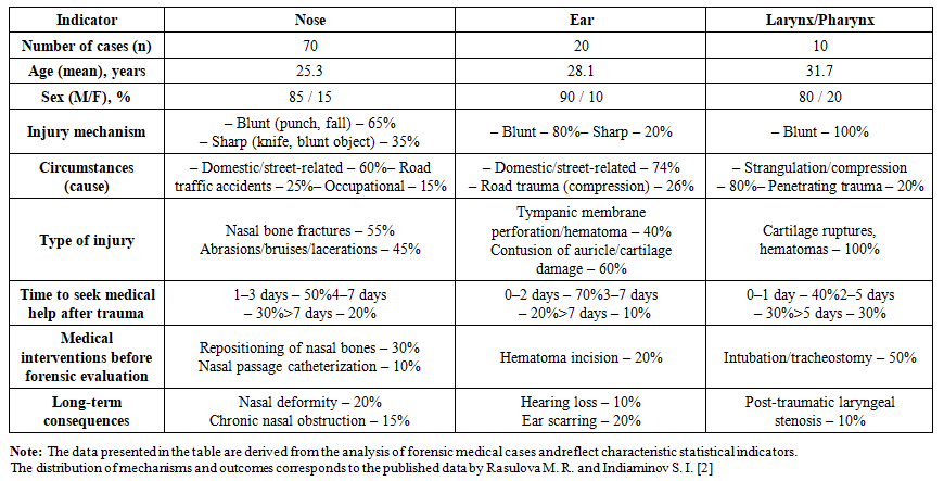 | Table 1. Clinical and expert data on ENT organ injuries (sample size n=100) |
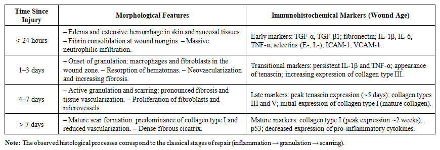 | Table 2. Morphological and immunohistochemical features at different stages of ENT organ wound healing |
4. Discussion
- The obtained results confirm that a combined approach - integrating clinical examination, instrumental methods, and morphological analysis - provides the most comprehensive understanding of the nature of ENT injuries and their consequences. The high frequency of domestic injuries reflects a typical criminogenic pattern: blows to the face, received in fights and conflicts, often lead to nasal bone fractures and contusions of the external ear. The proportion of nasal injuries exceeding 90% in the sample confirms the high vulnerability of this anatomical area.Instrumental diagnostics (CT, X-ray, ultrasound). Facial skeleton radiography is still used in forensic practice, but its sensitivity is limited due to the overlap of anatomical structures. Computed tomography allows for the detection of hidden fractures and combined injuries of the paranasal sinuses and orbits, as well as three-dimensional reconstruction for surgical planning. The advantage of CT is its high sensitivity and the ability to visualize even minimal displacements in detail. Meanwhile, ultrasound examination of the nasal bones remains an alternative when CT is contraindicated (pregnancy, pediatric patients), demonstrating high diagnostic reliability.Objectification of nasal breathing. Active anterior rhinomanometry (AARM) has proven to be a reliable method for verifying nasal breathing disorders. In injuries accompanied by swelling or deformation, the patient's subjective sensations may not correspond to the objective picture. AARM allows for the quantitative assessment of airflow resistance in the nasal passages and thereby establishes the degree of impairment. This is especially important when examining persistent consequences, such as chronic congestion that impedes breathing. The methodology is characterized by high reproducibility and objectivity, making it applicable in forensic practice.Assessment of injury severity. In the expert assessment of ENT injury severity, the nature of the damage, the duration of functional loss, and the presence of persistent consequences are taken into account. For nasal injuries, the main criteria are the duration of respiratory disorders and the presence of deformities; for ear injuries - the degree of hearing loss; for laryngeal injuries - the severity of respiratory and phonation disorders. Such a comprehensive approach allows for more accurate qualification of health damage and justification of the expert opinion.Photo documentation and expert protocols. Mandatory photo documentation significantly increases the reliability of expert conclusions. It not only records the external signs of injury but also serves as evidence in court proceedings, supplementing written descriptions. Modern expert examination algorithms involve the phased implementation of all diagnostic procedures - from initial examination to morphological analysis and immunohistochemistry.Morphology and immunohistochemistry. Morphological examination of damaged tissues remains the gold standard in determining the duration and stage of injury. In addition to standard histology, immunohistochemical methods allow for the determination of inflammatory marker expression (IL-1β, IL-6, TNF-α) and repair markers (fibronectin, TGF-β, various types of collagens). Using these data in conjunction with the morphological picture makes it possible to clarify the timing of the injury with an accuracy of up to a day. This is especially valuable during examinations with unclear medical history and the absence of reliable information about when the injuries were sustained.
5. Conclusions
- The conducted research confirmed the need for a comprehensive approach to the forensic medical assessment of ENT injuries, including clinical, instrumental, and laboratory methods. Computed tomography and ultrasound examination provide high accuracy in detecting fractures of the nasal and temporal bones, including in cases that are difficult to diagnose radiologically. Objective functional assessment methods, such as anterior active rhinomanometry, allow for quantitative characterization of the degree of nasal breathing impairment and use of the obtained data for expert interpretation.Morphological studies with histological and immunohistochemical analysis enable reliable determination of the timing of injuries and the stage of the reparative process. The use of specific markers of inflammation and tissue scarring significantly increases the accuracy of determining the age of trauma, especially in cases with unclear medical history. Photographic documentation of injuries at all stages of expert examination enhances the evidentiary value of the materials and increases the reproducibility of conclusions.The developed clinical and morphological scales for assessing the severity of ENT injuries, based on a set of criteria (the nature of the injury, the duration and persistence of functional disorders), can be recommended for implementation in practice. The combination of applied methods makes forensic medical diagnostics of these injuries more objective, accurate, and compliant with modern standards of expert activity.