-
Paper Information
- Next Paper
- Previous Paper
- Paper Submission
-
Journal Information
- About This Journal
- Editorial Board
- Current Issue
- Archive
- Author Guidelines
- Contact Us
American Journal of Medicine and Medical Sciences
p-ISSN: 2165-901X e-ISSN: 2165-9036
2025; 15(10): 3708-3712
doi:10.5923/j.ajmms.20251510.92
Received: Oct. 6, 2025; Accepted: Oct. 23, 2025; Published: Oct. 31, 2025

Occurrence Types and Pathomorphological Characteristics of Salivary Gland Tumors in the Population of Khorezm Region
 Abstract
 Reference
 Full-Text PDF
 Full-text HTML
Bektemur Bakhromovich Sultanov1, Rasulbek Khasanovich Karimov2, Mexribon Xadjimuratovna Xadjimuratova1
1Assistant, Department of Pathological Anatomy, Urgench Branch of Tashkent Medical Academy, Urgench, Uzbekistan
2PhD, Associate Professor, Department of Oncology, Urgench Branch of Tashkent Medical Academy, Urgench, Uzbekistan
Correspondence to: Bektemur Bakhromovich Sultanov, Assistant, Department of Pathological Anatomy, Urgench Branch of Tashkent Medical Academy, Urgench, Uzbekistan.
| Email: |  |
Copyright © 2025 The Author(s). Published by Scientific & Academic Publishing.
This work is licensed under the Creative Commons Attribution International License (CC BY).
http://creativecommons.org/licenses/by/4.0/

Salivary gland tumors are rare but clinically significant neoplasms of the head and neck, representing approximately 3-6% of cases worldwide. Their morphological diversity and variable biological behavior make them diagnostically challenging, particularly in populations where regional data remain scarce. Objective: To evaluate the pathomorphological characteristics of salivary gland tumors among patients from the Khorezm Region of Uzbekistan. Methods: Malignant tumors predominated, especially undifferentiated carcinoma and adenocarcinoma. Benign tumors were rare, with only one pleomorphic adenoma and one monomorphic adenoma. Histopathological examination revealed specific features: solid sheets of atypical cells with nuclear pleomorphism in undifferentiated carcinoma, irregular glandular structures in adenocarcinoma, biphasic epithelial-stromal morphology in pleomorphic adenoma, and uniform epithelial proliferation in monomorphic adenoma. Results: Malignant tumors predominated, especially undifferentiated carcinoma and adenocarcinoma. Benign tumors were rare, with only one pleomorphic adenoma and one monomorphic adenoma. Histopathological examination revealed specific features: solid sheets of atypical cells with nuclear pleomorphism in undifferentiated carcinoma, irregular glandular structures in adenocarcinoma, biphasic epithelial-stromal morphology in pleomorphic adenoma, and uniform epithelial proliferation in monomorphic adenoma. Conclusions: In the Khorezm Region, salivary gland tumors demonstrated a higher proportion of malignant cases compared to global trends. Conventional histopathology of surgically resected specimens remains the gold standard for classification and prognostic evaluation.
Keywords: Salivary gland tumors, Histopathology, Pleomorphic adenoma, Monomorphic adenoma, Adenocarcinoma, Undifferentiated carcinoma, Surgical specimens, Khorezm Region, Uzbekistan
Cite this paper: Bektemur Bakhromovich Sultanov, Rasulbek Khasanovich Karimov, Mexribon Xadjimuratovna Xadjimuratova, Occurrence Types and Pathomorphological Characteristics of Salivary Gland Tumors in the Population of Khorezm Region, American Journal of Medicine and Medical Sciences, Vol. 15 No. 10, 2025, pp. 3708-3712. doi: 10.5923/j.ajmms.20251510.92.
Article Outline
1. Introduction
- Salivary gland tumors constitute a rare but clinically significant category of head and neck neoplasms, accounting for approximately 3–6% of cases worldwide. These tumors display remarkable morphological heterogeneity, ranging from benign entities such as pleomorphic and monomorphic adenomas to malignant forms including adenocarcinomas and undifferentiated carcinomas. Histopathological examination remains the cornerstone for accurate diagnosis of salivary gland tumors. Microscopic evaluation of tumor architecture, cellular morphology, and stromal components provides critical information for classification and treatment planning. The use of high-quality histological slides and photomicrographs allows for detailed assessment and documentation of tumor features. While epidemiological data on salivary gland tumors are widely reported in Western countries, studies focusing on Central Asian populations, particularly in Uzbekistan, remain scarce. The Khorezm Region represents a unique demographic setting where comprehensive pathomorphological analysis of salivary gland tumors has not been sufficiently addressed. The aim of this study was to investigate the pathomorphological characteristics of salivary gland tumors in 48 patients diagnosed in the Khorezm Region between 2023 and 2025, based on microscopic morphology and histological evaluation, supported by illustrative photomicrographs. Pleomorphic adenomas are among the most common benign tumors of the salivary glands, with pleomorphic adenoma being the most frequent subtype [1,4]. Pleomorphic adenoma (benign mixed tumor) is composed of epithelial and myoepithelial cells and is characterized by extensive morphological diversity due to the admixture of different tissue elements [2,5]. It accounts for 45–75% of all salivary gland tumors and, although seen in both sexes, occurs slightly more often in females [8]. The majority of pleomorphic adenomas arise in the parotid gland (up to 84%) [8]. Oncocytic adenoma (oncocytoma) is a relatively rare benign tumor, representing only 0.4–1% of all salivary gland tumors [5]. It consists of large epithelial cells with abundant eosinophilic granular cytoplasm, rich in mitochondria [5]. Oncocytomas most frequently occur in the parotid gland and usually affect patients over the age of 50 [5]. Morphologically, oncocytomas differ from pleomorphic adenomas: oncocytomas are composed of uniform layers of oncocytes surrounded by a well-defined capsule, while pleomorphic adenomas show mixed cellular and stromal components [6].
2. Materials and Methods
- Tumor types and localizationHistopathological diagnoses comprised: Surgical specimens
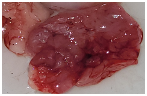 | Picture 1. Surgically resected specimen of salivary gland tumor. Histological appearance of adenocarcinoma, stage I, localized in the left parotid gland (3 cases; Nos. 849-b, 841-b, 482-b). The tumor shows irregular glandular and tubular structures lined by atypical epithelial cells with mild pleomorphism (H&E stain) |
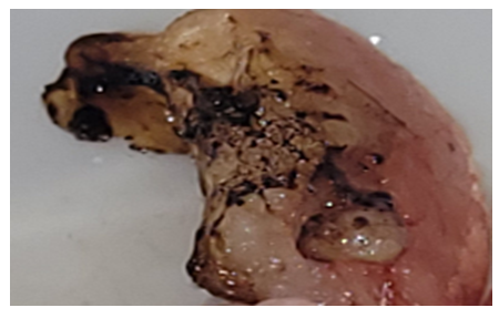 | Picture 2. Surgically resected specimen of salivary gland tumor. Histological appearance of pleomorphic adenoma (mixed tumor), localized in the left parotid gland (1 case; No. 2755). The tumor demonstrates biphasic morphology with epithelial and myoepithelial components embedded in a heterogeneous myxoid–chondroid stroma (H&E stain) |
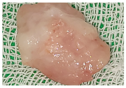 | Picture 3. Surgically resected specimen of salivary gland tumor. Histological appearance of monomorphic adenoma, localized in the left submandibular gland (1 case; No. 1101). The lesion shows uniform epithelial cell proliferation arranged in solid and trabecular patterns with sharp demarcation from the surrounding tissue (H&E stain) |
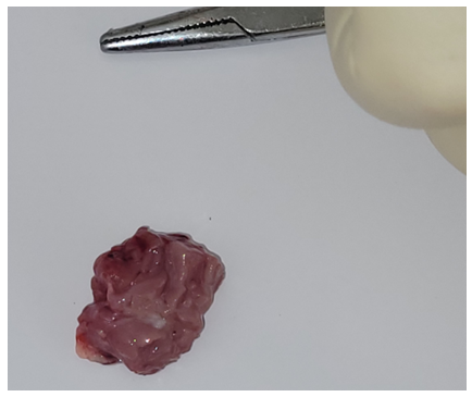 | Picture 4. Surgically resected specimens of salivary gland tumors. Histological appearance of undifferentiated carcinoma (4 cases), localized in the right palatal mucosa (Nos. 1297, 1298) and mandible (Nos. 1383, 1384). The tumor is composed of solid sheets and nests of atypical epithelial cells with a high nuclear–cytoplasmic ratio, marked nuclear pleomorphism, hyperchromasia, and absence of glandular differentiation (H&E stain) |
3. Results
- A total of 48 patients with salivary gland tumors were examined between 2023 and 2025. The cohort included 45 males and 3 females. The most frequent malignant neoplasms were undifferentiated carcinoma (4 cases) and adenocarcinoma, stage I (3 cases). Benign tumors included pleomorphic adenoma (1 case) and monomorphic adenoma (1 case).Histopathological examination revealed distinct morphological patterns characteristic of each tumor type. Representative photomicrographs are presented below.
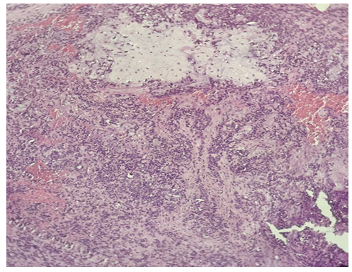
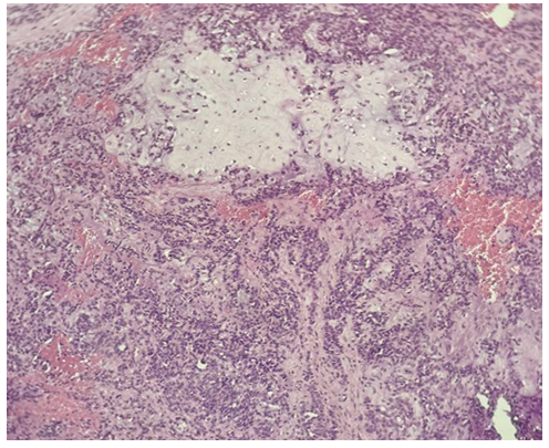
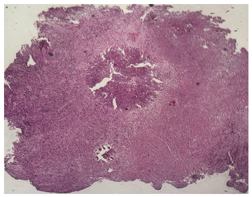 | Figure 1. Undifferentiated carcinoma (H&E stain, ×10) |
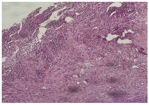 | Figure 2. Undifferentiated carcinoma (mandibular localization, H&E stain, ×10) |
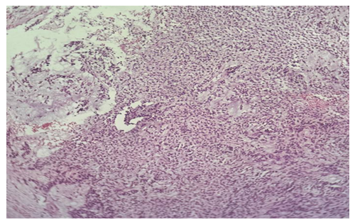
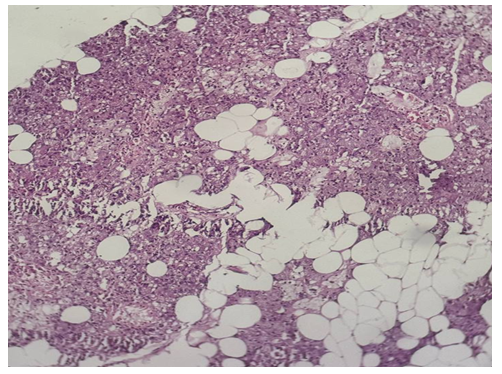 | Figure 3. Monomorphic adenoma (left submandibular gland, H&E stain, ×20) |
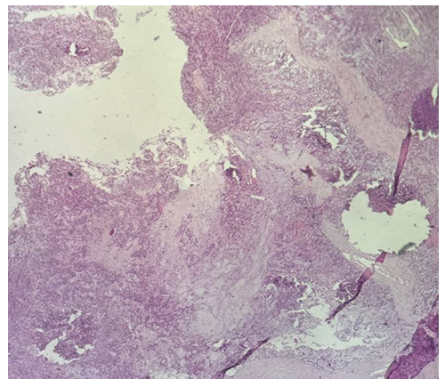 | Figure 4a. Pleomorphic adenoma |
 | Figure 4b. Pleomorphic adenoma |
 | Figure 4c. Pleomorphic adenoma |
 | Figure 4d. Pleomorphic adenoma |
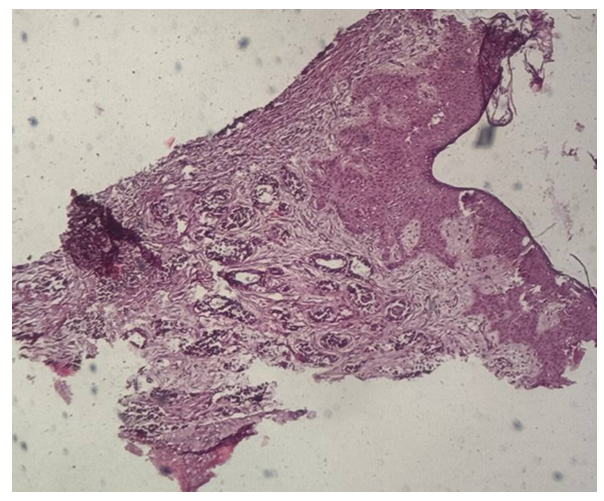 | Figure 5a. Adenocarcinoma, stage I |
4. Results and Discussion
- This study highlights the predominance of malignant tumors among salivary gland neoplasms in the Khorezm Region. The relatively high proportion of undifferentiated carcinoma (4/48) contrasts with global data, where pleomorphic adenoma typically dominates. These tumors demonstrated aggressive histological features, consistent with poor prognosis reported in international series.Adenocarcinoma, stage I was the second most frequent malignant tumor, showing early glandular differentiation but atypia consistent with malignant behavior. Literature estimates adenocarcinomas to represent 10-15% of malignant salivary gland tumors, aligning with our findings. Benign lesions were rare: only one pleomorphic adenoma and one monomorphic adenoma. Pleomorphic adenoma, despite being globally the most common benign salivary tumor, accounted for only 2.1% in our series. Monomorphic adenoma was likewise rare, consistent with reported global incidence.These results indicate a regional variation, possibly related to demographic, ecological, or healthcare access factors. Importantly, our analysis confirms that classical histopathology alone remains highly reliable in diagnosing salivary gland tumors, even in the absence of immunohistochemistry.
5. Conclusions
- 1. Salivary gland tumors in the Khorezm Region showed a predominance of malignant forms, mainly undifferentiated carcinoma and adenocarcinoma.2. Undifferentiated carcinoma demonstrated solid growth and marked atypia, confirming aggressive biological potential.3. Adenocarcinoma, stage I, displayed early malignant features with irregular glandular morphology.4. Pleomorphic and monomorphic adenomas were rarely identified, contrasting with global patterns.5. Morphological evaluation using conventional histology remains the cornerstone of diagnosis and provides reliable classification.