-
Paper Information
- Next Paper
- Previous Paper
- Paper Submission
-
Journal Information
- About This Journal
- Editorial Board
- Current Issue
- Archive
- Author Guidelines
- Contact Us
American Journal of Medicine and Medical Sciences
p-ISSN: 2165-901X e-ISSN: 2165-9036
2025; 15(10): 3695-3697
doi:10.5923/j.ajmms.20251510.88
Received: Oct. 8, 2025; Accepted: Oct. 22, 2025; Published: Oct. 31, 2025

Investigation of Immunohistochemical Changes in the Kidneys of 6-Month-Old White Outbred Rats Following 70% Acetic Acid-Induced Injury of the Gastrointestinal Tract
 Abstract
 Reference
 Full-Text PDF
 Full-text HTML
Muxammadiyeva Farida Ruzimurodovna
Department of Anatomy, Clinical Anatomy (OHTA), Bukhara State Medical Institute named after Abu Ali Ibn Sino, Bukhara, Uzbekistan
Correspondence to: Muxammadiyeva Farida Ruzimurodovna, Department of Anatomy, Clinical Anatomy (OHTA), Bukhara State Medical Institute named after Abu Ali Ibn Sino, Bukhara, Uzbekistan.
| Email: |  |
Copyright © 2025 The Author(s). Published by Scientific & Academic Publishing.
This work is licensed under the Creative Commons Attribution International License (CC BY).
http://creativecommons.org/licenses/by/4.0/

Even in the era of rapid development of information technology, mass media and social networks, the problems related to the consequences of acetic acid poisoning, which is widely used in kitchens and is used in everyday life for various purposes, from food storage to cleaning, remain relevant. Despite the widespread use of vinegar, high concentrations of acetic acid are a dangerous chemical, and cases of poisoning from it can become a serious health threat.
Keywords: Kidney,Acetic acid, Nephron
Cite this paper: Muxammadiyeva Farida Ruzimurodovna, Investigation of Immunohistochemical Changes in the Kidneys of 6-Month-Old White Outbred Rats Following 70% Acetic Acid-Induced Injury of the Gastrointestinal Tract, American Journal of Medicine and Medical Sciences, Vol. 15 No. 10, 2025, pp. 3695-3697. doi: 10.5923/j.ajmms.20251510.88.
1. Introduction
- The kidney is one of the organs that undergoes intense functional stress throughout a person's life and is a complex organ responsible for filtering waste products from the blood through urine production. It holds an increasingly important place in modern research.Additionally, the kidneys perform other vital functions, including maintaining homeostasis, regulating blood pressure, osmotic pressure, and acid-base balance. Reviewing their functions is undoubtedly important for advancing ideas about the significance of the kidney and its scienc [1,2,4].It has been proven that in adult and fetal kidneys, stress at the tissue level leads to morphological changes describing an "urgent adaptation" between interstitial relations, nephron segments, and different types of nephrons (subcapsular and juxtamedullary). These structural changes include signs of intensified filtration processes, alterations in blood filling of vessels in various zones of the kidney, similar tubulo-interstitial reactions, and disruption of the epithelial layer of nephron tubules [3,6,7].The development of reactive changes in the kidney, like in other organs, is a response to stress. H. Selye defines stress as "a nonspecific response of living matter to any demand, manifested by true morphological changes and adaptations in various organs [9]."Toxic nephropathy" refers to damage to the glomerular structures and tissues of the kidney. This condition primarily occurs due to impairment of the kidney’s detoxification (neutralization of harmful substances) and excretion (elimination) functions. Such damage happens under the influence of nephrotoxins that are either exogenous (originating from outside) or endogenous (produced within the body).The main representatives of nephrotoxins with an exogenous source are medicinal drugs, low-quality alcoholic products, and acetic acid. Poisoning with acetic acid occurs not only due to its specific tropic effect but also through its general toxic-stressor impact. Its specific effects on various internal organs have been well studied. In response to stressor exposure, stress-realizing hormones can be produced not only by the adren [8].Specific features of hemoglobinuric nephrosis have been identified in accidental or suicidal poisoning with acetic acid. But the degree of damage to the kidney parenchyma is more obvious if it is acute or chronic before poisoning. Stress - It is known that poisoning of various etiologies is accompanied by stress, but despite being toxic, the role of the stress factor in the pathogenesis of poisoning is very small. Stress has its own characteristics. In cases of accidental poisoning, there is no stress on the body before taking the toxic substance. In cases of suicide, poisoning also occurs during the anxiety phase in a state of passion or against the background of long and strong stress that causes the patient to become depressed and induces him to commit suicide [5].
2. Materials and Methods
- The research was conducted at the Department of Anatomy and Clinical Anatomy (OHTA) of the Bukhara State Medical Institute named after Abu Ali ibn Sina. The study involved 94 white outbred rats of both sexes. The rats were housed in standard metal cages (5 rats per cage) under controlled ambient conditions (24 ± 2°C) and a 12-hour light/dark cycle. They were maintained in quarantine for one week and only included in the experiment after confirming the absence of any somatic or infectious diseases. Throughout the study, the behavior and physiological status of the rats in both the control and experimental groups were closely monitored. To assess the morphological and morphometric characteristics of kidney development during postnatal ontogenesis, three age-based subgroups were established: 1-month-old, 3-month-old, and 6-month-old rats.
3. Results and Discussion
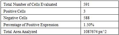
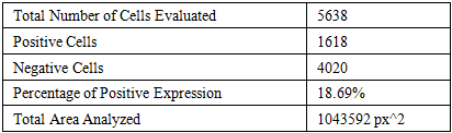
- E-cadherin is a pivotal protein responsible for maintaining epithelial cell-cell adhesion and is predominantly localized at the cell membrane. It plays a crucial role in preserving the integrity and polarization of epithelial cells, contributing significantly to the structural cohesion of organ tissues. E-cadherin is also involved in processes such as cell migration, invasion, and apoptosis. A decrease in E-cadherin expression is often associated with invasive characteristics, as observed in carcinoma cases.In six-month-old white outbred rats subjected to 70% acetic acid-induced burns of the gastrointestinal tract, followed by a treatment phase, the expression of E-cadherin was detected in kidney tissues. This suggests the preservation or potential restoration of epithelial cell-cell junctions.Severe chemical injuries to the gastrointestinal tract, particularly due to acetic acid exposure, result not only in local damage but also in systemic alterations. Such conditions can lead to endotoxicosis, adversely affecting distant vital organs, especially the kidneys. Therefore, investigating immunohistochemical changes in kidney tissues is essential for a comprehensive understanding of post-burn pathogenesis and for developing effective therapeutic strategies.
|
|
4. Conclusions
- Control Group Findings: In 6-month-old white outbred rats, the expression of the E-cadherin marker in kidney tissue was observed to be 10.12%. This indicates a moderate expression level, suggesting that epithelial cell integrity is maintained in the control group. Post-Acetic Acid Burn Findings: Following burns induced by a 70% acetic acid solution, the E-cadherin expression in kidney tissue decreased to 9.46%. This reduction may reflect early epithelial cell damage or alterations in cell-cell adhesion mechanisms. Post-Treatment Findings: After subsequent treatment, the E-cadherin expression increased to 12.59%. This elevation suggests a potential recovery or adaptation of epithelial cells, possibly indicating a response to injury and an attempt to restore cellular integrity.These findings underscore the dynamic nature of E-cadherin expression in response to gastrointestinal tract burns and subsequent treatment in 6-month-old white outbred rats. The observed changes highlight the importance of E-cadherin as a marker for epithelial cell integrity and its potential role in the pathophysiology and recovery processes following such injuries.