-
Paper Information
- Next Paper
- Previous Paper
- Paper Submission
-
Journal Information
- About This Journal
- Editorial Board
- Current Issue
- Archive
- Author Guidelines
- Contact Us
American Journal of Medicine and Medical Sciences
p-ISSN: 2165-901X e-ISSN: 2165-9036
2025; 15(10): 3655-3657
doi:10.5923/j.ajmms.20251510.79
Received: Sep. 27, 2025; Accepted: Oct. 22, 2025; Published: Oct. 31, 2025

Morphofunctional and Functionally Diagnostic Parallels of the Cecocolonic Sphincters in Children
 Abstract
 Reference
 Full-Text PDF
 Full-text HTML
Raupov Farkhod Sayidovich
Bukhara State Medical Institute named after Abu Ali Ibn Sino, Bukhara, Uzbekistan
Correspondence to: Raupov Farkhod Sayidovich, Bukhara State Medical Institute named after Abu Ali Ibn Sino, Bukhara, Uzbekistan.
| Email: |  |
Copyright © 2025 The Author(s). Published by Scientific & Academic Publishing.
This work is licensed under the Creative Commons Attribution International License (CC BY).
http://creativecommons.org/licenses/by/4.0/

It is well known that the sphincter apparatuses of the digestive tract are functionally active regions that play a crucial role in regulating the passage of food and chyme. The presence of sphincters divides the entire digestive tract into separate cavities (such as the oral cavity and stomach) or into sections within the same organ (such as the intestine). Each of these parts is characterized by its own osmotic pressure, intracavitary pressure, microflora, and specific role in maintaining internal homeostasis. The sphincters of the digestive system ensure the forward movement of contents while preventing reflux.
Keywords: Resection, Intestines, Children, Sphincter
Cite this paper: Raupov Farkhod Sayidovich, Morphofunctional and Functionally Diagnostic Parallels of the Cecocolonic Sphincters in Children, American Journal of Medicine and Medical Sciences, Vol. 15 No. 10, 2025, pp. 3655-3657. doi: 10.5923/j.ajmms.20251510.79.
1. Introduction
- Colon resection is a surgical procedure involving the removal of part of the colon. This operation must be performed with extreme care and responsibility, as complications occurring during or after surgery can cause significant harm to a child’s health. The importance of the large intestine extends beyond digestion, fecal formation, and excretion; it also plays roles in mineral metabolism and absorption of the liquid part of the chyme. Moreover, colonizing microorganisms synthesize certain vitamins and may contribute to additional postoperative complications [3,5,6].Surgical correction of colon pathologies in most cases is a necessary intervention, performed to treat congenital or acquired diseases of the gastrointestinal tract (GIT) in children. Such procedures must be approached cautiously, since any intraoperative or postoperative complication can seriously affect the patient’s health and life [2,8,12,16]. Despite advancements in colon resection techniques, postoperative complications remain a subject of ongoing debate among domestic and international experts. This highlights the need to refine surgical methods by considering the anatomical and physiological characteristics of the colon [1,4,8,10]. During surgical treatment of colon pathologies, such as abdominoperineal proctoplasty (STD) or transanal endorectal colon reduction (TNENTC), wide resections, intestinal shortening, or tension-related reductions often lead to a loss of normal intestinal physiology. In such cases, physiological bends (anorectal, rectosigmoid, sigmoid–descending) and sphincters (Bern–Piragova–Moutier, Rossi–Moutier, Bally) may be disrupted or removed, leading to fecal incontinence [1,9,17]. According to L.L. Kolesnikov, a “sphincter” is a specially organized muscular structure (smooth or striated) that regulates the amount and duration of communication between compartments (segments or parts) of a hollow organ [13,15]. Purpose of the Study. To study and compare the morphofunctional and radiological characteristics.
2. Materials and Methods
- To identify colon pathologies, irrigography and irrigoscopy were performed on 48 children aged 1 to 14 years. When interpreting the results, attention was given to the shape, location, and lumen diameter of the colon, the elasticity and extensibility of its wall, the relief of the mucous membrane, the function of the Bauhin’s valve, and the functional condition of different parts of the colon, including the sphincters. A 50% reduction in the colonic lumen at sphincter sites was interpreted as an anatomical narrowing and served as a radiological marker. The collected data allowed for the anatomical and functional interpretation of the colon sphincters’ X-ray appearance.
3. Results and Discussion
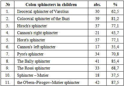
- According to several authors [14], physiological narrowing throughout the colon corresponds to the location of physiological sphincters. At the junction of the ileum and cecumlies the ileocecal sphincter of Varolius; at the border between the cecum and the ascending colon - the colocecal sphincter of Buzi; between the middle and upper thirds of the ascending colon - the Hirsch sphincter. Three sphincters are identified in the transverse colon: the right Cannon sphincter (near the hepatic flexure), the Horst sphincter (in the middle third), and the left Cannon sphincter (near the splenic flexure). The descending colon contains the Pyre and Bally sphincters, while the sigmoid colon includes the Rossi–Moutier sphincter (middle third) and the O’Bern–Pirogov–Moutier sphincter (distal third) (Figure 1, 2).
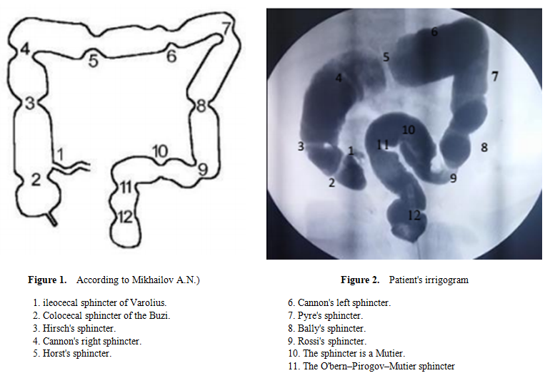 | Figure 1-2 |
|
4. Conclusions
- Thus, the detectability of colonic sphincters during irrigography varies. This variability is due to changes in the diameter of the lumen in sphincter zones as the intestinal contents move, which affects the frequency of their radiological visualization.