
-
Paper Information
- Next Paper
- Previous Paper
- Paper Submission
-
Journal Information
- About This Journal
- Editorial Board
- Current Issue
- Archive
- Author Guidelines
- Contact Us
American Journal of Medicine and Medical Sciences
p-ISSN: 2165-901X e-ISSN: 2165-9036
2025; 15(10): 3486-3489
doi:10.5923/j.ajmms.20251510.43
Received: Sep. 11, 2025; Accepted: Oct. 7, 2025; Published: Oct. 25, 2025

Morpho-Histochemical Evaluation of Thymic Mast Cells Under Energy Drink Influence
 Abstract
 Reference
 Full-Text PDF
 Full-text HTML
Rakhmonova Kamilla Erkinovna
Bukhara Branch of the Specialized Scientific and Practical Center of Oncology and Radiology, Bukhara, Uzbekistan
Correspondence to: Rakhmonova Kamilla Erkinovna, Bukhara Branch of the Specialized Scientific and Practical Center of Oncology and Radiology, Bukhara, Uzbekistan.
| Email: |  |
Copyright © 2025 The Author(s). Published by Scientific & Academic Publishing.
This work is licensed under the Creative Commons Attribution International License (CC BY).
http://creativecommons.org/licenses/by/4.0/

The aim of the study was to perform a morpho-histochemical assessment of thymus mast cells under the influence of an energy drink. The study object was the thymus tissues of animals from the control and experimental groups. Histochemical staining with toluidine blue was used to identify mast cells, allowing for the assessment of granulation degree and morphological condition of the cells. A comparative analysis of the quantitative and qualitative characteristics of mast cells between control samples and samples obtained from animals that received an energy drink was performed. The data obtained indicate the possibility of modifying the structure and functional state of the thymus mast cells under the influence of exogenous stimulants.
Keywords: Rat thymus, Energy drinks, Medulla, Cortex, Lymphocytes, Macrophages, Dendritic cells, Hassall's corpuscles, Mast cells
Cite this paper: Rakhmonova Kamilla Erkinovna, Morpho-Histochemical Evaluation of Thymic Mast Cells Under Energy Drink Influence, American Journal of Medicine and Medical Sciences, Vol. 15 No. 10, 2025, pp. 3486-3489. doi: 10.5923/j.ajmms.20251510.43.
1. Introduction
- Mast cells are granulocytes rich in biologically active substances such as histamine and heparin, which play a key role in immune defense, inflammation, angiogenesis and regulation of vascular permeability [1]. These cells were first described by Paul Ehrlich in 1878 based on their unique ability to metachromatic staining - staining different from the color of the dye itself [2]. Metachromatic staining with toluidine blue is one of the most common and accessible methods for detecting mast cells in tissues. It is based on the interaction of aniline dyes with sulfated glycosaminoglycans (mainly heparin) in mast cell granules, as a result of which the dye changes color, acquiring a violet-red hue (metachromasia), while the background remains blue (orthochromatic). The accuracy of the method depends on the pH of the solution, the duration of staining, the type of tissue and fixation [4].Modern studies have shown that various biologically active substances affect the functional state of mast cells. Thus, it was found that caffeine and catechin are able to reduce the degree of mast cell degranulation in a dose-dependent manner, stabilizing their condition. At the same time, low concentrations of catechin synergistically enhance the effect of caffeine - they almost completely inhibit mast cell exocytosis. Despite extensive study of individual components of such drinks, the effect of complex exogenous stimulants, in particular energy drinks, on tissues of lymphoid origin, including the thymus, remains poorly understood. It is known that the effect of energy drinks on the liver and kidneys of animals includes the development of degenerative changes, inflammatory infiltration and oxidative stress, but the effect on immune components, such as thymus mast cells, has been practically unstudied [5]. The morphofunctional state of the central organ largely determines the activity of secondary (peripheral) structures of immunogenesis and the severity of protective reactions of the whole organism. At present, knowledge of the structural features of the organs of the immune system and the determination of the onset of differentiation of immunocompetent cells at different stages of the ante- and postnatal periods of ontogenesis allows us to understand the processes of formation of immunological functions characteristic of these organs [3].The purpose of the studyOur research aim is to substantiate the significance of the study, disclose the methodological basis and review existing knowledge about the regulation of mast cells under the influence of exogenous stimulants.
2. Materials and Methods
- The object of the study were white rats. In the vivarium of the Bukhara Medical Institute named after Abu Ali Ibn Sina, 66 rats were examined in this group. The thymus of the rats was taken for the studies. An energy drink was used as a product in the experiments. The solution is administered intragastrically using a metal tube once a day in a total dose of 10 g / kg of body weight for 2 months. Rats of the experimental group aged 1 month reached 3 months of age, rats aged 3 months reached 5 months of age. Control animals received equal volumes of water intragastrically.The rats were divided into 3 groups (n = 209): I - animals of the control group aged 3 months (n = 34) and 5 months (n = 35); in the II experimental group, 3-month-old (n=35) and 5-month-old (n=33) infants consumed an energy drink for 2 months at a dose of 10 g/kg/day, the III group received EN and flaxseed oil for 2 months at a dose of 8 ml/kg/day, 3-month-old (n=34) and 5-month-old (n=38).Microscopic examination was used to study the morphological parameters of the thymus. Morphometric studies of animals were carried out on the basis of the conclusion of the Ethics Committee of the Ministry of Health of the Republic of Uzbekistan. No. 1560/13/02/25.The isolated organ was fixed in 10% neutral formalin solution, then the blocks were placed in an automated tissue processing station KD-TS3D1 Automatic Tissue Processor. The tissue was dehydrated, embedded in paraffin and thinly sectioned at 4 μm thickness using a Semi Automatic Rotary Microtome KD-3358. Histological staining of the tissue was performed with hemotaxillin-eosin dye using a KD-RS2 Automatic Slide Stainer. The obtained thymus sections were documented microscopically using a Nikon eclipse E200 MV research microscope using a LEICA ICC50 E color camera. The staining technique for the histochemical study was toluidine blue staining for mast cell detection. Toluidine blue is a basic metachromatic dye widely used in histochemistry to detect mast cells due to their ability to accumulate heparin and other acidic glycosaminoglycans in granules. Staining allows these cells to be distinguished from other structures due to metachromasia (dye color change).
3. Results and Discussion
- In the thymus, mast cells are localized mainly in the perivascular zones and connective tissue septa. The functional characteristics of mast cells allow us to identify pathological, physiological, allergic reactions that implement innate or adaptive immunity. Histochemical reaction with toluidine blue staining of 3-month-old rats of the control group. When staining the thymus tissue, mast cells are located chaotically, the number is from 6-9 in one field of vision, small in size, the structure is uneven with basophilic-granular inclusions, the nuclei are rounded and the location of mast cells in the organ parenchyma is mainly under the capsule and in trabeculae. The tissue stroma consists of loose connective tissue, the parenchyma is colored light blue. Figure 1 shows an image of the structure of the thymus of rats in the control group of 3 months of age. Histochemical reaction with toluidine blue staining of thymus tissue of 5-month-old rats in the control group showed that the thymus tissue is stained blue. In the organ parenchyma there are large granular mast cells 4-6 with moderate granularity of the cytoplasm, oval nuclei, the surface of the cells is uneven with a stellate membrane. Figure 2 shows mast cells of the thymus of rats at 5 months of age in the control group.
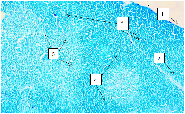 | Figure 1. Thymus structure of 3-month-old rats in the control group. 1-thymus capsule; 2-thymus trabecula; 3-single mast cells with a rounded shape, clear contours located in loose connective tissue; 4-thymus cortex with densely located lymphocytes; 5-medulla is lighter with large lymphocytes; Method: toluidine blue staining. ok. 10 x ob. 10 |
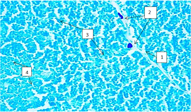 | Figure 2. Mast cells in the thymus of 5-month-old rats in the control group. 1-thymus trabecula; 2-mast cells; 3-cortex; 4-medulla; Method: toluidine blue staining. ok. 20 x ob. 40 |
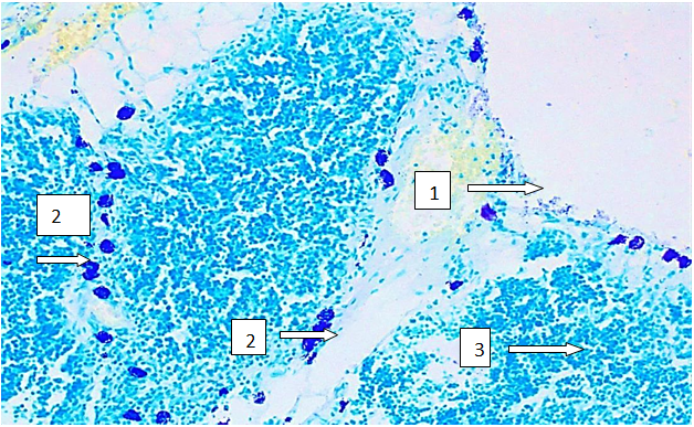 | Figure 3. Increased mast cells in the thymus stroma in the experimental group of 3-month-old rats. 1-thymus capsule; 2-mast cells with increased granularity and granularity in the cytoplasm; 3-thymus parenchyma; Toluidine blue staining technique. Ok. 10x ob. 20 |
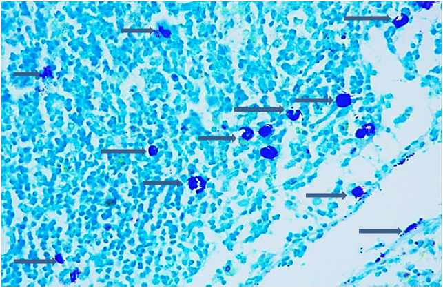 | Figure 4. Thymus of 5-month-old rats, experimental group. Mast cells, stained dark purple with increased granularity in the cytoplasm, with an unclear outline of the nuclei and fine-grained granules scattered along the periphery of the tissue. Staining with toluidine blue. ok. 10x ob. 40 |
4. Conclusions
- Histochemical approaches to assessing the regulation of mast cells in the thymus tissue in the control and experimental groups were varied. In the control group, the number of mast cells was calibrated within 3-9 in the field of view, and their structural granularity was preserved. In the experimental group, the number of mast cells increased sharply and reached 8-20 in the field of view, the structure of mast cells was disrupted and granularity in the cytoplasm increased. In the experimental group at the age of 3 months, the greatest increase in mast cells was observed - 40.8%, and at the age of 5 months - 24.4% in relation to the control group. In the group with the correction of linseed oil at the age of 3 months, a decrease in the number of mast cells from 6-10 was observed, which were stained purple, basophilic granularity of the cytoplasm, which may indicate a decrease in the activity of degranulation, at the age of 5 months in this group, the parenchyma of the tissue was stained blue, mast granular cells decreased and the number varied from 7-10, that these changes are probably due to the antioxidant and membrane-stabilizing effect of the components of linseed oil.