
-
Paper Information
- Next Paper
- Previous Paper
- Paper Submission
-
Journal Information
- About This Journal
- Editorial Board
- Current Issue
- Archive
- Author Guidelines
- Contact Us
American Journal of Medicine and Medical Sciences
p-ISSN: 2165-901X e-ISSN: 2165-9036
2025; 15(10): 3480-3485
doi:10.5923/j.ajmms.20251510.42
Received: Sep. 16, 2025; Accepted: Oct. 10, 2025; Published: Oct. 25, 2025

Reproductive Tract Microbiota Dysbiosis and Hormonal Imbalance: Interrelation in the Pathogenesis of Infertility
 Abstract
 Reference
 Full-Text PDF
 Full-text HTML
Jurayeva A. Zh., Shukurov F. I.
Tashkent State Medical University, Tashkent, Uzbekistan
Correspondence to: Shukurov F. I., Tashkent State Medical University, Tashkent, Uzbekistan.
| Email: |  |
Copyright © 2025 The Author(s). Published by Scientific & Academic Publishing.
This work is licensed under the Creative Commons Attribution International License (CC BY).
http://creativecommons.org/licenses/by/4.0/

Background. Infertility in reproductive-aged women is a major global health issue, with microbial dysbiosis and hormonal imbalance playing key roles in its pathogenesis. Objective. To evaluate the relationship between localization of reproductive tract microbiota dysbiosis and hormonal profile in infertile women. Materials and Methods. A total of 120 infertile women aged 20–40 years were enrolled. Four groups were formed: vaginal–cervical dysbiosis (n=30), endometrial–tubal dysbiosis (n=30), ovarian dysbiosis (n=30), and a healthy control group (n=30). Microbiota composition was assessed by 16S rRNA sequencing, while serum hormones (FSH, LH, E₂, progesterone, AMH, prolactin) were measured using the Immulite 2000 XPI immunoassay system. Results. Ovarian dysbiosis was associated with increased FSH (10.6 mIU/mL) and LH (8.1 mIU/mL), decreased AMH (1.12 ng/mL), estradiol (142 pg/mL), progesterone (8.7 ng/mL), and elevated prolactin (20.8 ng/mL) (p<0.001). Endometrial–tubal dysbiosis correlated with low progesterone, while vaginal–cervical dysbiosis was linked to hyperprolactinemia. Conclusion. The closer the dysbiosis is localized to the ovaries, the more pronounced are hormonal disturbances, ovarian reserve depletion, and reduced fertility.
Keywords: Reproductive system, Microbiota, Dysbiosis, Hormonal imbalance, Infertility, Ovarian reserve
Cite this paper: Jurayeva A. Zh., Shukurov F. I., Reproductive Tract Microbiota Dysbiosis and Hormonal Imbalance: Interrelation in the Pathogenesis of Infertility, American Journal of Medicine and Medical Sciences, Vol. 15 No. 10, 2025, pp. 3480-3485. doi: 10.5923/j.ajmms.20251510.42.
Article Outline
1. Introduction
- Infertility is considered one of the most pressing challenges facing global healthcare systems in women of reproductive age. According to the World Health Organization (WHO), approximately 15% of reproductive-age couples worldwide experience various forms of fertility impairment, with more than half of these cases attributed to female health factors [1,2]. The main causes of infertility include ovulatory dysfunction, endometrial dysfunction, tubal-peritoneal factors, as well as endocrine and immunological imbalances [3–5].In recent years, research on the female reproductive tract microbiota has been rapidly advancing. While earlier studies were primarily focused on the vaginal microbiota, current investigations highlight the importance of cervical, endometrial, tubal, and even ovarian microbiota in determining fertility potential [6–8]. Disturbance of microbial balance (dysbiosis) has been widely associated with reduced endometrial receptivity, disrupted folliculogenesis, impaired immuno-hormonal homeostasis, and diminished implantation success [9–11].Molecular and clinical studies have demonstrated a close interrelationship between microbiota alterations and hormonal imbalance. Specifically, an overrepresentation of conditionally pathogenic species in the ovarian microbiota has been linked to diminished ovarian reserve, elevated follicle-stimulating hormone (FSH) levels, and decreased anti-Müllerian hormone (AMH) concentrations [12,13]. Dysbiosis within the endometrial and tubal microbiota has been correlated with decreased progesterone levels and impaired endometrial receptivity [14,15]. Likewise, alterations in the vaginal and cervical microbiota have been associated with hyperprolactinemia, potentially leading to luteal phase hormonal imbalance [16].There is growing evidence that the closer the localization of microbiota dysbiosis is to the ovary, the more pronounced the hormonal disturbances and disruption of the hypothalamic–pituitary–ovarian axis. This condition has been linked to decreased ovarian reserve, ovulatory dysfunction, and impaired reproductive capacity [17–20]. Therefore, simultaneous evaluation of microbiota status and hormonal homeostasis is of critical importance for a deeper understanding of infertility pathogenesis, as well as for the development of individualized therapeutic and preventive strategies.
2. Purpose of the Research
- The aim of this study was to evaluate the relationship between the localization of microbiota dysbiosis and the hormonal profile in infertile women of reproductive age, and to determine the role of these factors in the pathogenesis of infertility.
3. Materials and Methods
- This prospective cross-sectional study was conducted at the Gynecology Clinic of Tashkent State Medical University between 2022 and 2024 and included 120 infertile women of reproductive age (20–40 years). Inclusion criteria were age between 18 and 40 years, inability to conceive after at least 12 months of unprotected sexual intercourse, and infertility confirmed clinically and by laboratory testing. Exclusion criteria comprised recent use of antibiotics or hormonal therapy, sexually transmitted infections, and severe somatic or oncological diseases. Based on the anatomical site of microbiota dysbiosis, participants were divided into four groups: Group I – vaginal and cervical canal dysbiosis (n=30), Group II – endometrial and tubal dysbiosis (n=30), Group III – ovarian dysbiosis (n=30), and a control group of healthy women without dysbiosis (n=30).Biological samples were collected under aseptic conditions using sterile instruments. Vaginal and cervical swabs, endometrial aspirates, fallopian tube lavages, and follicular fluid samples were obtained during diagnostic and therapeutic gynecological procedures. For the control group, only non-invasive vaginal–cervical swabs were collected during routine examinations. All participants provided written informed consent prior to sample collection. Ethical approval was obtained from the Institutional Review Board of Tashkent State Medical University (Approval No. 07/2024).Serum levels of follicle-stimulating hormone (FSH), luteinizing hormone (LH), estradiol (E₂), progesterone, anti-Müllerian hormone (AMH), and prolactin (PRL) were measured using the automated immunochemiluminescence analyzer Immulite 2000 XPI (Siemens, Germany). For microbiological evaluation, DNA was extracted using the Qiagen DNA Mini Kit (Qiagen, Germany) according to the manufacturer’s protocol. The V3–V4 hypervariable regions of the 16S rRNA gene were amplified using universal primers 341F (5′-CCTACGGGNGGCWGCAG-3′) and 806R (5′-GACTACHVGGGTATCTAATCC-3′), and the products were sequenced on the Illumina MiSeq platform (Illumina, USA) with paired-end reads (2 × 300 bp). PCR reactions were carried out in 25 µL volumes containing 10 ng of template DNA. The amplification protocol included an initial denaturation at 95°C for 3 minutes, followed by 30 cycles of 95°C for 30 seconds, 55°C for 30 seconds, 72°C for 45 seconds, and a final extension at 72°C for 5 minutes. PCR products were purified using the AMPure XP magnetic bead system (Beckman Coulter, USA), and library quality was verified with a Qubit 4.0 fluorometer (Thermo Fisher Scientific, USA).Raw sequencing data were processed using the QIIME2 pipeline, with quality filtering, trimming, denoising, and clustering of reads into operational taxonomic units (OTUs) at 97% similarity. Taxonomic assignment was carried out using the SILVA reference database (v.138). Alpha-diversity was evaluated using the Shannon index and Chao1 richness, while beta-diversity was assessed using Bray–Curtis dissimilarity and principal coordinate analysis (PCoA). Relative abundance at the phylum, genus, and species levels was calculated, and dysbiosis was defined as deviation from control microbial profiles.All procedures were approved by the Institutional Review Board (Approval No. 07/2024) and performed in accordance with the Declaration of Helsinki. No invasive procedures were carried out in healthy volunteers; only non-invasive sampling was used.Statistical analysis was performed using SPSS version 26.0. The Kolmogorov–Smirnov test was applied to assess data normality. Group differences were evaluated using Student’s t-test and the χ² test, while correlations were analyzed with Pearson’s correlation coefficient. A two-tailed p value of <0.05 was considered statistically significant.
4. Results and Discussion
- The hormonal profile of all participants was evaluated by measuring serum levels of follicle-stimulating hormone (FSH), luteinizing hormone (LH), estradiol (E₂), progesterone, anti-Müllerian hormone (AMH), and prolactin (PRL). Analysis revealed statistically significant differences between the study groups.In addition to hormonal evaluation, microbiota analysis was performed across all studied groups. Sequencing results revealed significant differences in microbial composition and diversity depending on dysbiosis localization. Alpha-diversity (Shannon and Chao1 indices) decreased progressively from the vaginal–cervical to ovarian sites, while beta-diversity analysis (Bray–Curtis dissimilarity, PCoA) showed distinct clustering of microbiota communities by anatomical site. The dominant genera included Lactobacillus, Gardnerella, Prevotella, and Atopobium, with an increased relative abundance of conditionally pathogenic taxa in the ovarian dysbiosis group. These findings are presented in Supplementary Table S1 and Figures 1–3.
|
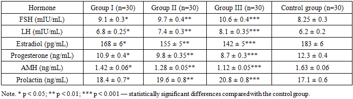
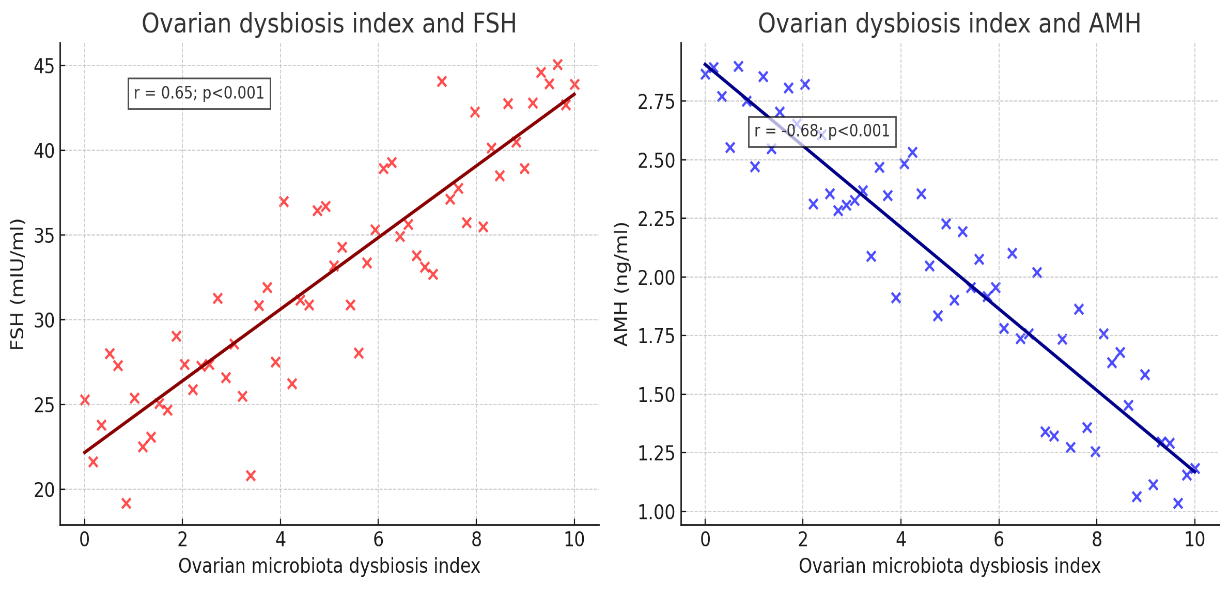 | Figure 1. Correlation between ovarian dysbiosis and hormonal parameters |
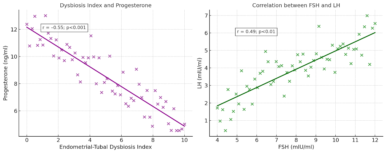 | Figure 2. Correlation between endometrial/tubal dysbiosis and hormonal parameters |
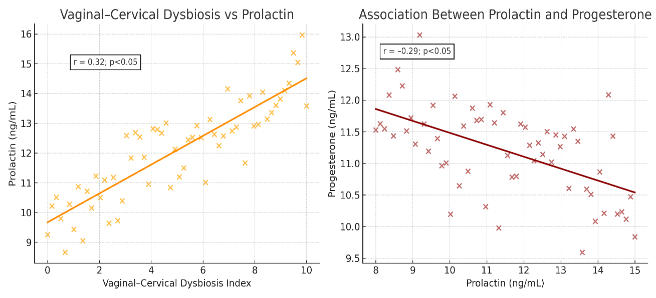 | Figure 3. Correlation between vaginal–cervical dysbiosis and hormones |
5. Discussion
- The findings of this study confirm that the localization of microbiota dysbiosis within the female reproductive tract is closely associated with specific patterns of hormonal imbalance in infertile women. We demonstrated that ovarian dysbiosis showed a strong positive correlation with elevated FSH levels and a strong negative correlation with AMH, indicating diminished ovarian reserve and altered hypothalamic–pituitary–ovarian axis regulation. These observations are in line with recent studies suggesting that microbial colonization of the ovaries may contribute to impaired folliculogenesis and reduced fertility potential [17].Endometrial and tubal dysbiosis demonstrated a moderate negative correlation with progesterone levels, reflecting luteal phase insufficiency and decreased endometrial receptivity. This supports previous evidence that the endometrial microbiota directly influences implantation success or failure and plays a critical role in the maintenance of reproductive function [15,18]. Similarly, disturbances in the uterine microbial environment have been linked to infertility, implantation failure, and chronic endometritis, emphasizing the pathogenic significance of microbiota alterations in this region [5,19].In addition, vaginal and cervical microbiota dysbiosis showed a weak but significant positive association with prolactin levels, combined with an inverse correlation with progesterone. This aligns with reports that vaginal microbiota imbalance contributes to reproductive dysfunction, pregnancy complications, and may modulate systemic endocrine balance [9,16,20]. Collectively, these results highlight that the closer dysbiosis is localized to the ovaries, the more severe the hormonal disturbances become, manifesting in reduced estradiol and progesterone levels and increased gonadotropin activity.The novelty of our study lies in the integrative evaluation of microbiota localization and hormonal profiles in infertile women, representing one of the first comprehensive investigations of this type in the Central Asian region. While previous research has described associations between vaginal, uterine, or ovarian microbiota and infertility [13,14], our data provide new insights into the stepwise relationship between the anatomical site of dysbiosis and its hormonal consequences. This integrated perspective strengthens the concept that reproductive tract microbiota is a key determinant of endocrine homeostasis and reproductive potential.Clinically, these findings emphasize the need to incorporate microbiota assessment into the diagnostic work-up of infertility, alongside hormonal and echographic evaluation. Identifying the localization of dysbiosis may help stratify patients by risk of ovarian reserve decline, luteal phase insufficiency, or impaired endometrial receptivity. Furthermore, the data suggest that microbiota-targeted therapies, when combined with hormonal correction, could form the basis of individualized and more effective infertility treatment strategies [6,15].
6. Conclusions
- The study demonstrated that dysbiosis of the female reproductive tract microbiota is closely associated with hormonal imbalance, with endocrine alterations becoming more pronounced as the localization of dysbiosis approaches the ovaries. In women with ovarian dysbiosis, significant increases in FSH (10.6 mIU/ml) and LH (8.1 mIU/ml) levels were observed, while AMH (1.12 ng/ml), estradiol (142 pg/ml), and progesterone (8.7 ng/ml) levels were decreased, accompanied by elevated prolactin (20.8 ng/ml) (p<0.001). Endometrial-tubal dysbiosis was associated with reduced progesterone levels (9.8 ng/ml), whereas vaginal-cervical dysbiosis was linked to elevated prolactin concentrations (18.4 ng/ml). These findings highlight the progressive relationship between microbiota dysbiosis and hormonal imbalance. The closer the dysbiosis is localized to the ovaries, the more severe the endocrine disturbances and decline in fertility become. This provides a rationale for individualized diagnostic and therapeutic strategies integrating microbiota and hormonal assessment.