
-
Paper Information
- Next Paper
- Previous Paper
- Paper Submission
-
Journal Information
- About This Journal
- Editorial Board
- Current Issue
- Archive
- Author Guidelines
- Contact Us
American Journal of Medicine and Medical Sciences
p-ISSN: 2165-901X e-ISSN: 2165-9036
2025; 15(10): 3474-3479
doi:10.5923/j.ajmms.20251510.41
Received: Sep. 16, 2025; Accepted: Oct. 10, 2025; Published: Oct. 25, 2025

Diagnostic Value of Sonographic and Doppler Parameters in Infertile Women with Endometrial Dysfunction
 Abstract
 Reference
 Full-Text PDF
 Full-text HTML
Jalolova G. S., Shukurov F. I.
Tashkent State Medical University, Tashkent, Uzbekistan
Correspondence to: Shukurov F. I., Tashkent State Medical University, Tashkent, Uzbekistan.
| Email: |  |
Copyright © 2025 The Author(s). Published by Scientific & Academic Publishing.
This work is licensed under the Creative Commons Attribution International License (CC BY).
http://creativecommons.org/licenses/by/4.0/

Introduction. Infertility affects 10–15% of reproductive-age couples, with endometrial dysfunction being a significant contributing factor. Morphological and functional alterations reduce endometrial receptivity and impair implantation outcomes. Objective. To evaluate the diagnostic value of sonographic and Doppler parameters in infertile women with endometrial dysfunction. Materials and Methods. A prospective study included 120 women: Group I – 40 infertile women of early reproductive age, Group II – 50 infertile women of late reproductive age, and Control – 30 healthy women. Endometrial thickness, echogenicity, structure, and blood flow indices (RI, PI) were assessed. Results. Endometrial thickness was 6.2±0.21 mm and 5.7±0.20 mm in patients vs. 9.1±0.28 mm in controls (p<0.001). Hypoplasia was present in 100%, increased echogenicity in 90–92%, stromal fibrosis in 72.5–76%. Elevated RI (≥0.65) was observed in 42.5% and 54%, PI (≥1.4) in 37.5% and 48%. A strong negative correlation was found between endometrial thickness and RI (r=–0.61; p<0.001). Conclusion. Sonographic and Doppler parameters provide reliable diagnostic markers for endometrial dysfunction, supporting prediction of implantation outcomes and personalized infertility treatment strategies.
Keywords: Endometrial dysfunction, Infertility, Sonography, Doppler, Receptivity, RI, PI
Cite this paper: Jalolova G. S., Shukurov F. I., Diagnostic Value of Sonographic and Doppler Parameters in Infertile Women with Endometrial Dysfunction, American Journal of Medicine and Medical Sciences, Vol. 15 No. 10, 2025, pp. 3474-3479. doi: 10.5923/j.ajmms.20251510.41.
Article Outline
1. Introduction
- Infertility is one of the most pressing issues in modern gynecology and reproductive medicine, and according to various epidemiological data, it is observed in approximately 10–15% of couples of reproductive age [1,2]. Among the main causes of infertility, in addition to ovulatory dysfunctions, tubal-peritoneal factors, and endocrine disorders, particular importance is attributed to endometrial dysfunction [3,4]. The endometrium, through normal cyclic changes and the development of receptivity, represents the key tissue in preparing for pregnancy. When its physiological function is impaired, implantation failure and infertility may occur [5].Endometrial receptivity, particularly during the post-ovulatory phase, is ensured by secretory transformation and adequate blood circulation. Morphofunctional alterations in these processes can hinder the establishment of pregnancy [6,7]. According to the World Health Organization (WHO) and the FIGO classification, endometrial dysfunction is recognized as an independent cause of infertility, with its prevalence ranging from 30% to 60% across different countries [8].Clinically, infertility associated with endometrial dysfunction is closely linked to menstrual irregularities, anovulation, echographic hypoplasia, the loss of endometrial receptivity markers, as well as repeated instrumental interventions [9,10]. Furthermore, chronic endometritis and subclinical inflammation aggravate functional alterations in the endometrium [11,12]. As a result, hypoplasia, stromal fibrosis, pathological echogenicity, and disturbances in vascular structures are frequently observed in the endometrium [13,14].At present, although hormonal and immunomodulatory therapies are employed in the treatment of infertility associated with endometrial dysfunction, their effectiveness remains limited, with some studies reporting success rates not exceeding 20–30% [15,16]. Therefore, a more in-depth evaluation of endometrial morphology and blood circulation, particularly through echographic and Dopplerometric techniques, has become of critical importance [17].In recent years, ultrasound and Dopplerometric methods have been increasingly applied in the evaluation of endometrial receptivity. Parameters such as endometrial thickness, structural characteristics, echogenicity, and blood flow indices (resistance index – RI, pulsatility index – PI) are recommended as reliable markers of pregnancy preparedness and implantation success [18–20]. It has been established that an endometrial thickness of ≥8 mm is associated with a higher probability of pregnancy, whereas elevated RI and PI values are linked to reduced receptivity [21]. Moreover, in infertile women with endometrial dysfunction, echographic hypoplasia and impaired blood flow have been reported to decrease implantation efficiency by 3–4 times [22].In this regard, an in-depth investigation of the diagnostic value of echographic and Dopplerometric parameters in infertile women with endometrial dysfunction represents an important step in elucidating the pathogenesis of infertility, developing individualized preventive measures, and advancing innovative therapeutic approaches.
2. Purpose of the Research
- The aim of the study was to evaluate echographic and Dopplerometric indicators in infertile women with endometrial dysfunction and to determine their diagnostic significance.
3. Materials and Methods
- This study had a prospective clinical design and included a total of 120 women of reproductive age. Based on clinical and instrumental parameters, participants were divided into three groups: Group I – 40 infertile women aged 18–35 years with endometrial dysfunction (early reproductive age); Group II – 50 infertile women aged 36–40 years with endometrial dysfunction (late reproductive age); and the comparison group – 30 healthy women of reproductive age, whose health status was confirmed through clinical and laboratory examinations.Inclusion criteria were defined as: age between 18 and 40 years, inability to achieve pregnancy for at least 12 months, and presence of endometrial dysfunction confirmed by hormonal and echographic evaluations. Exclusion criteria included severe somatic or oncological diseases, tubal-peritoneal infertility, and congenital anomalies of the reproductive organs.Echographic examinations were performed in the luteal phase of the menstrual cycle using a transvaginal probe (7.5 MHz frequency). Endometrial thickness (mm), structural features, echogenicity, and the presence of the “triple-line” sign were assessed. Additional parameters such as the condition of the functional-glandular layer, signs of hypoplasia, stromal fibrosis, micropolyposis, and perimetrial changes were recorded.Dopplerometric assessment was conducted by evaluating blood flow in the spiral arteries and subendometrial circulation. The resistance index (RI) and pulsatility index (PI) were measured, with deviations from normal values considered at RI ≥0.65 and PI ≥1.4. In patients with reduced endometrial perfusion, the frequency of morphological changes (hypoplasia, fibrosis, pathological echogenicity) was comparatively analyzed.All laboratory and instrumental studies were performed according to standard protocols. The hormonal profile (FSH, LH, estradiol, prolactin, AMH) was assessed using the immunochemiluminescence method. The study was conducted in accordance with WHO and FIGO clinical recommendations.Statistical analysis was carried out using Microsoft Excel and SPSS Statistics version 26.0. Mean values (M±m) were calculated, and differences between groups were evaluated using Student’s t-test and the χ² test. Pearson’s method was applied to determine correlation coefficients. Risk factors were assessed using odds ratios (OR) and 95% confidence intervals (CI). A p-value of <0.05 was considered statistically significant.
4. Results and Discussion
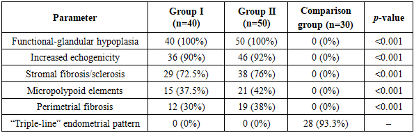
- According to the study findings, all patients with infertility associated with endometrial dysfunction (Group I – 40 women, Group II – 50 women) demonstrated a significant reduction in endometrial thickness during the luteal phase of the cycle. The mean values were 6.2 ± 0.21 mm in Group I and 5.7 ± 0.20 mm in Group II, which were markedly and statistically significantly lower compared with healthy women in the comparison group (9.1 ± 0.28 mm; p<0.001). This observation once again confirms that physiological endometrial thickness plays a decisive role in enhancing implantation and pregnancy potential.Echosonographic analysis revealed a high prevalence of clinically significant pathological changes in all infertile women with endometrial dysfunction. In particular, functional-glandular layer hypoplasia was detected in 100% of patients in both Group I (40/40) and Group II (50/50). Increased endometrial echogenicity was identified in 36 patients (90%) in Group I and 46 patients (92%) in Group II; in most cases, this alteration was associated with stromal fibrosis, inflammation, and trophic changes. Stromal fibrosis and sclerosis were observed in 29 women (72.5%) in Group I and 38 women (76%) in Group II. Micropolypoid formations and vascular “depressions” were noted in 15 women (37.5%) in Group I and 21 women (42%) in Group II. In addition, perimetrial fibrosis was documented in 12 women (30%) in Group I and 19 women (38%) in Group II.These findings indicate that distinct pathological changes in the morphological and functional state of the endometrium may reduce the degree of readiness for implantation and thereby contribute to the persistence of infertility.In the comparison group of healthy women (n = 30), endometrial thickness, structure, and echogenicity fully corresponded to physiological norms; no pathological changes were identified on echosonographic analysis. In 28 women (93.3%), a “triple-line” endometrial pattern was observed, while no cases of micropolyposis, fibrosis, or pathological echogenicity were detected (Table 1).
|
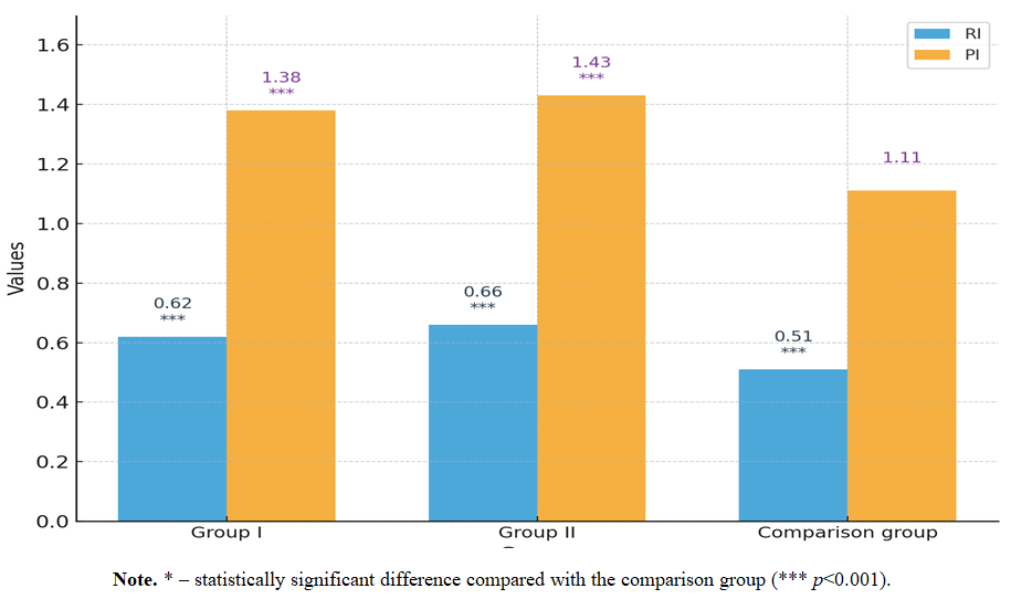 | Figure 1. Dopplerometric parameters in infertile women with endometrial dysfunction |
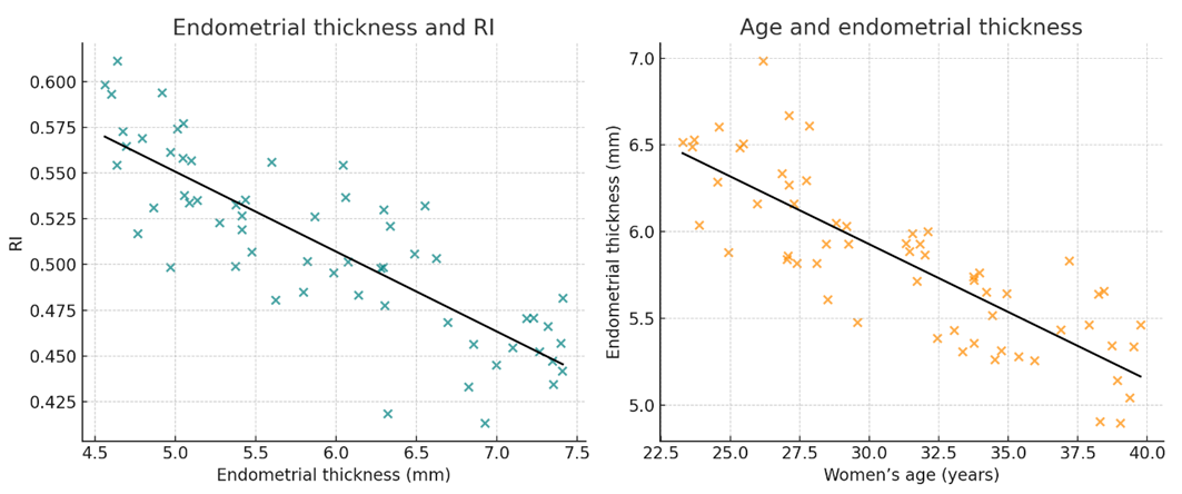 | Figure 2. Correlation between endometrial thickness, vascularization, and resistance index (RI) in infertile women with endometrial dysfunction |
5. Discussion
- The present study confirms that infertile women with endometrial dysfunction display a high prevalence of pathological echographic and Dopplerometric findings, including reduced endometrial thickness, increased echogenicity, hypoplasia, stromal fibrosis, and impaired vascularization. These observations are in agreement with earlier reports, where morphological alterations of the endometrium were shown to compromise implantation potential and reproductive outcomes (Wang et al., 2020; Motovilova et al., 2024).Importantly, our data highlight that elevated resistance index (RI) and pulsatility index (PI) values are strongly associated with poor perfusion and reduced receptivity. This finding supports previous Doppler-based studies, which identified abnormal uterine and subendometrial blood flow as predictors of implantation failure (Volovsky & Seifer, 2024). However, by demonstrating a strong inverse correlation between RI and endometrial thickness (r = –0.61; p<0.001), as well as an age-dependent decline in endometrial morphology and vascularization (r = –0.54; p<0.01), our results provide additional evidence of the integrative role of hemodynamic and morphological markers in predicting fertility outcomes.Recent advances in reproductive biology have introduced molecular and genomic approaches for assessing endometrial receptivity. Transcriptomic profiling (Díaz-Gimeno et al., 2011), microRNA-based biomarkers (Burns et al., 2023), and endometrial microbiota characterization (Ni et al., 2019; Fan et al., 2021) offer complementary strategies to identify implantation failure. Nevertheless, these technologies remain costly and not widely available. In contrast, sonographic and Dopplerometric assessments are inexpensive, reproducible, and can be integrated into routine gynecological practice. Our findings thus underscore the clinical utility of these non-invasive tools in settings where advanced molecular diagnostics are not accessible.The novelty of this study lies in the combined evaluation of echographic and Dopplerometric markers in a prospective cohort of infertile women stratified by reproductive age. By establishing clear associations between morphological changes, vascular parameters, and fertility potential, our work bridges the gap between traditional ultrasound assessment and emerging molecular approaches. This integrative perspective may aid in developing individualized treatment strategies, including patient stratification before assisted reproductive technology (ART) cycles.In summary, the present results strengthen the evidence that combined sonographic and Doppler evaluation of the endometrium is a reliable diagnostic approach to predict implantation outcomes. When integrated with modern molecular research, these parameters may serve as a cornerstone for precision medicine in reproductive care.The novelty of our study lies in the fact that it is the first prospective clinical investigation in the Central Asian region to comprehensively evaluate both echographic and Dopplerometric markers in infertile women with endometrial dysfunction. By integrating morphological assessment with hemodynamic parameters and performing correlation analysis, our work provides unique region-specific insights and strengthens the global evidence base on endometrial receptivity.
6. Conclusions
- This study confirms that infertile women with endometrial dysfunction exhibit marked pathological echographic and Dopplerometric changes, including reduced endometrial thickness, hypoplasia, stromal fibrosis, increased echogenicity, and impaired vascularization. Elevated RI and PI values were closely linked to poor perfusion and diminished receptivity, while correlation analysis highlighted the inverse association between endometrial thickness and RI, as well as age-related declines in morphology and blood flow.Taken together, these findings demonstrate that integrated sonographic and Doppler evaluation provides reliable, non-invasive markers for predicting implantation success and tailoring individualized infertility management. Early and accurate assessment of endometrial status is therefore essential for implementing innovative, personalized strategies to improve fertility outcomes.